Clostridium piliforme Infection: Tyzzer’s Disease
Epizootics of Tyzzer's disease have been observed in Syrian hamsters in various parts of the world. The causative agent, C. piliforme, is a spore-forming bacillus that multiplies only within cells.
The organism has a wide host range, including all of the species covered in this book, but isolates from various host species have been found to have host specificity.Diagnosis
Clostridium difficile should be recoverable on anaerobic culture and its presence confirmed by PCR, but detection of toxin is essential to confirm cause. Due to the importance of this disease in humans, there are a number of commercially available assays for detection of C. difficile A and B cytotoxins, using fresh intestinal contents or feces. Differential diagnoses include Tyzzer's disease, salmonellosis, and enteropathogenic E. coli infections.
Cecal Mucosal Hyperplasla of Unknown Etiology
Spontaneous cases of cecal hyperplasia have been observed in suckling and weanling hamsters. Diarrhea, runting, and high mortality were associated with the disease. At necropsy, ceca are congested, contracted, and opaque due to mucosal thickening (Fig 3.10).
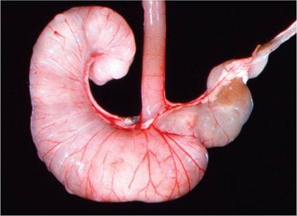
FIG. 3.10. Cecal mucosal hyperplasia in a hamster. This syndrome is probably the aftermath of clostridial enteropathy. Source: Barthold et al. 1987. Reproduced with permission from American Association for Laboratory Animal Science.
Epizootiology and Pathogenesis
Hamsters may become infected by contact with affected animals or by contaminated bedding, which remain infectious with environmentally resistant spores for many months. Predisposing factors, such as poor sanitation, intestinal parasitism, and inappropriate feeding practices, play a role in precipitating clinical outbreaks of the disease. Weanling hamsters are most often affected.
In hamsters inoculated with infected liver homogenates, organisms, and lesions are detectable in the mucosa of the small and large intestine by 3 days postinoculation, and multiple lesions and bacilli may be present in the liver by day 6-8 postexposure.Pathology
At necropsy, there is a variable distribution of lesions. In some epizootics, lesions may be confined to either the liver or the intestinal tract. Multifocal hepatic necrosis is evident in some cases. Intestinal lesions, when evident grossly, usually involve the lower ileum, cecum, and colon and are associated with diarrhea and soiling of the perineum. Affected areas are edematous and dilated, with fluid contents. Microscopically, there are foci of hepatocellular necrosis with leukocytic infiltration. Intracellular bundles of bacilli are usually best demonstrated at the periphery of hepatic lesions. When lesions are present in the intestinal tract, there is edema of the lamina propria, with polymorphonuclear leukocyte infiltration and effacement of the mucosal architecture. There may be extension of the inflammatory process into the underlying muscular layers. Typical bacilli are usually demonstrable within enterocytes in the region and in hepatocytes adjacent to necrotic foci (see Gerbil Chapter 4, “Clostridium piliforme Infection”). Focal granulomatous myocarditis, with conspicuous pale bulging nodules, has been associated with Tyzzer's disease in this species.
Diagnosis
Definitive diagnosis of acute Tyzzer's disease entails the demonstration of the typical intracellular bacilli in affected cells (enterocytes, hepatocytes, and myocytes), using Warthin-Starry or Giemsa stains. Differential diagnoses include clostridial enterotoxemia, salmonellosis, coliform enteritis, and Campylobacter enteritis. Both serologic and fecal PCR assays are commercially available, and are typically used for colony surveillance. PCR can be used to discriminate between rabbit and rodent C. piliforme, emphasizing the need to use the appropriate assay for the affected host species.
More on the topic Clostridium piliforme Infection: Tyzzer’s Disease:
- Clostridium piliforme Infection: Tyzzer’s Disease
- Clostridium piliforme Infection: Tyzzer’s Disease
- Clostridium difficile Infection
- TYZZER'S DISEASE
- Clostridium difficile, Clostridium perfringens, and Clostridium spiroforme: Clostridial Enteropathy
- Clostridium difficile and Clostridium perfringens: Clostridial Enteropathy
- Characteristics of Infection and Disease
- Characteristics of Infection and Disease
- Fusobacterium necrophorum Infection: Schmorl’s Disease, Necrobacillosis
- Corynebacterium bovis Infection: Coryneform Hyperkeratosis; Scaly Skin Disease
- Immunology of Paratuberculosis Infection and Disease