Histopathology
Despite the frequency with which otitis externa occurs in veterinary patients, minimal attention has been paid in the veterinary literature to the histologic features of normal and abnormal ear canals.
Most of what is known is derived from a sparse collection of early anatomic studies from the 1950s,27 1960s,28-30 and a few more recent publications. Indeed, only four articles addressing this topic have appeared in peer-reviewed journals since 1980.31-34 In the available literature, the majority of work is focused on the canine ear, with remarkably little scientific investigation of the histopathology of the feline ear. Familiarity with the normal histoanatomy and abnormal pathologic changes associated with otitis is useful for veterinarians seeking better understanding of the causes of otitis, progression of disease, and therapeutic intervention.Normal Histoanatomy
External Canal
The external ear canal consists of two telescoping tubes of cartilage lined by normal squamous epithelium (analogous to epidermis) and normal subepithelium (analogous to dermis). The epithelial lining transitions with normally haired skin at the opening to the pinna. This tissue continues along the entire length of the vertical and horizontal canal, where it intersects with the tympanic membrane, forming a closed- ended tube. Similar to normal haired skin, the lining of the ear canal contains hair follicles, sebaceous glands, modified apocrine glands, vessels, lymphatics, nerves, collagen fibers, elastin fibers, and other cellular components common to dermis and epidermis. Several important variations from normal skin are worth noting.
The epithelium is stratified squamous epithelium (like normal skin) but is much thinner than that found on most areas on the body, consisting of only two to three cell layers (Figure 3-6).28 In normal epidermis the terminally differentiated keratinocytes are shed directly into the environment.
If this occurred in the blind-ended tube of the external canal, the lumen would fill with dead cells and debris. Because the ear canal does not contain cilia to evacuate the accumulated debris, living epithelial cells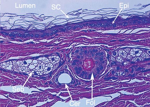
Figure 3-6
Photomicrograph of normal vertical ear canal. Lumen of the ear canal is visible at the top. SC, Stratum corneum, normal “basket-weave” appearance of healthy end-differentiated epithelial cells; Epi, epithelium, one to two cell-layer thick normal epithelial layer; Seb, sebaceous glands, small normal cluster of sebaceous gland cells associated with every hair follicular unit; Cer, ceruminous gland, single cross-section of modified apocrine gland associated with every hair follicular unit, Fol, follicle, cross-section of hair follicle with actively growing hair shaft visible in center.
migrate centripetally from a point of origin on the tympanum, up and out of the ear canal, depositing debris, cerumen, and desquamated corneocytes onto the pinnae.35 In the normal ear, this process of epithelial migration keeps the tympanum and ear canal free of debris.
Hair follicles are sparsely distributed throughout the entire length of the ear canal. Most breeds have simple follicles with single hair shafts, compared with compound primary and secondary follicles found elsewhere in haired skin.28,32 In addition to being sparse and simple, hair follicles in the ear canal are also miniaturized relative to haired skin (Figure 3-7). Cocker spaniels are the exception; this breed has been shown to have in the ear canal predominantly compound follicles that occupy a larger cross-section of tissue than non-spaniel breeds.32
Each hair follicle is associated with two types of adnexal glands: (1) sebaceous glands and (2) ceruminous glands. Sebaceous glands are similar in appearance to those found in haired skin. These glands supply neutral lipids to the lumen of the ear canal via a duct that opens in the hair follicle.
The lipids coat the hair shaft, spread to the surface, and diffuse into the stratum corneum, contributing to the normal barrier function of the epithelium. On histopathologic specimens, sebaceous glands are easily recognized by the characteristic clustering of round to polygonal cells with pale foamy cytoplasm and a small, centrally located nucleus (Figure 3-8). Variation in sebaceous gland concentration occurs along the length of the canal. In general, sebaceous glands are most prominent in the tissue toward the opening of the ear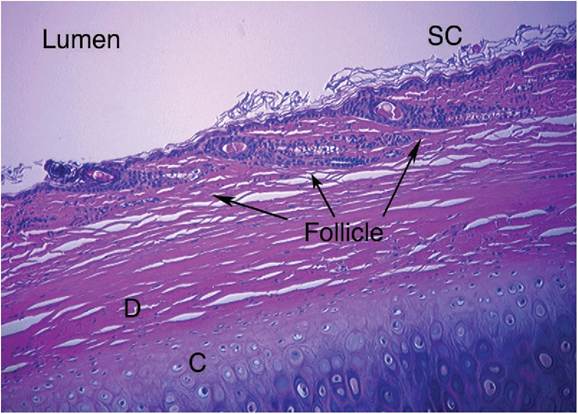
Figure 3-7
Photomicrograph demonstrating multiple small hair follicles that line the length of the external ear canal. Note how thin the section of tissue is between the lumen of the canal and the cartilage boundary. SC, Stratum corneum; D, dermis; C, cartilage.
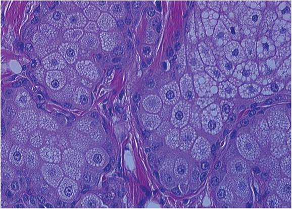
Figure 3-8
Photomicrograph close-up of sebaceous gland tissue.
canal and become less concentrated in proximal tissues closer to the tympanic membrane.32
Ceruminous glands are modified apocrine, or sweat, glands that also have ducts opening into the hair follicle. Typically they are positioned deeper in the dermis than sebaceous glands.32 In contrast to sebaceous glands, ceruminous glands are most abundant near the tympanum, decreasing in size toward the opening onto the pinna.32 Ceruminous glands are easily recognized as a single layer of cuboidal to columnar glandular epithelium surrounding a large central lumen. Ceruminous glands are actually coiled, so on cross-section a single gland may appear to be multiple adjacent glands as the slide transects the same lumen multiple times (Figure 3-9).
In the normal ear canal the ratio of sebaceous glands to ceruminous glands varies depending on location. As one moves farther away from the tympanic membrane, the ratio transitions from predominantly ceruminous glands in the horizontal canal to predominantly sebaceous glands in the vertical canal.32 On the epithelial surface, sebaceous secretions mix with apocrine secretions to form a thin, tenacious layer of cerumen.
Since sebaceous secretions are thicker and contain primarily neutral lipids, while ceruminous glands produce a thinner secretion containing phospholipids and acid mucopolysaccharides, the variation in gland ratios may influence cerumen composition in the horizontal canal compared with the vertical canal.34Cerumen is vital for the normal function of the ear canal and protection of the tympanic membrane. Cerumen traps debris and pathogenic organisms, contains immunoglobulins and antimicrobial peptides that provide local protection, and forms a barrier to transepithelial water loss, maintaining a moist, pliable tympanic membrane and epithelium.
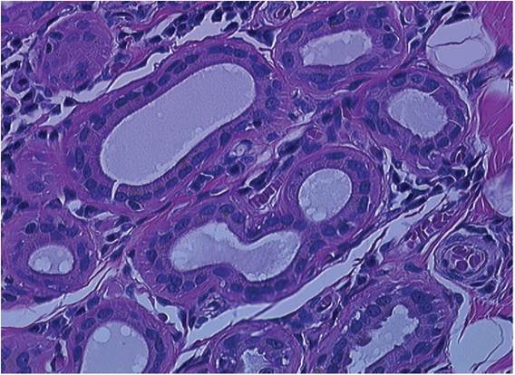
Figure 3-9
Photomicrograph close-up of ceruminous gland tissue. Note the multiple cross-sections of a single, coiled gland give the appearance of multiple adjacent glands.
Beneath the cerumen and epithelium and between follicles, the tissue of the ear canal is quite similar to the interfollicular dermis of the skin. This area contains numerous large collagen bundles, elastin fibers, blood vessels, capillaries, fibroblasts, mast cells, and other cells of the immune system. Below this layer are the auricular and annular cartilage tubes that define the boundaries of the ear canal.
Tympanic Membrane
The tympanic membrane consists of two sections: the pars tensa and pars flaccida. The pars tensa is a tough, semitransparent, multilayered tissue stretched taut across a fibrocartilaginous ring. The ring is in turn attached to the skull. Histopathologically, the pars tensa consists of four distinct layers. The first layer is continuous and equivalent to the epithelial surface of the external canal. The stratified squamous epithelium is only a few layers thick. Epithelial migration originates from a germinal center in this layer. The second stratum is roughly equivalent to the dermis, containing a thin layer of fibroblasts, fine nerves, and blood vessels.
Hair follicles and glandular tissue are totally absent from this layer. Next is a fibrous intermediate layer, which connects to the fibrocartilaginous ring. This is the thickest layer of the pars tensa. The first of the auditory ossicles, the malleus, is embedded in this layer. The arrangement of fibers is orderly, to maintain strength and maximize transmission of vibrations. The fourth layer consists of a single sheet of respiratory epithelial cells overlying a thin lamina propria. Centrally, the cells of squamous morphology become more cuboidal toward the margins and finally columnar as the layer becomes continuous with the lining of the tympanic cavity. Unlike normal respiratory epithelium, this layer contains no cilia or goblet cells.27,28,36,37The pars flaccida is a soft, pliable portion of the tympanum located dorsal to the pars tensa. This tissue contains a loose arrangement of collagen fibers covered by a thin layer of epithelium. Unlike the pars tensa, the pars flaccida is highly vascular and gives rise to the capillary branches that supply the germinal epithelium of the pars tensa. With acute inflammation, such as that associated with atopy, the pars flaccida may become severely erythematous and edematous.
Cocker Spaniels
American Cocker Spaniels differ substantially from other breeds in normal histoanatomy. In the study by Stout-Graham et al, Cocker Spaniels with normal ear canals were found to exhibit increased ceruminous gland tissue relative to non-spaniel breeds (Greyhounds, mixed).32 In the same study, Cocker Spaniels were found to have compound hair follicles rather than simple follicles seen in other breeds; furthermore, Cocker Spaniel hair follicles were more densely packed.