Homeostasis
The 19th-century French physiologist Claude Bernard (1965) coined the term milieu interieur, which referred to the relatively constant internal environment, that is, extracellular fluid, in which cells live.
Walter Cannon (1932), a 20th-century American physiologist, later coined the term homeostasis, meaning "unchanging" internal environment. While the concept of homeostasis is fundamental to understanding physiology, the term is better understood as a relatively steady state that is maintained within an animal despite a wide range of environmental conditions. In this way, various internal conditions, such as plasma glucose, electrolyte concentrations, or body temperature, are maintained within narrow limits through homeostatic mechanisms.Homeostasis is maintained at all levels of life. Individual cells, for example, control their internal environment via selectively permeable membranes. These membranes will allow selective movement across the membrane based on such factors as pH, size, or whether there is a specific transport system for that compound. Whole animals maintain their internal environment by a host of behavioral and physiological mechanisms. A behavioral method of regulation may include moving from a sunny area to a shady area to decrease body temperature, whereas a physiological method may involve an increase in sweating or panting to accomplish the same goal.
Homeostatic regulatory mechanisms
Elaborate regulatory mechanisms exist to maintain homeostasis. Homeostasis is maintained by the actions of the nervous and endocrine systems that communicate changes in the internal and external environment. The two systems work in conjunction to make relatively rapid or slow changes, respectively. The nervous system responds to immediate, short-term needs, as seen in a reflex arc in which an animal withdraws its foot after stepping on a sharp object.
In contrast, the endocrine system generally elicits responses that last 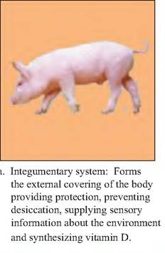

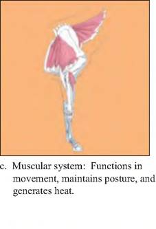
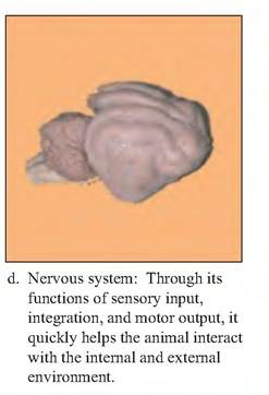
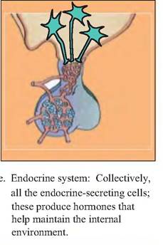
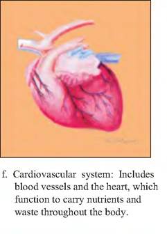
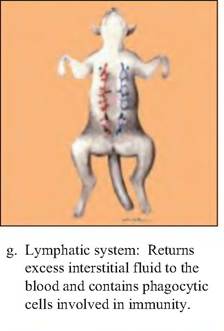
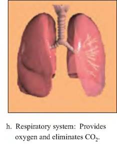
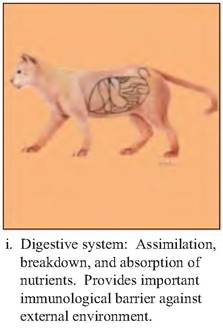
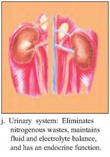
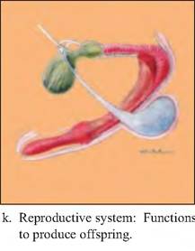
Fig. 1.3. Organ systems. The body consists of 11 major organ systems that are shown above along with examples of their components.
hours or days such as the release of insulin in response to a rise in blood glucose levels.
When regulation occurs at either the cellular, tissue, organ, or organ system level, it is termed autoregulation. For example, the presence of tryptophan in the small intestine will cause the local release of cholecystokinin (CCK) that will cause the pancreas to secrete enzymes. Extrinsic regulation, on the other hand, involves the coordinated action of both the nervous and endocrine systems. Such regulation occurs, for example, during prolonged stress where there is release of norepinephrine, epinephrine, and corticosteroids from the paired adrenal glands. This results in an increase in blood pressure and a change in blood flow such that there is an increase to the skeletal muscle and a decrease to the digestive tract.
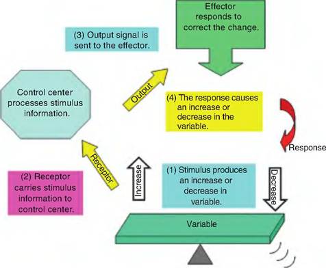
Fig. 1.4. Feedback systems. (1) A stimulus causes a change in a variable (i.e., plasma glucose, blood pressure, and heart rate). (2) A receptor senses the change in the variable and sends that information to the control center. (3) The control center compares the level of the variable to a set point and then initiates appropriate responses to change the variable. (4) The actions of the effectors bring about a change in the variable.
Chapter 1
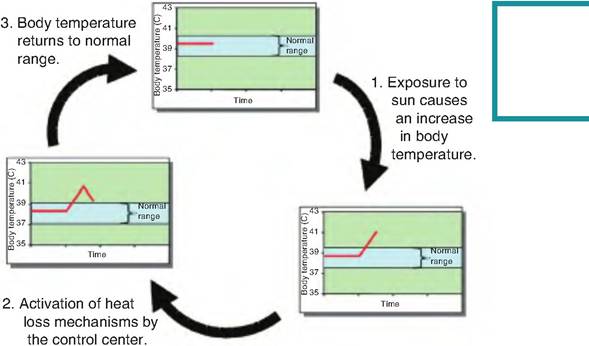
Fig. 1.5. Negative feedback systems. (1) A stimulus causes a change in a variable, in this example, an increase in body temperature. (2) Information regarding the increase in body temperature is carried to the control center, which activates appropriate heat loss mechanisms to decrease body temperature toward the set point. (3) As a result of the heat loss mechanisms, body temperature is returned to the set-point value, and homeostasis is maintained.
The factor being regulated is the variable. The regulatory mechanisms involve a receptor, a control center, and an effector. The receptor is a neuron that senses a change in the environment, called a stimulus. In response to the stimulus, the receptor carries an afferent (away) signal to the control center. The control center has a set point around which the variable is maintained. When the input signal is outside of the range of the set point, an appropriate response is elicited to correct the variable. An efferent (toward) signal is then sent to the effector. The effector induces a change in the controlled variable to bring it back to the set point (Fig. 1.4).