Hyperplasia
Hepatocellular hyperplasia/nodular regeneration
The liver is capable of significant regenerative effort. In the canine liver, nodular regeneration and hyperplasia are commonly observed in older patients.
Nodular hyperplasia is uncommonly encountered in the cat (Cullen & Popp, 2002). Cytologically, hyperplasia/regeneration is noted as mild to moderate hepatocellular atypia including increased cell size, altered nuclear to cytoplasmic (N:C) ratio, and bi- and trinucleation (Stockhaus et al., 2002). In extreme cases, atypia can be marked, and differentiation of hyperplastic from neoplastic processes may be challenging (Figures 9.20a–c). Finding atypical hepatocytes juxtaposed with cytologically normal hepatocytes in hyperplastic cases is more consistent with hyperplasia than neoplasia (Stockhaus et al., 2002). Often, distinction of hyperplasia from neoplasia is dependent on evaluation of lobular architecture, requiring a biopsy specimen and histopathology. Hyperplasia will arrange into normal bilayer plates while neoplastic liver nodules will display a more disorganized architecture disrupting the typical lobular organization (Figures 9.21a–c). 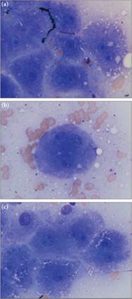
Figures 9.20a–c Liver aspirate from a Siberian Husky who was undergoing chemotherapy (CCNU). Ultrasonographic evaluation found nodules in the liver. (a) Bile is present as coarse green–black granular pigmented material in the cytoplasm and canalicular spaces. The hepatocytes frequently are binucleate with variably-sized nucleoli. (b) Rarely, large trinucleate hepatocytes with anisonucleoliosis are found. (c) Considerable size and shape variability in both nuclei and nucleoli are present. The cellular atypia is likely associated with the toxic effects of the therapeutic regimen (Wright–Giemsa, 1,000? magnification).
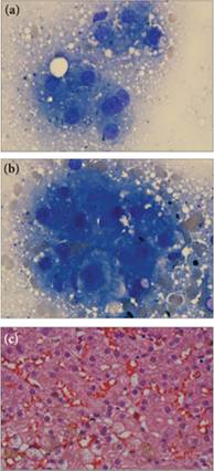
Figures 9.21a–c (a, b) Frequent binucleation, mild to moderate anisocytosis, and anisokaryosis with few hepatocytes containing multiple nucleoli are present. The black granular material in 9.21b is consistent with bile (Wright–Giemsa, 1,000? magnification). (c) Histologic evaluation of this patient’s liver confirmed the frequent binucleation, anisocytosis, and anisokaryosis. Although these changes are present, hepatic plates are organized. The histologic diagnosis is nodular hyperplasia. The pale brown material present between the hepatocytes is largely cell associated and consistent with bile. A few bile casts located between hepatocytes were noted (not imaged) (Wright–Giemsa, 500? magnification).