Inclusion Body Nephritis
Over the course of 40 years, the authors have encountered multiple sporadic incidents and referrals in which renal tubules have epithelial nuclei with marginated chromatin and prominent, homogeneous eosinophilic inclusions.
This is typically accompanied by adjacent interstitial infiltrates of lymphocytes in immunocompetent mice (Fig. 1.122). Ultrastructurally, the inclusions contain flocculent electron-lucent material. Numerous inclusions without interstitial infiltrates have been noted in globally immunodeficient Rag1 null mice (C. Brayton, unpublished). The inclusions are reminiscent of virus inclusions, but affected mice are negative for polyoma virus, K virus, and adenovirus.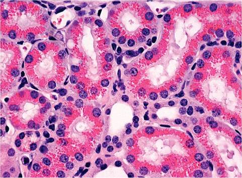
FIG. 1.121. Kidney of a mouse with histocytic sarcoma in distant tissues. Renal tubular cells are filled with eosinophilic hyaline bodies.
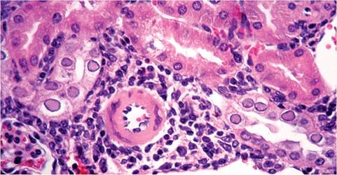
FIG. 1.122. Inclusion body nephritis in a mouse. Note the prominent intranuclear inclusions within tubular epithelium and interstitial infiltration of lymphocytes.
More medical literature on Medic.Studio
More on the topic Inclusion Body Nephritis:
-
Infectious diseases -
Internal diseases -
Obstetrics and Gynaecology -
Pediatrics -
Veterinary medicine -
-
Conflictology -
Ecology -
Economy -
Finance -
History -
Law -
Medicine -
Philosophy -
Religious studies -