Infectious Agents
There is a wide range of infectious agents which can cause disease. Some exist as normal organisms on the animal or in its environment and only cause disease when in an unusual site or when the immunity of the animal is compromised (i.e.
reduced). A good example of this is the bacterium Escherichia coli, most commonly known as E. coli. It is present in the intestine of all cattle, where it usually causes no problems. However, if it gains access to the udder it can cause quite severe disease. This is especially true in the early lactation animal, whose udder defences against infection are often poor. However, for some other infections, whenever they are present disease occurs. A good example of this would be the foot-and-mouth virus.Infectious agents may be subdivided into the following categories:
• Bacteria
• Viruses
• Protozoa
• Fungi
• Worms
• Ectoparasites
Bacteria
These are single-celled organisms which contain all the components needed for a separate existence. A typical bacterium is shown in Figure 1.1. It is a single cell, and consists of a thick outer polysaccharide structure, the cell wall, inside of which there is a protein membrane enclosing the cytoplasm and the nuclear material. The nuclear material contains the genetic components, the DNA (deoxyribonucleic acid), and by a complex arrangement of molecules, DNA functions as the regulator for all cell processes, determining the size and shape of the cell and the activities that take place within its cytoplasm. These ‘activities' are the processes of reproduction and growth. The whole bacterium may be enclosed by a gelatinous capsule, a thick membrane which renders the bacterium more resistant to phagocytosis (i.e. being engulfed by white blood cells). Bacteria are therefore individual discrete units of life.
Given ideal conditions of warmth, nutrients and moisture most of them can also multiply outside the animal's body.
Under adverse conditions some bacteria can turn themselves into a very resistant spore form, which can survive for many years. The classic example is that of anthrax, whose spores can persist in the soil for up to 40 years. Bacteria absorb nutrients for their growth from their immediate surroundings (e.g. blood, milk or body tissue) and excrete waste products. It is often these waste products which cause disease. The ‘waste' is then known as a toxin and the animal is said to be suffering from a toxaemia. Typical examples of toxaemia are acute E. coli mastitis and severe uterine infections.Bacteria are the major cause of mastitis, they are commonly involved in respiratory disease and they are the cause of the clostridial group of diseases such as blackleg, tetanus and anthrax. Bacteria commonly form pus and are also responsible for conditions such as navel ill, calf diphtheria and abscesses. Bacteria are killed by antibiotics, with different antibiotics being needed to kill the different species of bacteria. This is explained in more detail in the treatment section.
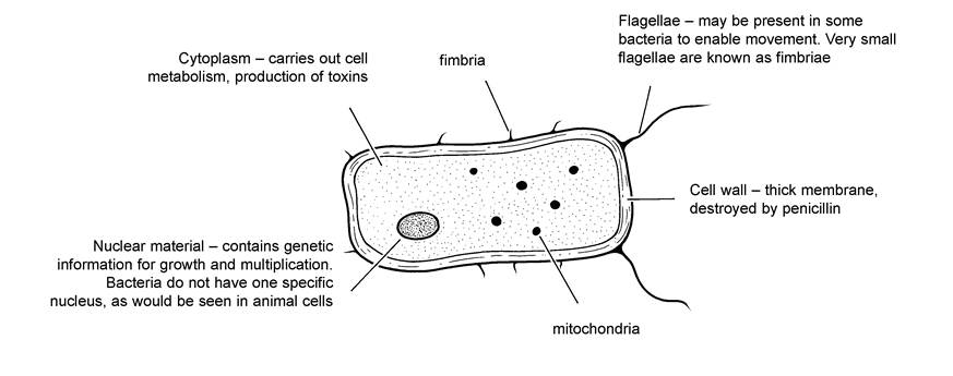
Figure 1.1. Atypical bacterial cell. Even the largest bacteria (e.g. anthrax) are only 0.005 mm long. They multiply by dividing into two; and under favourable conditions this may occur every 30 minutes, so that one bacterium could produce 17 million offspring in 12 hours!
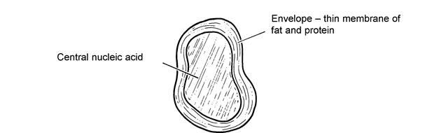
Figure 1.2. Avirus particle. Avirus is very much smaller than a bacterium. It uses the processes of metabolism within the animal cell for its own multiplication and growth, and as such it cannot live a separate existence away from the animal. This is very different from bacteria.
Viruses
Viruses are much smaller than bacteria and in fact they may even infect and cause disease in bacteria. They cannot be seen with a normal light microscope; electron microscopy is required.
They consist simply of central nuclear material, which may be DNA or RNA (ribonucleic acid), and this is surrounded by a capsule of fat and protein (Figure 1.2).Because viruses have no cytoplasm or proper nucleus, they cannot carry out their own metabolic functions of growth and reproduction and they are therefore unable to multiply outside the animal's living cells. For their survival away from the animal, and hence their transfer from one animal to another, viruses must be protected, for example in sputum (pneumonia viruses), milk (foot-and-mouth virus) or blood (EBL virus).
Once inside the animal, viruses inject their own RNA or DNA into the animal cell and then use the metabolic processes within the cytoplasm of that cell for their own purposes of multiplication and growth. When a cell is packed full of viruses, it bursts and virus particles are released to penetrate and infect adjacent animal cells. It is the bursting of these cells which generally causes the detrimental effects on the animal and hence the signs of disease, although some viruses (e.g. those causing teat warts or EBL) induce a proliferation or excessive multiplication of animal cells to produce a tumour.
Both bacteria and viruses may be very specific in the site they choose to infect. For example, certain groups will grow only in the respiratory tract - and these cause the clinical signs of a cold, influenza or pneumonia. Others can live only in the intestine and they will cause scouring. It is by this mechanism that we associate particular strains of bacteria or viruses with specific diseases. Different strains of bacteria and viruses vary considerably in shape and size, in the same way that the different species of animal or bird are so variable.
Viruses cause a wide range of disorders including foot-and-mouth and diseases of the teat skin, and they are often the primary cause of calf pneumonia. Whereas antibiotics will kill bacteria, there is no specific drug to kill viruses.
This is one reason why many virus diseases are controlled by vaccination. Mycoplasma, ureaplasmas and rickettsia are organisms which have some characteristics of bacteria and some of viruses.Protozoa
Protozoa are also single-celled organisms, although they are larger and more complex than bacteria and may have a free-living existence. Examples include Babesia and Trypanosoma, which live in the blood of cattle and cause redwater, and coccidia and Cryptosporidia, both of which live in the intestine and cause scouring. Other protozoal diseases of cattle include Neospora, a cause of abortion, Theileria which causes East Coast fever in southern Africa, and Besnoitia which causes skin disease and abortion.
The treatment of protozoal infections requires specific therapy. For example, there is one drug specifically used against Babesia (imidocarb) and another against coccidiosis (e.g. amprolium). There is no specific treatment against Cryptosporidia.
Fungi (Yeasts and moulds)
Fungi are very simple members of the plant kingdom. Yeasts are commonly found in the environment and primarily cause disease when they enter an unusual site, e.g. the udder, where they can cause a chronic mastitis. They do not respond to antibiotics and they need special therapy, e.g. iodine (see Chapter 7). In fact yeasts may grow in the oily ‘carrier’ used in bovine intramammary mastitis tubes. There are some specific fungal diseases of cattle, namely ringworm, and other diseases where a common environmental fungus invades an unusual site. A good example of this is abortion caused by the fungus Aspergillus.
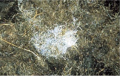
Plate 1.1. Aspergillus mould growing on silage. This led to five abortions when fed to dry cows.
Aspergillus may be seen as a blue-grey mould growing on silage (Plate 1.1), and if eaten by a pregnant
cow it can lead to abortion.
Worms (Helminth parasites)
Cattle can be affected by a wide range of helminth or endoparasitic worms.
In low numbers worms cause no problems, although if allowed to multiply they can cause serious disease. As with bacteria and viruses, different worms live in different parts of the body. Examples include nematodes such as the lungworm (Dictyocaulus viviparus), the stomach worm Ostertagia and the intestinal worms Nema- todirus and Oesophagostomum. There is even a worm (Thelazia) which lives in the eye! Tapeworms (cestodes) can also occur and are found in the intestine. Flukes (trematodes) are related parasites and live in the liver.Most worms have a direct life cycle, that is eggs laid by adult worms are passed in cattle faeces, develop into mature larvae on the ground and are then ingested by grazing animals. Other helminth parasites may have an indirect life cycle, for example the liver fluke spends part of its life in the host animal and part in a snail.
An anthelmintic is the general name given to drugs which are used to treat worms (i.e. wormers). The same drug will often treat all species of lungworm and gutworm, although different products are usually required to treat liver fluke. Anthelmintics are often subdivided into white drenches and clear drenches. The white drenches are the benzimidazole group of compounds, examples of these being oxfendazole and fenbendazole. Clear drenches include levamisole and the avermectin range of products, e.g. ivermectin, doramectin and moxidectin. Each drug has a slightly different spectrum of action and length of activity, so make sure that you have read the manufacturer’s instructions before use.
Ectoparasites
An ectoparasite is the name given to an organism which lives on the body surface of an animal (intestinal worms are known as endoparasites). The range of different ectoparasites on cattle includes lice, mange, flies, maggots and ticks. Some have a direct life cycle (lice and mange) where all stages of the life cycle can be found on the same animal. Others, such as ticks, are more complex and part of their life cycle is spent off the animal.
More on the topic Infectious Agents:
- INFECTIONS OF LABORATORY MICE: EFFECTS ON RESEARCH
- Software Agents
- Chapter 14 ROUTINE TASKS AND DEALING WITH POISONS
- I INFECTION CONTROL ^xiv ^69 ^107 ^140
- Vasculitic Syndromes
- Bibliography for viral infections
- VIRUSES MAINTAINED BY HOSTS WITH CLINICALLY ACTIVE INFECTIONS
- Immunodeficiency
- SELECT DEFINITIONS AND CONCEPTS
- Reovirus Infection