INFECTIOUS CAUSES
Skin diseases can be produced by bacterial or viral infections. Sometimes the skin changes are only part of the overall disease syndrome, for example calf diphtheria (Chapter 2), which may produce a swelling on the face and/or on the tongue, and similarly for wooden tongue in older animals.
Lumpy Jaw
This is an infection of the jaw bone caused by the bacterium Actinomyces bovis. The lower jaw on one side may very slowly develop a swelling. If you examine it carefully you can feel that the swelling is extremely hard and that it is firmly attached to, and even part of, the bone. Some say that injecting antibiotics (penicillin and streptomycin) into the lump in the
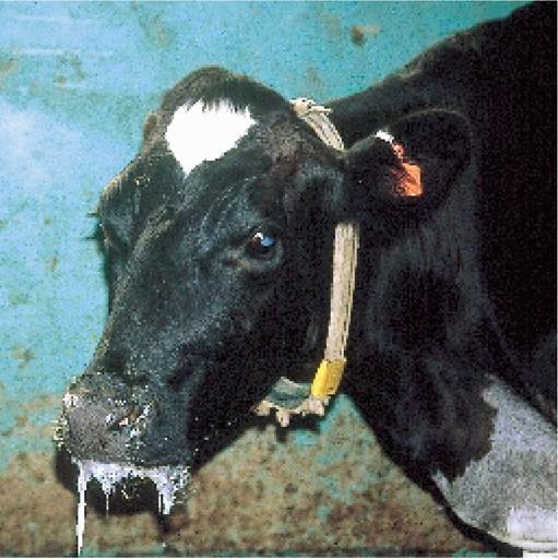
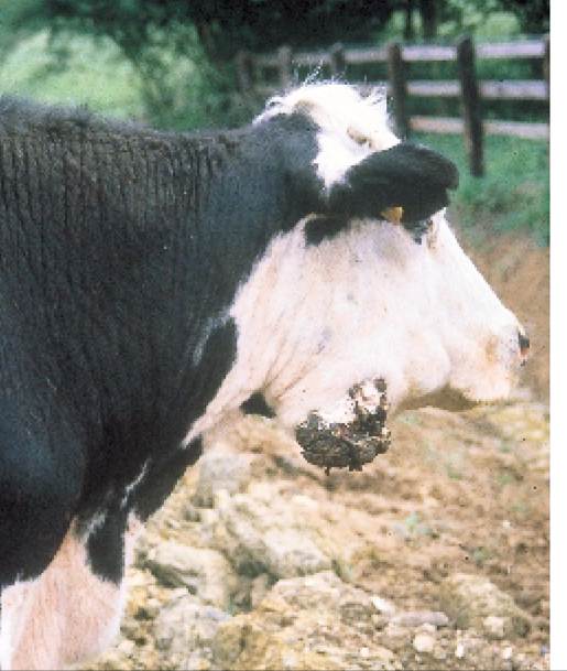
Plate 10.9. Cow drooling. This could be due to wooden tongue, lumpy jaw, a tooth abscess or even foot-and-mouth, so careful examination is required.
early stages may effect a cure. As the condition progresses, the roots of the molar tooth become displaced, eating and chewing the cud are painful and the cow begins to drool (Plate 10.9) and lose weight. The Hereford steer in Plate 10.10 is an advanced case. At this stage treatment is hopeless.
Severe knocks and bruising can cause a similar reaction in the jaw bone, but these will eventually heal, so you need to get your vet to examine it carefully before deciding to cull the animal.
Prevention is described under wooden tongue.
Plate 10.10. Lumpy jaw. This neglected case is unlikely to recover.
Wooden Tongue
This is sometimes confused with lumpy jaw. It is caused by a different organism, the bacterium Actinobacillus Iignieresii which invades the soft tissues of the mouth. The tongue is the favourite site, although sometimes the cheek or oesophagus may be affected, and I knew one farm where animals developed large, discharging lumps in their skin at sites all over the body.
This is a disease which is often easier to feel than to see. The hardening is on the raised portion at the back of the tongue (Plate 10.11), but you must use a mouth gag (Chapter 14) before putting your hand that far into the mouth; otherwise you might lose your fingers - as I almost did once (Figure 9.20B)!
As the infection progresses, the tongue becomes hard and swollen. The animal is reluctant to eat, it drools and loses weight and often there is a secondary swelling in the throat as shown in Plate 10.12. If the oesophagus is affected, chronic bloat or cud regurgitation (Plate 6.6) may occur due to interference with rumination.
Treatment
Unlike lumpy jaw, wooden tongue responds to treatment very well. Traditionally iodine was used, giving an initial ‘loading’ dose of sodium iodide intravenously, followed by potassium iodide by mouth. Antibiotics are now the treatment of choice, and the treatment may need to be prolonged, e.g. penicillin and streptomycin injection daily for seven to ten days.
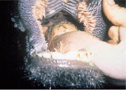
Plate 10.11. Wooden tongue. The back of the tongue is hard and thickened. This is best appreciated by palpation.
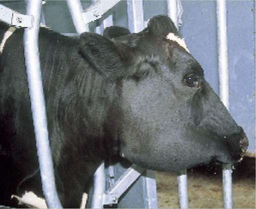
Plate 10.12. Aswelling beneath the jaw, as in this animal, could be an indication of wooden tongue.
Prevention
Both lumpy jaw and wooden tongue gain entry via abrasions in the mouth, lumpy jaw perhaps beside a loose tooth. Both organisms are found in the soil and outbreaks of disease may be associated with feeding potatoes or other foods heavily contaminated with earth and small stones, the stones leading to the abrasions which allow the entry of infection.
Jaw Abscesses
This is a common condition in cattle of all ages, and leads to a hard swelling at the angle of the jaw bone. The lesion is clearly seen in the cow in Plate 10.13. Infection most probably originated from a penetration wound at the back of the pharynx (Figure 2.1), in other words from inside the mouth, but the pus then accumulates under the skin.
Sometimes the abscess bursts on its own, but usually it has to be lanced, drained and flushed with antiseptic solution. Antibiotic cover may be needed.Malignant Oedema (Necrotic Cellulitis)
This is another disease which leads to a swelling of the face, but it is much more serious and unless treatment is
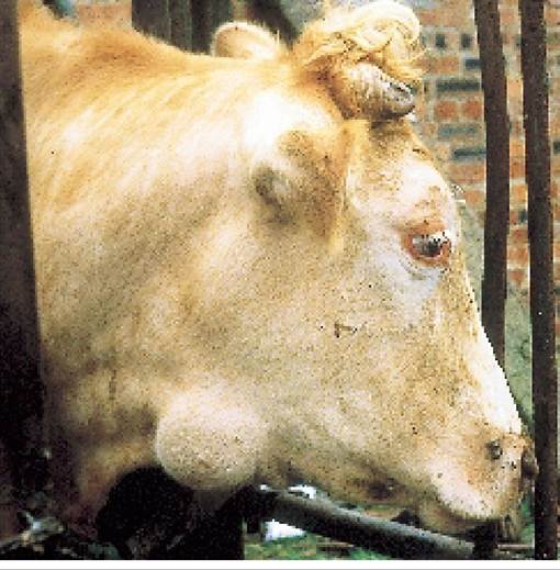
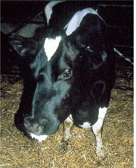
Plate 10.14. Malignant oedema (necrotic cellulitis) is an acute clostridial infection of tissues under the skin.
Plate 10.13. Jaw abscesses commonly result from penetration of the inside of the mouth (the pharynx) by sticks or other sharp objects.
given quickly the animal may die. The cause is an infection under the skin and hence its alternative name of necrotic cellulitis. The disease is produced by a bacterial infection, Clostridium septicum, and is caused by any skin damage, such as sticks or stones in the feed, drenching gun injury or external trauma, which allows entry of the infection. Other species of Clostridia are also sometimes involved.
Care must be taken not to confuse this with wooden tongue or blaine. With necrotic cellulitis cows are much more seriously ill. They have a high temperature (41-42°C) and often only one side of the face is swollen. Plate 10.14 shows an advanced case, with swelling of the face, drooling and swelling of the brisket. Penicillins are the treatment of choice, but although this cow was dosed at a continuous high level for seven to ten days, infection spread down the front legs and she had to be culled. Anti-inflammatory drugs help counteract concurrent toxaemia.
Warts
Warts are skin tumours caused by a virus infection. They most commonly affect the head, neck (Plate 10.15), belly and teats (Plate 7.45). They may also be found on the penis of young bulls (Plate 10.16). Young animals, one to two years old, are most commonly affected, especially when they are group housed and in close confinement.
Flies have been implicated in the spread of infection. When neck warts are large and pendulous like those in Plate 10.15, they often develop a secondary bacterial infection and start to smell.Some animals are so badly affected that the warts have to be removed surgically. Removal of a wart to prepare a vaccine may help, partly from the vaccine itself and partly because pulling off the wart may release some virus into the blood, generate immunity and result in a self-cure. (In some countries a licence may be required for a vaccine.) In most animals which are only mildly affected, the warts will eventually fall off without causing any problems. Teat warts are discussed in Chapter 7.
Skin Tumours
Occasionally younger cattle develop small lumpy swellings of the skin over the whole body. This could be a cancer (tumour) known as a lymphosarcoma, as shown in Plate 10.17. There is no treatment and the animal should be culled.
Skin TB
If you live in an area where TB is a problem and annual testing is a regular feature, you will have already seen skin TB. A typical example is shown in Plate 10.18. The small hard lumps are under the skin, whereas the TB test reaction occurs in the skin, as it is an intra-dermal test. The swellings of skin TB are enlarged lymph nodes and that is why they are often found in lines. They may also be seen on the legs (especially the front legs) and chest. Skin TB may be produced by non-pathogenic bacteria which are from the same family as TB. In this instance they can
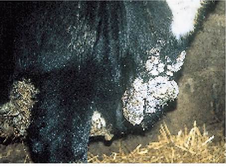
Plate 10.15. Skin warts are caused by a virus infection and are most commonly seen in younger animals.
Plate 10.16. Warts on the penis, a common problem in younger group-housed bulls.
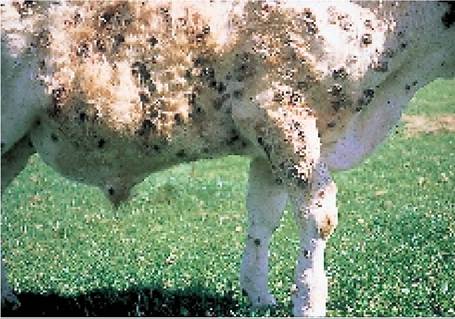
Plate 10.17. Lymphosarcoma - multiple small skin tumours. (Courtesy J. Gallagher)
influence the development of the skin reaction in the TB test and as such interfere with its interpretation. However, other causes are possible and in the United States similar lymph node enlargement (i.e. skin TB) is associated with bovine immunodeficiency virus (BIV) (see Chapter 13).