Iron
With Romanowsky-type stains, hemosiderin appears as brown-black to gray-black aggregates in macrophages or extracellularly in the thick areas of marrow granules (Figure 19.16). Hemosiderin may also be noted as more dispersed finely granular green-brown pigment in individual macrophages.
Prussian blue stain is useful to highlight iron in cytology or histology preparations. In humans, absence of stainable iron is only confidently reported when at least seven marrow particles in a sample lack stainable iron, which often requires evaluation of more than one slide (Chauffaille et al., 2019). Iron stores may be better assessed in marrow biopsy sections since they contain more hematopoietic cells than aspirates but decalcification can also reduce stainable iron (Riley et al., 2009). Lack of coarse iron in marrow may be associated with but is not necessarily indicative of iron deficiency anemia in humans and dogs (Barron et al., 2001; Thomason & Almiski, 2009; Pawsat et al., 2023). A scoring system for iron in canine marrow aspirates has been reported with scores for stainable iron correlating well with other parameters of iron availability such as reticulocyte hemoglobin content (Pawsat et al., 2023). Iron is normally absent in feline marrow. 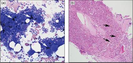
Figures 19.16a,b Iron (arrows) appears dark black on Wright stain (a) and brown-gold on H&E stained sections (b), both at 100 magnification.
Iron deficiency should be suspected from CBC indicators such as hypochromic reticulocytes, hypochromic microcytic or normocytic erythrocytes, increased erythrocyte fragmentation, and anemia, and should be confirmed by measurement of serum iron and ferritin concentration, iron binding capacity, and low iron scores on marrow slides. Decreases in erythrocyte volume and development of anemia occur relatively late in iron deficiency.