Large intestine (cecum, colon, and rectum)
Normal
The colon contains larger numbers of goblet cells relative to the small intestine (Figures 7.46a, b, 7.47a, b). Villi are absent, and crypts are longer than in the small intestine (Bacha & Bacha, 2012).
The rectum is lined by stratified squamous epithelium, which becomes progressively keratinized in the posterior portion as it approaches the anal canal. Apocrine anal glands occur in the submucosa and muscularis of the anal canal in both dogs and cats. Circumanal glands occur in the subcutis around the anus of the dog. These are modified sebaceous glands that resemble hepatocytes and thus are often referred to as ‘hepatoid’ glands. Diseases affecting these latter structures are described in Chapter 4.Sampling of the large intestine may be accomplished by FNA or impression smears of affected tissue or masses similar to other lesions along the GIT. Additionally, endoscopic brush cytology (described above) and rectal scraping may assist in the detection of certain disease conditions such as infiltrative neoplasms or intestinal infections (e.g. histoplasmosis or protothecosis). Some fecal parasites may also be identified via rectal scrapings (see ‘Intestinal Parasites’ below). As the intent of a rectal scrape is to sample a larger concentration of cells directly from the colonic mucosa, tools such as a moistened cotton swab, fecal loop, or spatula are often used to gently dislodge cells. Additionally, gentle scraping of the rectal mucosa with a gloved fingertip usually results in good exfoliation of cells. Although judicious use of gel lubricant is helpful for rectal palpation, it is important to remember that the presence of this material can obscure cellular details and render cytologic assessment useless (Figure 7.48). Therefore, it is generally recommended to avoid the use of lubricants when obtaining a sample by rectal scrape.
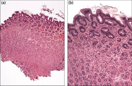
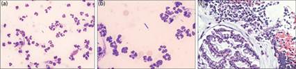
Figures 7.46a,b Histologic sections of normal colonic epithelium from a dog obtained by colonoscopic biopsy (H&E; a, 40? magnification; b, 100? magnification).
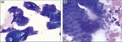
Figures 7.47a,b Cytology of normal colonic epithelium from a dog (Wright–Giemsa, a, 400? magnification; b, 1,000? magnification).
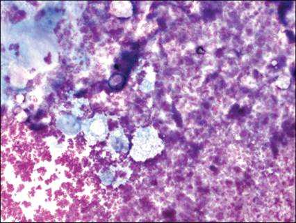
Figure 7.48 Rectal scraping from a dog. The cytologic constituents of the sample are obscured by the presence of amorphous, eosinophilic globular material consistent with gel lubricant (Wright–Giemsa, 500? magnification).
Hyperplastic lesions
Colonic hyperplasia is described uncommonly in animals and typically associated with primary inflammatory disease. The formation of colorectal polyps has been recognized as a cause for large bowel diarrhea in dachshunds in Japan (Uchida et al., 2016). As hyperplastic epithelium cannot be differentiated from either normal colonic epithelium or adenomatous polyps, definitive diagnosis requires biopsy and histopathologic interpretation (see Figures 7.47).
Inflammatory lesions
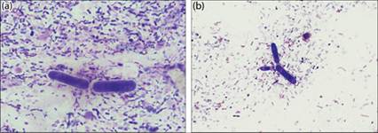
Recent studies have speculated that chronic diarrhea in dogs is more often associated with lifestyle and behavioral (e.g. pica) factors than specific pathogens (Berset-Istratescu et al., 2014). Campylobacter spp. (Figure 7.49) and Clostridium spp. (Figure 7.50) have both been cultured from the feces of dogs with and without diarrhea; therefore, the identification of these organisms in feces does not always correlate with the clinical status of the patient (Rossi et al., 2008; Weese, 2011; Berset-Istratescu et al., 2014).
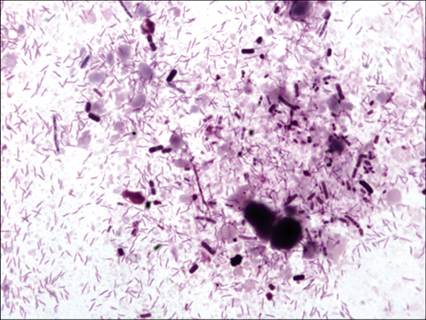
Figure 7.49 Fecal smear from a dog with diarrhea.
Apparent overgrowth of spiral-shaped bacteria consistent with Campylobacter spp. (Wright–Giemsa, 1,500? magnification).
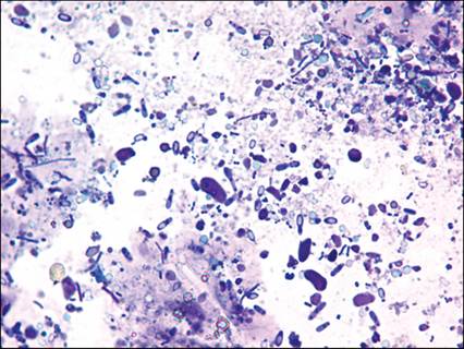
Figure 7.50 Fecal smear from a dog with diarrhea. Apparent overgrowth of bacterial rods consistent with Clostridium spp. (Wright–Giemsa, 1,500? magnification) (courtesy Anne Barger).
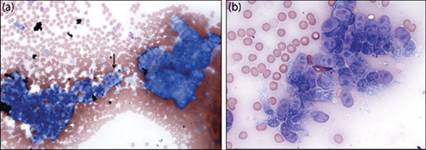
Although chronic inflammatory GI disorders frequently cause multifocal GI lesions, primary colitis may be seen. Similar to other forms of CE, hereditary conditions have been documented in dogs, including histiocytic ulcerative colitis in Boxers (Figure 7.51a–c) and French Bulldogs (German et al., 2003; Manchester et al., 2013; Sims et al., 2022) or eosinophilic granulomas in Rottweilers (Figure 7.52). Idiopathic colitis can also affect cats, although breed association is not described (Figure 7.53). Mixed inflammatory infiltrates may be seen on direct aspiration of focal lesions or rectal scraping. However, final, definitive diagnosis requires histopathology.
Figures 7.51a–c Colon, canine. Cytology from a rectal scrape from a 1-year-old Boxer dog with chronic hematochezia and large bowel diarrhea due to ulcerative colitis. (a) Mixed inflammatory cells including neutrophils and macrophages are present. (b) Intralesional bacterial rods are also found (Wright–Giemsa: a, 500? magnification; b, 1000? magnification). (c) Histology of the lesion shows hyperplastic epithelium associated with the inflammatory population (H&E, 400? magnification).
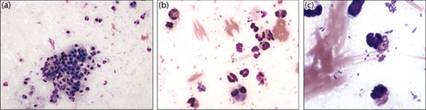
Figures 7.52a–c Colon, canine. Cytology from a rectal scrape from a dog with eosinophilic colitis. (a) Clusters of uniform colonic epithelial cells is found among scattered eosinophils with few neutrophils.
(b) Eosinophils are more readily identified at higher magnification. (c) Canine eosinophil granules are often variably sized and may coalesce (Wright–Giemsa: a, 300? magnification; b, 700? magnification; c, 1,000? magnification).
Figures 7.53 Colon, feline. Cytology from an aspirate from a 10-year-old cat with chronic large bowel diarrhea due to idiopathic eosinophilic colitis. Mast cells can be found admixed with eosinophils in several slides, which may mimic underlying mast cell neoplasia (Wright–Giemsa: 500? magnification).
Figures 7.54a,b Fecal sample from a French Bulldog with chronic large-bowel diarrhea. The sample contains numerous large cigar-shaped yeast organisms, consistent with C. guttulatus, among mixed bacteria. Organisms are found individually, (a) in short chains, and (b) branching in the sample (Wright–Giemsa, 1,000? magnification).
Fungal and ‘pseudofungal’ infections
Many fungal and algal organisms are associated with large intestinal lesions. Overgrowth of normal commensal and ingested organisms such as Cyniclomyces guttulatus may occur, resulting in diarrhea (Figure 7.54a, b). Histoplasma capsulatum, as described above, is commonly associated with chronic enteritis and, more often, colitis in infected individuals (Bromel & Sykes, 2005). Histoplasma spp. organisms may be identified on rectal scrapes or aspiration of mass lesions in the large intestine or associated lymph nodes, as dissemination is common (Figure 7.27, Case 3). Rarely, other pathogenic fungi such as Cryptococcus spp. can be found via rectal scrapes and/or fecal analysis (Figures 7.55a). Biopsies are also diagnostic for identifying organisms in infected tissues (Figure 7.55b).
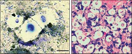
Figures 7.55a,b Direct fecal smear from a dog with systemic cryptococcosis.
(a) Encapsulated, narrow-based budding organisms consistent with Cryptococcus are seen (Wright–Giemsa, bar = 20 μm). (b) Histologic section of colon from the same dog with systemic cryptococcosis. Numerous encapsulated organisms are seen in the colonic mucosa and submucosa (H&E, 1,000? magnification). (From Graves TK, Barger AM, Adams B et al. (2005) Diagnosis of systemic cryptococcosis by fecal cytology in a dog. Vet Clin Pathol 34(4):409–412. Reprinted with permission.)
As described for algal lesions found in the small intestinal tract, GI disease, particularly colitis, is more common in dogs infected with Prototheca zopfii (Masuda et al., 2021). Rectal scrapes may be useful for identifying Prototheca spp., but definitive identification is often difficult as the organism is pleomorphic and may appear similar to commensal yeast in feces (Figures 7.56a–b). Organisms vary in size, depending on the species and stage of development, ranging from 1.3 to 13.4 μm in diameter. They have thin nonstaining cell walls, basophilic granular cytoplasm, small centrally placed nuclei, and reproduce by endosporulation. While morphologic features of the organisms may provide a preliminary diagnosis of protothecosis, definitive diagnosis and speciation require molecular analysis.
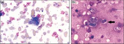
Figures 7.56a–b Rectal scrapes from two dogs with chronic colitis due to Prototheca infection. (a) Small numbers of oval organisms with thin clear cell walls and granular basophilic contents are found among a background of erythrocytes. The flocculant magenta material is consistent with gel-lubricant. (b) Organisms are variable in size. Note the presence of endosporulation (arrow) (Wright–Giemsa, 100? magnification) (7.56a courtesy Mary Christopher; 7.56b courtesy William Vernau).
Intestinal parasites
Rectal scrapings and fecal analysis are frequently performed for the diagnosis of parasites in dogs and cats.
The list of infective organisms is long, with the sensitivity of detection being based on the mode of acquisition (Table 7.1). Rectal scrapings and direct smears of feces are often useful for detecting protozoan pathogens such as Giardia and Tritrichomonas spp. (Figures 7.57, 7.58, respectively), while other organisms may require more refined flotation, sedimentation, and centrifugation methods. Occasionally, parasite ova may be identified on stained smears of feces (Figure 7.59).Table 7.1 Selected parasites causing signs of gastrointestinal disease in dogs and cats
| Organism | Common name | Examples in dogs and cats | Diagnosis |
| Nematodes | Roundworms (aka: ascarids) | Toxocara canis, Toxascaris leonina | Sugar flotation |
| Hookworms | Ancylostoma caninum, Uncinaria stenocephala | Sugar flotation | |
| Whipworms | Trichuris vulpis | Sugar flotation | |
| Threadworm | Strongyloides stercoralis | Flotation (fresh feces) | |
| Esophageal worm | Spirocerca lupi | NaNO3 [specific gravity 1.36] or sugar flotation | |
| Protozoa | Flagellates | Giardia lamblia | Fluorescent antibody stain; fecal smear; stained smears or culture |
| Coccidia | Tritrichomonas foetus | Sugar flotation; PCR or culture | |
| Eimeria spp. | |||
| Isospora sp. | Sugar flotation | ||
| Trematodes Cestodes | Flukes Tapeworms | Nanophyetus salmincola | Sedimentation or fecal smear; flotation, but less reliable |
| Cestodes | Tapeworms | Taenia spp. Dipylidium caninum Echinococcus spp. | Flotation or identification of segments around anus PCR for speciation |
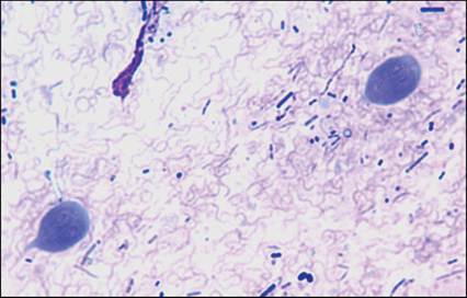
Figure 7.57 Fecal sample from a dog with Giardia spp. (Wright–Giemsa, 1,800? magnification) (courtesy Anne Barger)
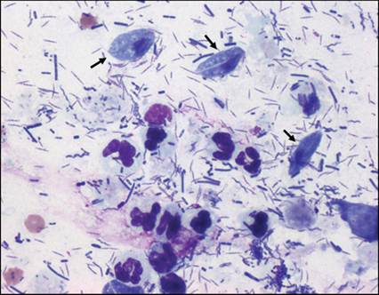
Figure 7.58 Fecal sample from a cat with Tritrichomonas foetus (arrows) (Wright–Giemsa, 800? magnification) (courtesy Mark Dunbar).
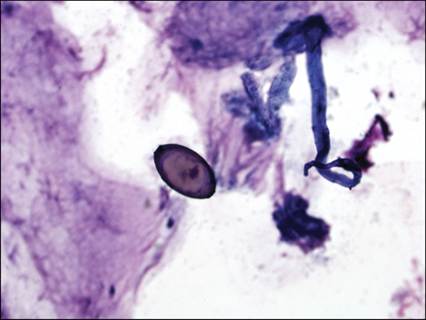
Figure 7.59 Rectal scrape from a dog with colitis. A large double-operculated ovum consistent with Trichuris vulpis is present (Wright–Giemsa, 200? magnification).
Neoplasms of the large intestines
Epithelial tumors
In the rectum, benign tumors are more frequently reported than at other locations within the GIT. Indeed, benign epithelial tumors appear to occur with equal frequency to malignant tumors in dogs. In cats, however, only a few reports of benign epithelial proliferations are found (Kehl et al., 2022). Benign epithelial tumors include adenomatous polyps, which are described as sessile, raised, pedunculated, single, or multiple mass lesions. Diagnosis of benign tumors by cytology can be difficult as differentiation from hyperplastic proliferation is often not possible. Therefore, the definitive diagnosis of such lesions usually necessitates surgical biopsy and histopathology (Valerius et al., 1997; Danova et al., 2006; Willard, 2012; Munday et al., 2017).
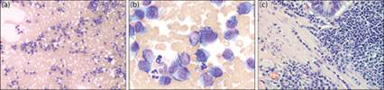
Adenocarcinomas of the intestines are generally more common in cats than in dogs. In dogs, however, they tend to be located in the colon and rectum. Unlike cats, where Siamese cats are overrepresented, a breed predisposition is not consistently seen in dogs; however, progression from benign polyps to adenocarcinoma is speculated (Slawienski et al., 1997; Saito et al., 2018). Cytologically, colonic carcinomas/adenocarcinomas are similar to tumors at other locations and are described more fully above (Figures 7.60a, b).
Figures 7.60a,b Cytology obtained from an FNA of a colonic lesion from a dog. (a) Two large aggregates of epithelial cells are present. At 200? magnification, criteria for malignancy are already apparent. Note the deep basophilia and multiple nucleoli (arrow). (b) Epithelial cells often maintain some semblance of normal shape but exhibit criteria for malignancy consistent with adenocarcinoma (Wright–Giemsa; a, 200? magnification; b, 600? magnification).
Stromal/spindle cell tumors
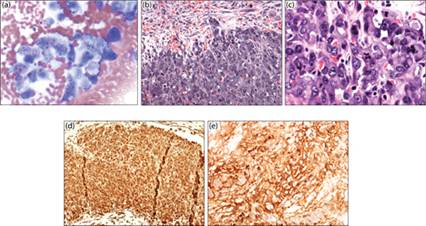
As previously stated, smooth muscle tumors (leiomyosarcoma, leiomyoma) appear to occur more commonly in the stomach and throughout the small intestine in dogs; however, GISTs are more commonly reported in the large intestines (Frost et al., 2003; Russell et al., 2007; Gillespie et al., 2011; Hayes et al., 2013). Only a few cases of large intestinal spindle cell tumors in cats are found in the veterinary literature (Rissetto et al., 2011; Kehl et al., 2022). As stated previously, differentiation between GISTs and smooth muscle tumors requires biopsy and immunohistochemical characterization. GISTs are positive for vimentin and KIT (Figures 7.61a, b) but negative for desmin, whereas smooth muscle tumors (Figures 7.62a–d) are KIT negative but more consistently positive for SMA and desmin (Frost et al., 2003).
alt=fig7.61.jpg>
Figures 7.61a,b Gastrointestinal stromal tumor in the cecum of a dog. (a) Histology section of the tumor shows interlacing neoplastic spindle cells adjacent to normal tissue (H&E, 100? magnification). (b) Tumor cells show marked reactivity for KIT expression (HRP polymer anti-rabbit IgG with Nova Red chromogen and hematoxylin counter stain, 100? magnification).
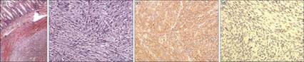
Figures 7.62a–d Histologic section of a leiomyosarcoma in the colon of a dog. (a) A large expansile mass is found invading into the submucosa (H&E, 40? magnification). (b) Tumor cells are spindle-shaped and found in streams (H&E, 200? magnification). (c) Tumor cells are positive for smooth muscle actin (HRP polymer anti-rabbit IgG with Nova Red chromogen and hematoxylin counter stain, 200? magnification). (d) Tumor cells are negative for KIT expression (HRP polymer anti-rabbit IgG with Nova Red chromogen and hematoxylin counter stain, 200? magnification).
Other sarcomas may arise de novo in the colon or metastasize from other locations, although these appear to be uncommon in the literature. Rare cases of histiocytic sarcomas, hemangiosarcomas, and fibrosarcomas have been described in the large intestine of both dogs and cats (Bonfanti et al., 2006; Shor et al., 2009; Rissetto et al., 2011; Kehl et al., 2022). The cytology of spindle cell tumors is described above. As they often appear both cytologically and histologically similar, special staining is usually necessary for definitive identification of the tissue of origin (Figures 7.63a–e).
Figures 7.63a–e Mass associated with the intestine of a dog. Disseminated neoplasia was identified post-mortem and definitively diagnosed as hemangiosarcoma by immunohistochemical evaluation. (a) Cytology of a large intestinal lesion showing markedly atypical spindle cells (Wright–Giemsa, 200? magnification). (b) Histology of the intestinal mass, which has effaced the submucosa at this location (H&E, 100? magnification). (c) Higher magnification of the mass demonstrates the anaplastic appearance of the neoplastic cells (H&E, 500? magnification). (d, e) Tumor cells are positive for vimentin (d) and von Willebrand expression (e).
Round cell tumors
Lymphoma
Lymphoma is considered the second most common tumor in the colon of cats (Rissetto et al., 2011) but appears to occur infrequently in the large intestine of dogs (Frank et al., 2007). In both species, colonic lymphomas are more often of B-cell lineage. In cats, B-cell large intestinal lymphoma is associated with a poorer long-term prognosis (Lingard et al., 2009; Moore et al., 2012). Contrary to cats, longer survival times are reported in dogs with colorectal lymphomas than in dogs with either gastric or small intestinal lymphoma (Frank et al., 2007; Van den Steen et al., 2012).
Similar to lymphoma in other sites, diagnosis is made by identifying a predominance of lymphoid cells, typically large lymphocytes, often with increased mitotic activity.
Plasma cell tumors
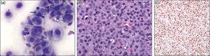
Extramedullary plasmacytomas are relatively common in dogs and may be associated with mucocutaneous junctions; however, only a few cases of intestinal localization (mainly colorectal) have been reported in dogs or cats (Ramos-Vara et al., 1998; Zikes et al., 1998; Danova et al., 2006; Kupanoff et al., 2006). The mucosa overlying the mass is often eroded or ulcerated and may be covered by fibrinonecrotic exudate. Although neoplastic plasma cells may be confined to the submucosa, infiltration into the lamina propria and muscle layers may also occur. Cytologically, the tumor cells are typical for plasma cell tumors at other sites and are often solitary; however, multifocal masses can occur (Figures 7.64a–c). Cells may be pleomorphic and binucleate and multinucleate cells are commonly noted, thus immunochemical staining may be necessary for full differentiation from other round cell tumor types (Figures 7.65a–c) (Kupanoff et al., 2006; Munday et al., 2017). Most studies indicate that these tumors are not aggressive and metastasis or progression to myeloma is rare.
Figures 7.64a–c Dog with multiple colonic plasma cell tumors and progression to myeloma syndrome. (a) FNA of one of the masses shows a relatively monomorphic population of round cells, among a background of erythrocytes (Wright–Giemsa, 200? magnification). (b) At higher magnification cells are discrete in appearance and characteristic for atypical plasma cells (Wright–Giemsa, 1000? magnification). (c) Histology of the mass shows expansion of the mucosa and submucosa by infiltrating plasma cells (H&E 400? magnification).
Figures 7.65a–c Plasma cell tumor in the distal rectum of a dog. (a) FNA of the mass shows a more pleomorphic population of round cells in this lesion. Golgi bodies (arrows) can still be discerned in several cells. Multinucleated cells are frequent (Wright–Giemsa, 500? magnification). (b) Histology of the same mass. Similar cellular atypia, including multinucleation, is noted in the biopsy sample (H&E, 500? magnification). (c) Neoplastic cells are positive for MUM1, a marker expressed by plasma cells (H&E, 500? magnification).
Mast cell tumors
Mast cell tumors are often localized to the large intestines of cats, but are rarely diagnosed in dogs (Ozaki et al., 2002; Rissetto et al., 2011; Sabattini et al., 2016). Similar to that noted in the small intestine above, these may occur as solitary mass lesions, but are often multifocal. Metastasis and paraneoplastic syndromes may be present at diagnosis in both species (Figure 7.66a–c).
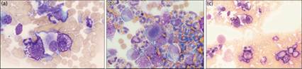
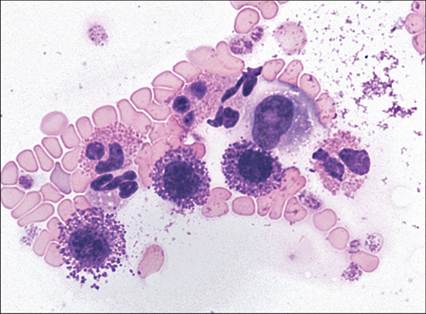
Figures 7.66a–c Colonic mast cell tumor in a dog with multifocal disease. (a) Cytology of the colonic mass is hemodilute with small clusters of well-granulated mast cells (Wright–Giemsa, 1000? magnification). (b) Abdominal fluid from the same patient. Large numbers of eosinophils are found among frequent atypical mast cells. (c) Splenic metastasis (Wright–Giemsa, 400? magnification).