Microscopic Anatomy of Tissues
OBJECTIVES
• define and describe the four types of tissues and their major subcategories
• describe the differences between simple, stratified, and pseudostratified layers of epithelial cells
• identify all forms of epithelial tissue, their component cells, and other structures, from prepared slides
• identify all forms of connective tissue (both connective tissue proper and special connective tissues), their component cells, and other structures
• identify the three forms of muscle tissue, their component cells, and other structures
• identify a nerve cell and its component parts
• locate the various tissue types in an animal’s body
MATERIALS
• compound light microscope
• immersion oil
• colored pencils
• prepared slides of the following types of epithelial cells: simple squamous (or use kidney section with glomeruli or “air sacs of the lungs”); simple cuboidal (or use kidney section with renal tubules); simple columnar (or use lining of small intestine); pseudostratified ciliated columnar (or use trachea); stratified squamous (non-keratinized, such as tongue); stratified cuboidal; stratified columnar (or use urethra, male); and transitional (or use lining of the urinary bladder)
• three-dimensional model of a glomerulus and Bowman’s capsule
• prepared slides of the following connective tissues: mesenchyme; loose irregular/areolar (or use loose smear or subcutis of intestinal tract); dense irregular (or use dermis of skin); dense regular (or use tendon or ligament); elastic (or use nuchal ligament); hyaline cartilage (or use trachea); elastic cartilage; fibrocartilage; reticular connective tissue; blood smear; and compact bone
• prepared slides of skeletal, cardiac, and smooth muscle (longitudinal sections)
• differential cell counter
Introduction
As stated in the previous chapter, a tissue is a group of cells of similar structure united in the performance of a particular function.
There are four primary tissue types: (1) epithelium,(2) connective tissue, (3) nervous tissue, and (4) muscle.
Each of these tissue types has distinctive structures, patterns, and functions. Each type can be further divided into subcategories, which we will cover in this chapter.To perform specific body functions, tissues are organized into organs. The intestinal tract absorbs nutrients through its lining cells, the heart muscle pumps blood throughout the body, and liver cells detoxify the waste products of the body. All these are examples of the special cellular functions that identify the nature of these organs. Because most organs contain numerous types of tissue, the arrangement of these tissues is critical to the structural shape
of the organ and how it functions. Only by understanding the normal cellular arrangement of tissues are we able to recognize the abnormal when it presents itself. Also, to understand the physiology of a particular organ, you need a mental picture and an understanding of how that organ is structurally arranged. You can appreciate how the skin acts as a barrier to microorganisms entering the body if you know the epithelial cell histology, and that the skin is thick and multilayered. Similarly, if you know that the lining of the respiratory system, where the alveoli of the lungs are located, is only one cell layer thick, you can understand that this is an easier portal for invasion.
Epithelial Tissue Histology
The epithelia, or epithelial tissues, are a special group of tissues designed to cover the external surface and line the internal surfaces (including the tubules and vessels) of an animal’s body. All epithelial cells are supported by a basement membrane that separates them from the underlying connective tissues. Cells are the principle component of epithelial tissue. The intercellular substance is sparse and only consists of a very thin layer of material, located between the cells, that helps hold them together. This is known as intercellular cement. Epithelial tissues always have a free surface, which faces the exterior of the body, the cavity of a hollow organ, the lumen of a tubular organ, or the lumen of a duct or vessel.
All cells of epithelial tissues that are only one cell layer thick line (touch) the free surface. In types of epithelial tissues that are multilayered, most of the cells do not touch the free surface. Instead, only the surface cells line the free surface. The free-sur- face cells may possess cilia, microvilli, or stereocilia.The functions of the epithelium are to protect the body; absorb, filter, excrete, and secrete substances; and detect sensations. The cell’s accessory structures also aid in their function. For example, the cilia attached to the epithelium lining the lungs help sweep away dust and other foreign particles. The following are characteristics that distinguish epithelia from other types of tissues.
1. Intercellular contacts: The cells fit closely together, forming membranes, or sheets of cells, and are bound together by intercellular cement and specialized junctions.
2. Surface cells: The membranes always have one free surface, called the apical (or free) surface. (Note: Unfortunately, in anatomy, as well as other disciplines of medicine, it is not uncommon to find two or three words meaning the same thing.) Either term is correct, and it is not uncommon to find two authors using different terms to mean the same thing. For example, in two A&P (anatomy & physiology)
books, both published in the year 2000, one author used the term apical surface, the other free surface.
3. Supported by connective tissue: Epithelial tissues are attached on the side opposite the free surface (the tissue side, also called the basal surface) to a basement membrane. The basement membrane is an amorphous material secreted by the basal epithelial cells (basal lamina) and connective tissue cells (reticular lamina) that lie adjacent to each other.
4. Avascularity: Generally speaking, epithelial cells have no blood supply; they are avascular. There are no capillaries that extend into the epithelium. Instead, epithelial cells are dependent on diffusion of nutrients from the underlying connective tissue.
The exception is glandular epithelium, which is very vascular.5. Regeneration: If epithelial cells are well nourished, they can easily regenerate themselves. This is an important characteristic considering where these cells are located and the trauma to which they are exposed.
One method of classifying epithelial tissues is based on the number of layers each possesses. Simple epithelium consists of a single layer of cells, whereas stratified epithelium contains two or more layers. The second major method of classifying epithelia is according to cell shape. Squamous cells are flat, thin, or scale-like. On cross section, they are usually only as tall as their nucleus. Cuboidal cells are cubelike, equal in height and width. Columnar cells are column-shaped, much taller than they are wide. These two classifications are combined to designate and classify most of the various types of epithelial tissue: simple squamous, simple cuboidal, simple columnar, stratified squamous, stratified cuboidal, and stratified columnar. Note that stratified epithelium is named for the shape of the cells on its apical surface, rather than those on their basal surface.
Two other types of epithelial cells are less easily classified. Pseudostratified epithelium is actually a form of simple columnar epithelium. All its cells attach to the basement membrane, but because the cells vary in height and some cells do not extend to the free surface, and since their nuclei lie at different levels above the basement membrane, it gives a false appearance of being stratified. Pseudostratified epithelium is usually ciliated on its free surface. Transitional epithelium is a variation of stratified squamous epithelium and is only found in the urinary bladder, proximal urethra, and distal ureters. When the bladder is not distended with urine, the cells are rounded. However, when the bladder is filling, these cells have the ability to change shape and slide over one another, allowing the organ to stretch.
The superficial cells are flattened (like true squamous cells)when the bladder is completely full. Therefore, transitional epithelium appears, in the distended form, as though the number of cell layers it contains has diminished.
The epithelial cells that form glands are highly specialized to remove compounds from the bloodstream and manufacture them into new substances, which they then secrete. Endocrine glands lose their ducts as they develop in the fetus and secrete their product into the blood or lymph directly. These glands produce hormones. Exocrine glands retain their ducts and secrete onto an epithelial surface. Exocrine glands can be found on the surface of the body (sweat and oil glands) or internally, such as those found in the pancreas, which secrete their products through the pancreatic ducts onto the epithelial surface of the duodenum (the first part of the small intestine).
EXERCISE 5.1 EPITHELIAL TISSUE HISTOLOGY
The procedure for this and subsequent exercises in this chapter is as follows:
1. When viewing the recommended types of tissues, first go to low power, focus, and orient yourself on the slide (i.e., find the free surface vs. the deeper structures of the tissue). Then, change to high power and view the necessary cellular detail to understand the morphology of the tissue. To see certain cells, such as blood cells, macrophages, mast cells, or plasma cells, it will be necessary to use the oil immersion lens.
2. For the types of tissues in these exercises, obtain the recommended slide(s) and locate all the things you are instructed to view and that are labeled in the diagrams and photomicrographs featured as figures throughout the chapter. Then, draw and label what you see in the space provided at the end of each section. Items you will be instructed to find and observe will include a certain type of cell, its nucleus and cytoplasm, the basement membrane, connective tissue fibers, and ground substance. You should find all items labeled in the drawings.
Structures to be identified are listed in colored bold print. Subsequent slides may also contain the same structures or other related structures, which may be listed in bold print or italics; they are of no less importance and therefore should also be identified. For instance, when viewing the epithelial slides, the basement membrane is only in colored bold under simple squamous epithelium. However, it is still important to try to find it in all the slides of epithelial tissue, even though it is difficult to see on certain slides.Simple Squamous Epithelium
Slides recommended: Slides labeled simple squamous epithelium (surface view), kidney, and/or air sacs of lungs.
Description: Simple squamous epithelium contains a single layer of flattened cells with rounded or oval central nuclei, and sparse cytoplasm. It is the thinnest of the epithelial tissues (Figure 5.1). Often it is hard to see the basement membrane, but it is always present on the opposite side of the free surface.
Location found: Simple squamous epithelial cells are found in the kidney, alveoli of the lungs, blood vessels, lining of the heart, lymphatic vessels, lining the thoracic and peritoneal cavities, and on the serosal surface of the abdominal and thoracic organs. The latter is also called the mesothelial surface, and the mesothelial cells are simple squamous in nature. The epithelial lining of the vascular system has a special name: endothelium, or endothelial cells.
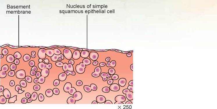
FIGURE 5.1: Cross section of simple squamous epithelial cells that make up the serosal surface of an organ (i.e., the covering layer).
Function: Because it is composed of a thin, single layer of cells, simple squamous epithelium permits passage of materials by diffusion and filtration in sites where protection is not necessary. For example, the mesothelial cells secrete a lubricating substance for the pleural and peritoneal cavities.
Slide: simple squamous epithelial cells (surface view)
This is a view of a sheet of epithelial cells looking both down on its surface and from the side. The cells appear to have asymmetrical shapes (Figure 5.2). All cells may or may not appear to have nuclei, depending on the level at which the cut was made. Making a cut so that each nucleus is visible within the cell is easier when the nuclei are in the same location in each cell or when the cells are short rather than tall.
Slide: kidney
There are several places to observe simple squamous epithelial cells easily. These include the kidney’s nephron (specifically Bowman’s capsule) and the capillaries (Figure 5.3). In the section of kidney you should easily find many large renal tubules. Look between these tubules and you will see a cross-sectional cut of numerous smaller tubules formed from very thin cells. These tubules are the peritubular capillary network of the kidneys. Each capillary is formed by two or three simple squamous epithelial cells linked in a circle.
In other parts of the kidney, note the rounded tufts of capillaries with a white space around them. On the slide they may appear as islands of tissue. Each is called a glomerulus. On the perimeter of the white space are the squamous epithelial cells that form Bowman’s capsule. The cells may be so flat that they only bulge where their nuclei are located. The white space is the inside of Bowman’s capsule, where the fluid filtered from the capillaries (called glomerular filtrate) enters the renal tubules. Also find these cells and this space on the plastic model of the nephron.
Slide: air sacs of the lungs
The cells that form the walls of the air sacs, or alveoli of the lungs, are made of simple squamous epithelial cells (Figure 5.4). On the slide you will note many large, white, irregularly shaped spaces.
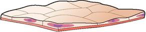
FIGURE 5.2: Flattened epithelial cells.
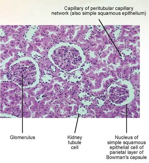
FIGURE 5.3: Kidney epithelial tissue (Bowman's capsule), showing simple squamous epithelium.
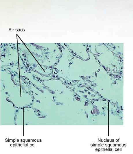
FIGURE 5.4: Lung epithelial tissue, showing simple squamous epithelium.
These are the alveoli, which are lined with a thin monolayer of simple squamous epithelial cells. Also look for small capillaries between the alveoli; these also are composed of simple squamous epithelium.
In the space below, using the slide that best represents simple squamous epithelial tissue, draw and label the parts of the tissue and cells.
Simple Cuboidal Epithelium
Slides recommended: simple cuboidal epithelium or kidney tissue.
Description: Simple cuboidal epithelium is composed of a single layer of cube-shaped cells with large, spherical, centrally placed nuclei (Figure 5.5). Cytoplasm is abundant. The basement membrane, on the opposite side of the free surface, can sometimes be observed.
Location found: This type of tissue can be found in the kidney tubules, ducts and secretory portions of small glands, and peripheral cells of the ovaries just deep to the serosal surface.
Function: Its function is secretion and absorption of fluid and electrolytes.
Slide: kidney or simple cuboidal epithelium
The best examples of simple cuboidal epithelial cells (Figure 5.6) are the cells that form the tubules of the nephron of the kidneys. In the section of kidney you should easily observe many large tubules, some oval and some more elongated. Because these tubules are convoluted in structure (look at the plastic model of the kidney’s nephron), on a cut section some will be perfect cross sections and others will be more oblique, giving the appearance of an elongated tubule. Because these cells are forming a tube, they are not perfectly square. Instead, they are usually a little narrower at the lumen of the tubule than at the basement membrane. Between the tubules are the capillaries mentioned previously.
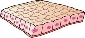
FIGURE 5.5: Simple cuboidal epithelial cells.
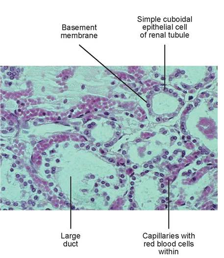
FIGURE 5.6: Simple cuboidal epithelial cells lining the tubules of the kidney.
Simple Columnar Epithelium
Slides recommended: Slides labeled simple columnar epithelium or small intestine (duodenum, jejunum, or ileum).
Description: Simple columnar epithelium consists of a single layer of tall cells with oval-shaped nuclei (Figure 5.7).The nuclei are located toward the base of the cell, rather than in the center or toward the free surface. Cytoplasm is abundant. The basement membrane, on the opposite side of the free surface, can sometimes be observed.
Location found: Non-ciliated simple columnar epithelium lines most of the digestive tract (stomach to anus), gallbladder, and the excretory ducts of some glands. Ciliated types line parts of the small tertiary bronchi of the lungs, the oviducts, and the uterus.
Function: This tissue’s function is the absorption of nutrients and fluids and the secretion of mucus, enzymes, and other substances. The ciliated types propel mucus (or reproductive cells) by ciliary action.
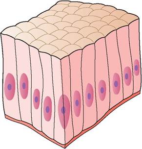
FIGURE 5.7: Simple columnar epithelial cells.
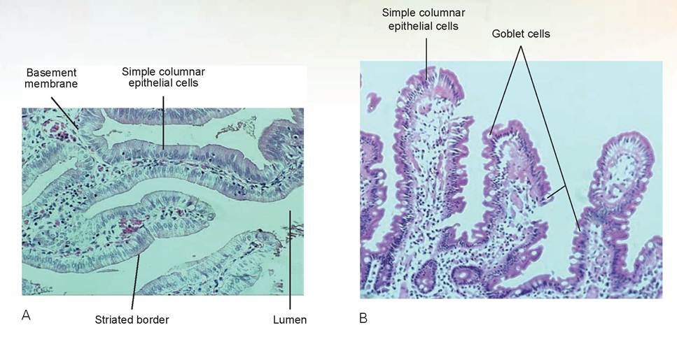
FIGURE 5.8: A. Simple columnar epithelial tissue from the small intestine. B. Similar epithelial tissue with goblet cells.
Slide: small intestine (jejunum) or simple columnar epithelium
The best examples of simple columnar epithelial cells are the cells that line the lumen of the intestinal tract (Figure 5.8A). Search the slide until you find the place where the tissue section perfectly bisects the intestinal lining, and the cells and their nuclei form a single row, side by side. The border of the free surface should appear as a thickened membrane; however, under oil immersion, if you look closely, you will note that it actually appears striated because it has a brush-like surface. This is due to the presence of microvilli and is called a striated border. Microvilli increase the surface area and absorptive capacity of the cells. As you look at the rows of columnar cells you will see oval areas between them. These are goblet cells (Figure 5.8B), which are mucus-secreting, unicellular glands. On some slides they may appear hollow, and on others they may contain a granular substance, which is mucus after processing.
In the space below, using the slide of simple columnar epithelial tissue, draw and label the parts of the tissue and cells.
Pseudostratified Columnar Epithelium
Slides recommended: Those labeled pseudostratified columnar epithelium or trachea.
Description: Pseudostratified columnar epithelium consists of a single layer of cells of varying heights (Figure 5.9). All cells are attached to the basement membrane, which is sometimes visible. Not all cells, however, reach the free surface. The nuclei may be seen at different levels, and some even appear to be stacked on top of one another. This gives the appearance that this tissue is stratified, but it is not—hence the name pseudostratified. Most of the cytoplasm is seen just below the free surface and above the nuclei. Goblet cells also are seen interspersed between these cells. Cilia are present on the free surface; therefore, this tissue is also called pseudostratified ciliated columnar epithelium.
Location found: The ciliated type of pseudostratified columnar epithelium lines the trachea and continues down the respiratory tract to the first part of the tertiary bronchi. The non-ciliated type is found in the male’s sperm-carrying ducts and the ducts of large glands.
Function: This tissue is active in secretion, particularly of mucus, and in propulsion of debris out of the respiratory system via ciliary action.
Slide: trachea or pseudostratified columnar epithelium
Note that some prepared slides labeled ciliated columnar epithelium are, in fact, pseudostratified ciliated columnar epithelium. Find the free surface of the section of trachea (Figure 5.10). This is best done by finding the cells with the small, hair-like cilia on the surface. The epithelial cells lining the respiratory (such as these from the trachea), digestive, and reproductive tracts are also known as
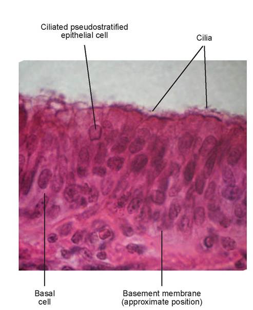
FIGURE 5.10: Pseudostratified ciliated columnar epithelial tissue from the trachea (not a perfect cross section, not unusual, so there appears to be more than one layer).

FIGURE 5.9: Pseudostratified ciliated simple columnar epithelial cells.
mucosal cells. Mucosal cells line mucous membranes. Note the numerous goblet cells present. On some slides you may note that the goblet cells are releasing some of the mucus from inside the cell onto the free surface.
In the space below, using the slide of pseudostratified ciliated columnar epithelial tissue, draw and label the parts of the tissue and cells.
Stratified Squamous Epithelium
Slides recommended: Those labeled stratified squamous, esophagus, or tongue.
Description: Stratified squamous epithelium is a thick epithelial surface composed of multiple cell layers (Figure 5.11). It is named for the appearance of the cells on the surface, which are squamous. Only the basal cells are attached to the basement membrane. The basal cells are cuboidal, or even columnar, and they are metabolically active. Mitosis occurs in the first few layers above the basement membrane and produces the cells to replace the cells of the more superficial layers as they die and slough off. The nuclei are easily seen in the lower layers of cells, but they tend to become smaller and disappear as they move toward the free surface where the cells are squamous.
Location found: The non-keratinized type of stratified squamous epithelium forms the moist lining of the mouth, esophagus, part of the stomach, the rumen, reticulum, omasum, and vagina. The keratinized type forms the epidermis of the skin.
Function: This tissue protects underlying tissues in areas subjected to abrasion.
Slide: tongue, esophagus, or stratified squamous epithelium
Locate the free surface of the tissue, where the cells appear more flattened or squamous in appearance (Figure 5.12). This is an example of non-keratinized stratified squamous tissue. Note that the cell layers go all the way to the free surface, where moisture is present. If it were keratinized, there would be two acellular layers of dry, cornified tissue, as is found in the epidermis of the skin. Considering the foods that contact the surface of these tissues, it is easy to understand why a thick, protective, mucosal surface is needed. The surface of the body also requires a thick layer of cells that are capable of regeneration to protect the body from dehydration, cellular desiccation, and the ubiquitous microorganisms of the external world. This is why the epidermis of the skin has extra layers of cornified tissue for protection. We will study the histology of keratinized stratified squamous epithelium in Chapter 6.
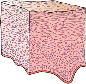
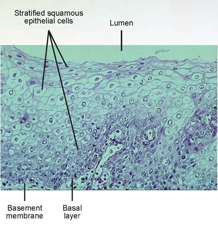
FIGURE 5.12: Stratified squamous epithelial tissue from mucous membranes in the oral cavity.
FIGURE 5.11: Stratified squamous epithelial cells.
In the space below, using the slide of stratified squamous epithelial tissue, draw and label the parts of the tissue and cells.
Stratified Cuboidal Epithelium
Slides recommended: Those labeled stratified cuboidal, salivary glands, or esophagus.
Description: Generally two layers of cuboidal cells form a stratified cuboidal epithelium (Figure 5.13).
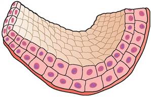
FIGURE 5.13: Stratified cuboidal epithelial cells.
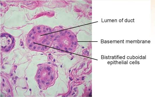
FIGURE 5.14: Stratified cuboidal epithelial tissue from the ducts of Sudoriporous glands.
Location found: This tissue is found in the largest ducts of sweat glands, mammary glands, and salivary glands, and in the glands found in the esophagus.
Function: Their purpose is protection and the transportation of fluid.
Slide: salivary glands, esophagus, or stratified cuboidal epithelium
Search for ducts containing two layers of cuboidal cells (Figure 5.14). They will have a relatively large elliptical lumen surrounded by the bilayered, cube-shaped cells.
In the space below, using the slide of stratified cuboidal epithelial tissue, draw and label the parts of the tissue and cell.

Stratified Columnar Epithelium
Slides recommended: Those labeled stratified columnar or urethra.
Description: Stratified columnar epithelium appears to resemble both stratified columnar and stratified cuboidal cells. The surface cells are columnar, but the basal cells and intermediate cells are more cuboidal (Figure 5.15).
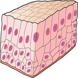
FIGURE 5.15: Stratified columnar epithelial cells.
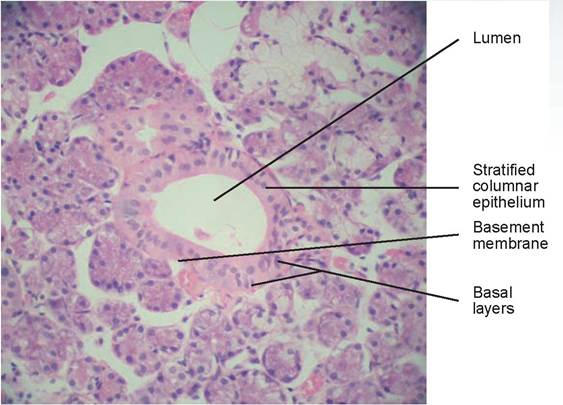
FIGURE 5.16: Stratified columnar epithelial tissue from the duct of a gland.
Location found: This tissue is found in areas of the urethra of male animals and in large ducts of some glands.
Function: It functions in protection, secretion, and transporting fluid.
Slide: urethra or stratified columnar epithelium
On the free surface, look for a multilayered mucosal epithelium of tall columnar cells with numerous layers of cuboidal-shaped cells beneath it, deep to the basement membrane (Figure 5.16). The nuclei of the superficial columnar cells are at the same level in each cell, and they are centrally located. The deeper nuclei and their cells appear more random in their distribution.
In the space below, using the slide of stratified columnar epithelial tissue, draw and label the parts of the tissue and cells.
Transitional Epithelium
Slides recommended: Those labeled transitional epithelium or urinary bladder.
Description: Transitional epithelium resembles both stratified cuboidal epithelium (when unstretched) and stratified squamous epithelium (when stretched) (Figure 5.17). The basal cells are cuboidal or slightly rounded. Remember, as stated previously, these cells have the ability to slide across each other and flatten out to become squamous-like when the bladder fills with urine and the wall stretches.
Location found: Transitional epithelium is found in the urinary bladder, and it lines the ureters and part of the urethra.
Function: It participates in the storage of urine by readily permitting distention of the urinary bladder, and it supports the transporting of urine to the bladder through the ureters and out of the body via the urethra.
Slide: urinary bladder or transitional epithelium
Find the surface cells that are dome-like, with centrally placed nuclei (in the unstretched urinary bladder, which is probably what your slide shows) (Figure 5.18). In contrast, find the smaller basal cells and the basement membrane to which they are attached.
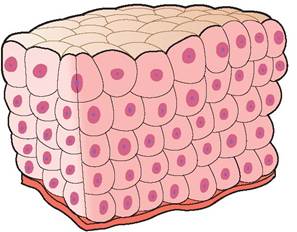
FIGURE 5.17: Transitional epithelial cells.
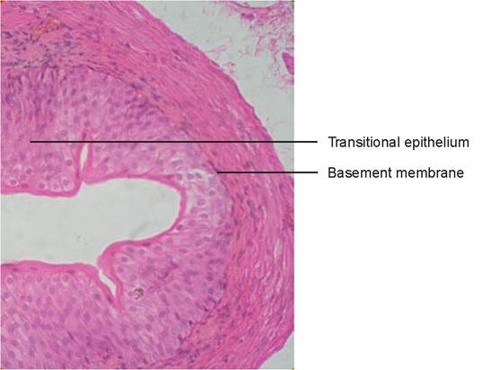
FIGURE 5.18: Transitional epithelial tissue, unstretched, from the urinary bladder.
EXERCISE 5.2 CONNECTIVE TISSUE HISTOLOGY
Connective tissue is found everywhere in the animal’s body. It binds tissues together, supports other types of tissues, and, in the case of blood, transports nutrients and oxygen. All connective tissues have three common characteristics: (1) they contain cells, which are their living component; (2) they contain fibers, which are produced by the cells; and (3) they contain a ground substance, which is composed largely of glycoproteins and glycosaminoglycans. In connective tissue, the latter two components compose the extracellular matrix, which predominates over the cellular elements. The ground substance forms a well-hydrated gel that fills the spaces between cells, fibers, and the vascular components of connective tissue. It varies in composition depending on the type of connective tissue, but generally it acts as a reservoir for interstitial fluid, providing a medium through which nutrients, oxygen, and metabolic by-products diffuse to and from cells between the connective tissue and the vascular system.
Connective tissue can be divided into three major types: embryonal, proper, and special connective tissues. Mesenchyme and mucous connective tissue are classified as embryonal connective tissue. Connective tissue proper (also known as proper connective tissue) includes the general types of connective tissue: loose and dense; regular and irregular; and reticular, elastic, and adipose. Special connective tissue includes tissues such as blood, cartilage, and bone.
There are three types of fibers found in connective tissue proper: (1) collagenous, (2) reticular, and (3) elastic. Collagenous fibers are composed of the protein collagen (which is actually the protein tropocollagen, which polymerizes to become collagen). Collagen is composed of strong, flexible fibers, which may be fine or coarse. They are characteristically unbranched and somewhat wavy, and they resist stretching. They stain pink with hematoxylin and eosin (H&E) stain. Reticular fibers are also composed of collagen; they are delicate, branching fibers possessing a coat of glycoproteins and proteoglycans. Silver-containing stains are used to differentiate these fibers from other fibers in connective tissue. Elastic fibers are formed from the protein elastin. They are typically fine fibers in areolar connective tissue, but in other places they can be coarse. They sometimes stain darker pink with H&E stains, or other special staining techniques also can be used to distinguish these fibers.
Fibroblasts are the most numerous type of cells found in connective tissue proper. They are responsible for producing the fibers and the ground substance.
Embryonal Connective Tissue, or Mesenchyme
Slides recommended: Those labeled either mesenchyme or embryonal connective tissue.
Description: This tissue is destined to become either connective tissue proper or special connective tissue (Figure 5.19 and 5.20). It has a soft, gel-like ground substance containing fibers and stellate (starshaped) mesenchymal cells.
Location found: These cells are found primarily in the embryo.
Function: Serves as a precursor to all other connective tissue types.
Slide: mesenchyme or embryonal connective tissue
Note the many stellate mesenchymal cells with small nuclei located within (Figure 5.20). The clear background between the cells and fibers is the fluid-like ground substance. Also notice how fine and sparse the fibers are in this slide.
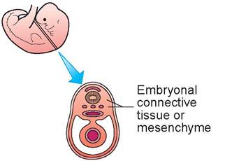
FIGURE 5.19: An embryo, showing where embryonal connective tissue is located.
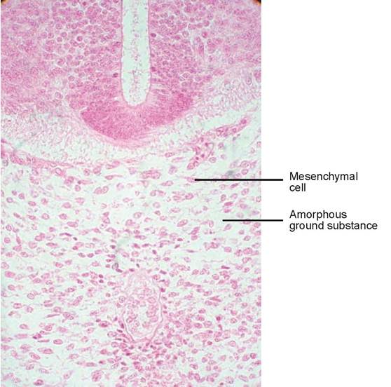
FIGURE 5.20: Embryonal mesenchymal tissue from a mammal.
Areolar or Loose Connective Tissue
Slides recommended: Those labeled areolar connective tissue.
Description: In areolar (loose) connective tissue, a gel-like ground substance predominates. All three types of fibers (collagenous, elastic, and reticular fibers) are present (Figure 5.21). There are many types of cells present, scattered about within the matrix. The majority of cells are fibroblasts, although macrophages (also known as histiocytes or tissue monocytes, which are white blood cells that have moved out of the bloodstream and into tissue), plasma cells (a type of tissue lymphocyte), and mast cells (cells that develop in the bone marrow in separate cell lines from white blood cells) can also be found. Other white blood cells, which include tissue eosinophils, neutrophils, basophils, and lymphocytes, can occasionally be seen, but they are in an unchanged state from what can be observed in blood.
Location found: Areolar connective tissue is found under the mucosal epithelium in most places of the body. It forms the lamina propria of mucous membranes, is found in the papillary layer of the dermis of the skin, is the connective tissue that attaches the epithelial cells of the serosa to organs, and surrounds capillaries.
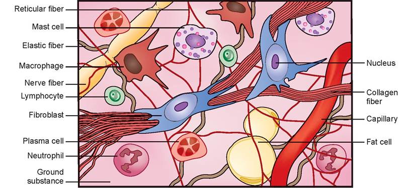
FIGURE 5.21: Areolar, or loose, connective tissue.
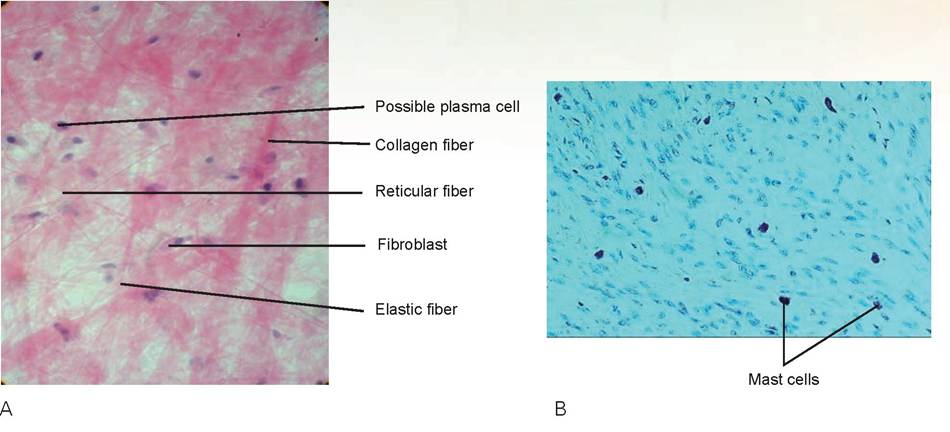
FIGURE 5.22: A. Areolar, or loose, connective tissue. B. Mast cells in areolar connective tissue (the dark granular cells).
Function: This tissue wraps and cushions organs, supports the mucosal epithelial cells, holds and conveys tissue fluids, and its immune cells help fight off infection by phagocytizing bacteria and other cells.
Slide: areolar connective tissue
Scan the slide and look for an area that contains the fibrous and cellular features found in this tissue: a fibroblast, which has visible cytoplasm and is fusiform in shape (is spindle shaped); thick collagen fibers; thin elastic fibers; hair-like reticular fibers; and, if you can find them, a mast cell; a macrophage; and a plasma cell (Figure 5.22A). Under H&E stain, the mast cell has many red granules, is irregularly shaped, and has a dark blue, round nucleus. Orcein or toluidine blue stains the mast cell’s granules purple and the nucleus blue (Figure 5.22B). The macrophage will be pinkish and elliptical to fusiform, with a dark blue nucleus. The plasma cell is slightly larger than the others, is elliptical, and has an eccentrically placed nucleus in which the stained chromatin appears almost spoke-like.
In the space below, using the slide of areolar connective tissue, draw and label the cells, fibers, and tissue parts.

Dense Irregular Connective Tissue
Slides recommended: Those labeled skin.
Description: Fibers predominate in dense irregular connective tissue. There are primarily irregularly arranged cords of collagen fibers, with some elastic fibers interspersed (Figure 5.23). The major cell type is the fibroblast.
Location found: This tissue makes up the dermis of the skin, the submucosa of the digestive tract, the capsule of organs and joints, the perichondrium around cartilage, and the periosteum.
Function: It supports the epidermis and provides the bulk of the strength for the skin, supports the joints, and provides strength and protection for bones and joints.
Slide: skin
Find the purple stained epidermal cells (which are stratified squamous epithelium); deep to these epithelial cells is where the dense irregular connective tissue is found (Figure 5.24). Note the pattern of abundant bands of thick, red collagen fibers. Note the irregular wavy pattern in which these bundles of collagen are weaved together. Also look for elastic fibers, which will appear thin. Locate the spindleshaped fibroblasts.
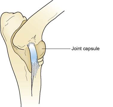
FIGURE 5.23: Fibrous joint capsule (from the elbow of a dog), where dense irregular connective tissue is located.
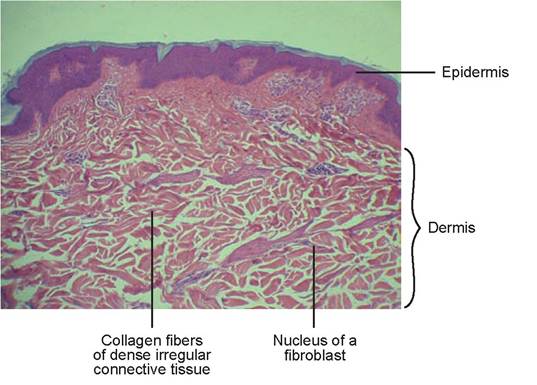
FIGURE 5.24: Dense irregular connective tissue located in the dermis.
Dense Regular Connective Tissue
Slides recommended: Those labeled tendon or ligament.
Description: Dense regular connective tissue is characterized by parallel collagen fibers. Few elastic fibers are present, and the major cell type is the fibroblast (Figure 5.25).
Location found: This tissue is found in ligaments, tendons, and aponeuroses.
Function: Tendons attach muscle to bone; ligaments attach bone to bone to support joints; and aponeuroses are sheets of tissue that give strength to the tissue to which they are attached. Dense regular connective tissue can withstand great tensile stress when force is applied in one direction.
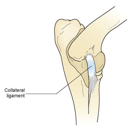
FIGURE 5.25: Ligament, where dense regular connective tissue is located.
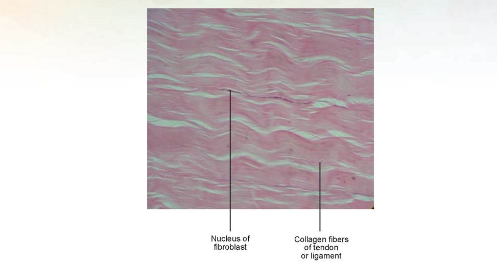
FIGURE 5.26: Dense regular connective tissue of a ligament.
Slide: tendon or ligament
Notice that the predominant feature of this slide is layer upon layer of parallel collagen fibers, which stain intensely pink (Figure 5.26). The nuclei of the fibroblasts are thin and elongated and found between the fibers; note that there are not very many in this tissue. Also, very little ground substance can be seen in this slide. The wave-like appearance is an artifact from processing; under natural conditions this tissue would be very linear in appearance.
In the space below, using the slide of dense regular connective tissue, draw and label the cells, fibers, and tissue parts.
Elastic Connective Tissue
Slides recommended: Those labeled ligamentum nuchae or mammal elastic connective tissue.
Description: Elastic connective tissue is characterized by numerous regularly and irregularly arranged elastic fibers (Figure 5.27). The major cell type is the fibroblast.
Location found: This tissue is found in the ligamentum nuchae, the large ligament that runs from the nuchal crest of the skull to the dorsal spinous processes of the thoracic vertebrae in grazing animals. It is also found in the vocal ligaments and in the walls of arteries and veins (Figure 5.28A).
Function: Elastic connective tissue supports the neck and head, and is responsible for phonation (the production of sound).
Slide: mammal elastic connective tissue
This slide shows the intensely pink-stained elastic fibers. Note that they lie parallel to each other. In comparison to a perfect slide of tendon or ligament, in which the collagen fibers line up almost perfectly parallel, elastic fibers tend to be wavier, are not as parallel, and generally contain more fibroblasts whose nuclei lie between the elastic fibers (Figure 5.28B).
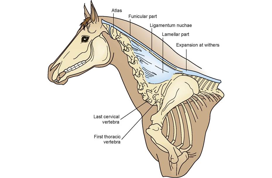
FIGURE 5.27: Ligamentum nuchae (from a horse), where elastic connective tissue is located.
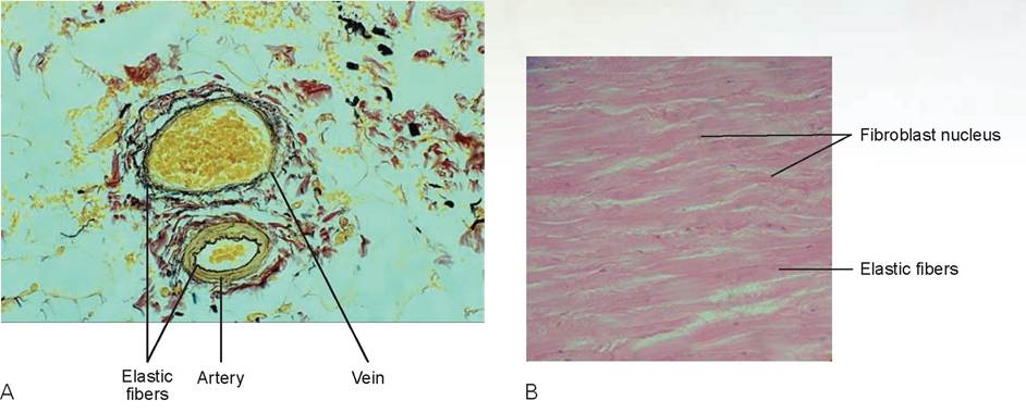
FIGURE 5.28: A. Elastic connective tissue in the walls of arteries and veins with a special stain to show the elastic fibers. B. Sheet of mammalian elastic connective tissue such as found in the ligamentum nuchae.
In the space below, using the slide of elastic connective tissue, draw and label the cells, fibers, and tissue parts.

Adipose Tissue
Slides recommended: Those labeled adipose tissue.
Description: Adipocytes, better known as fat cells, are predominant in adipose tissue (Figure 5.29). The nucleus of each cell is pushed to the side by the large intracellular vacuole containing the fat. White fat cells contain one vacuole (called monolocular), whereas brown fat contains multiple vacuoles (plurilocular). The matrix is similar to that of areolar connective tissue but is extremely sparse.
Location found: This tissue is found under the skin (subcutaneous fat), around the kidneys, within the omentum, in and around the mesentery of the digestive tract, around the kidneys (retroperitoneal fat), and in the mammary tissue. It also forms the marbling of muscle tissue.
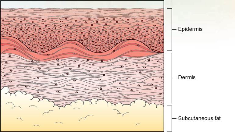
FIGURE 5.29: Subcutaneous fat, where adipose tissue is located.
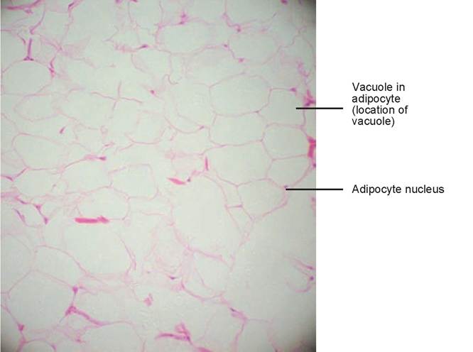
FIGURE 5.30: Mammal adipose tissue.
Function: Adipose tissue is used to store fuel for the body; it also insulates against heat loss and supports and protects organs.
Slide: adipose tissue
When viewing this slide, it may be necessary to dim your light or close down the condenser some to see the fat cells (Figure 5.30). Focus on an area of the slide where you can see clear definition of the cell walls and where the nuclei are visible. The large empty space between the cell walls is the vacuole that contains the fat. The nuclei are small and compressed by the large fat vacuoles of the cells.
Adipose Tissue
Reticular Connective Tissue
Slides recommended: Those labeled reticular connective tissue. This slide should be a special stained slide to demonstrate these cells and fibers.
Description: The fibers of reticular connective tissue form a network of fine reticular fibers, which are made of collagen and possess a coat of glycoproteins and proteoglycans (Figure 5.31). The fibers lie in a loose ground substance, and the reticular cells lie within the network.
Location found: This tissue is found in the liver, kidney, and lymphoid organs such as lymph nodes, spleen, and bone marrow.
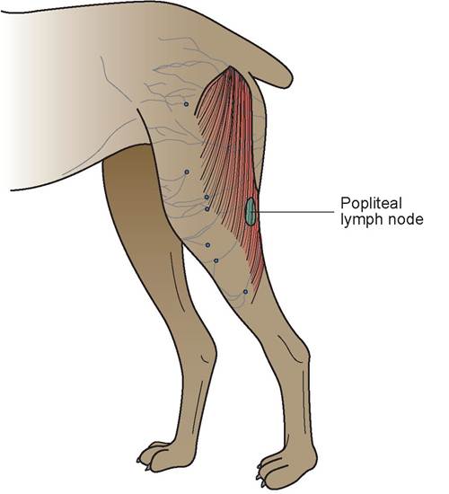
FIGURE 5.31: Popliteal lymph node of a dog, where reticular connective tissue is located.
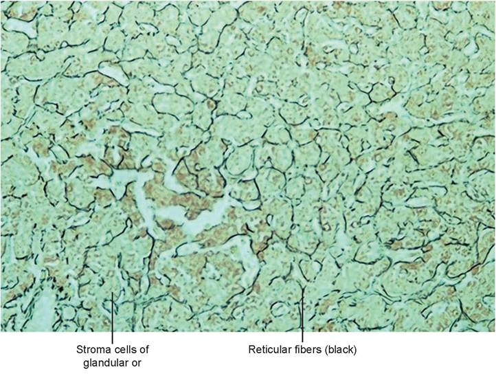
FIGURE 5.32: Mammal reticular tissue.
Function: The fibers of reticular connective tissue form the internal support structure, called the stroma, which supports the cells of these organs.
Slide: reticular connective tissue
Note the intensely dark-stained reticular fibers coursing between the bundles of cells. The reticular cells are smaller than the other cells, which also stain very dark, and they are adjacent to the fibers (Figure 5.32). The nuclei of the cells predominate, and there is little visible cytoplasm.
In the space below, using the slide of reticular connective tissue, draw and label the cells, fibers, and tissue parts.

EXERCISE 5.3 SPECIAL CONNECTIVE TISSUE
Special Connective Tissue: Cartilage
There are three basic types of cartilage: (1) hyaline, (2) elastic, and (3) fibrous, or fibrocartilage. Each type meets the same requirements as previously discussed types of connective tissue. However, the cells found in cartilage are called chondrocytes and chondroblasts (which are an earlier state destined to become chondrocytes; they produce the ground substance). The matrix is composed of an amorphous ground substance, which is rich in sulfated glycosaminoglycans, chondroitin sulfate, and hyaluronic acid. This is combined with an adhesion protein called chondronectin to form large molecules called proteoglycans, which are bound electrostatically to individual fibrils of collagen and elastin. The matrix is firm yet flexible, so it can bend when necessary yet support weight and resist tensile stress.
Hyaline Cartilage
Slides recommended: Those labeled hyaline cartilage or trachea.
Description: The ground substance makes up the bulk of hyaline cartilage tissue, and it can be separated into pale and darkly stained areas called the interterritorial matrix and territorial matrix, respectively (Figure 5.33).The latter, darker-staining areas contain a higher concentration of sulfated glycosaminoglycans. The chondrocytes (cartilage cells) are confined to small pockets called lacunae within the matrix. Often within a single lacuna are two newly divided cells, called an isogenous group, found with the matrix in a cluster of chondrocytes. The cartilaginous matrix is surrounded by a perichondrium. The outer portion of this is dense irregular connective tissue, and the inner layer is chondrogenic, containing cells with the capacity to become chondroblasts.
Location found: This tissue is found in the cartilages of the trachea, nose, and larynx. In addition, it covers the ends of the long bones in synovial joints, forms the costal cartilage of the ribs, and forms most of the embryonic skeleton.
Function: Hyaline cartilage composes the skeleton that supports the body of the growing fetus; forms a smooth surface for articularjoints; forms the structure of the costal ribs, nose, and larynx; and supports the structure of the trachea.
Slide: hyaline cartilage or trachea
Find the area of the slide where the cartilage is located. If you are viewing the trachea, it is the cross-sectional area of the tracheal rings (Figure 5.34). You should see a large, round mass of solid, pinkish-violet structure (the matrix) with numerous white holes (the lacunae) containing a cell within (a chondrocyte). The nucleus of the cell is dark, and the cytoplasm is very light. The collagen and elastic fibers are not visible because they blend imperceptibly within the matrix.
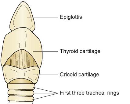
FIGURE 5.33: Ventral view of the larynx and trachea of a dog, where hyaline cartilage is found.
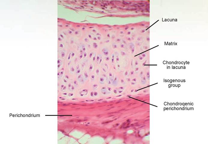
FIGURE 5.34: Mammal hyaline cartilage.
In the space below, using the slide of hyaline cartilage or trachea, draw and label the cells and tissue parts.
Elastic Cartilage
Slides recommended: Those labeled elastic cartilage.
Description: The matrix forms the bulk of elastic cartilage tissue, and it contains many of the same substances as hyaline cartilage except that it has a much larger amount of elastic fibers, for which it is named (Figure 5.35). The chondrocytes are inside lacunae located within the matrix.
Location found: This tissue is found in the cartilages of the epiglottis, parts of the larynx, and the pinna of the ear.
Function: It is able to support and maintain the shape of the structure while allowing great flexibility.
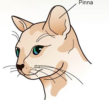
FIGURE 5.35: Pinna of a cat's ear, where elastic cartilage is found.
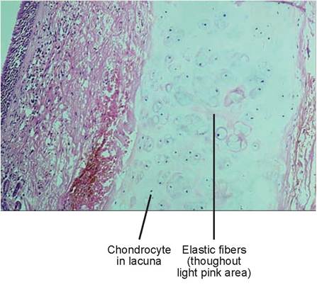
FIGURE 5.36: Mammal elastic cartilage from the pinna of an ear.
Slide: elastic cartilage
Find the area of the slide where the cartilage is located. The matrix is most recognizable because of the large, dark-stained purple area within its center (Figure 5.36). Upon close examination, small red elastic fibers may be seen at the periphery of the purple area and should appear to course transversely across the cartilage. The chondrocytes’nuclei and cytoplasm are viewable within the lacunae.
In the space below, using the slide of elastic cartilage, draw and label the cells, fibers, and tissue parts.
Fibrous Cartilage (Fibrocartilage)
Slides recommended: Those labeled fibrocartilage.
Description: Fibrous cartilage is distinctively different from the other cartilage types. It contains large amounts of fibrous connective tissue in the form of collagen, which is embedded in a ground substance
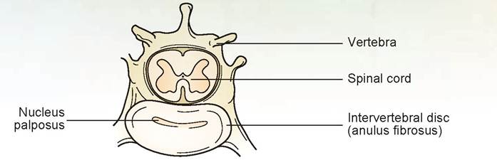
FIGURE 5.37: Intervertebral disc (found between vertebrae), where fibrous cartilage is located.
not unlike that of the other cartilages (Figure 5.37). The fibers are distributed linearly throughout the ground substance (i.e., they run in the same direction). The chondrocytes lie in the lacunae, which are also distributed linearly throughout the matrix.
Location found: Fibrocartilage is found in intervertebral discs, the pubic symphysis, discs of the knee joint, and within some tendons close to where they attach to bone.
Function: It has high tensile strength for support and to absorb compressive shock.
Slide: Bbrocartilage
Note the large amount of collagen fibers located within this cartilage. Fibrous cartilage might be easily confused with dense irregular connective tissue if not for the large number of lacunae present with their resident chondrocytes (Figure 5.38).
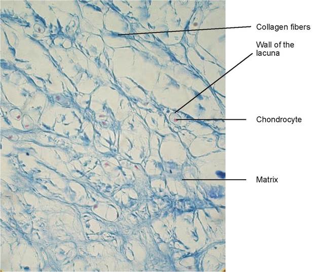
FIGURE 5.38: Mammal fibrocartilage.
Special Connective Tissue: Blood
This section will introduce you to the types of blood cells found in peripheral blood. Courses in clinical laboratory procedures, including hematology, will go into this subject matter in more depth.
Blood is classified as a connective tissue that is composed of cells in a fluid matrix. Plasma is the fluid portion, which is called serum when depleted of fibrinogen and other clotting factors. The formed elements include erythrocytes (red blood cells), leukocytes (white blood cells), and platelets (also known as thrombocytes). Blood cells and platelets usually are studied using stained blood smears. After a drop of blood is spread thinly on a glass slide and dried, it is stained with a Romanovsky-type stain such as Giemsa or Wright’s stain. On one end of the slide is a thin monolayer of cells that are more flattened and less crowded. This is the area in which cell morphology should be studied. Blood smears should be scanned using the high-power objective lens; then oil immersion can be used for studying specific cells in detail or for performing a differential cell count of the leukocytes. The differential cell count is done by counting the various cell types among the first 100 white blood cells encountered. The count is expressed as in the following example: segmented neutrophils: 66; band neutrophils: 3; lymphocytes: 18; monocytes: 9; eosinophils: 3; basophils: 1. Because these total 100 cells it also means they can be expressed as a percentage (e.g., of the total white blood cells, 66% were segmented neutrophils).
Description: Mammalian erythrocytes are the largest in the dog (7.0 μm) and smallest in the goat (4.1 μm). Mature red blood cells are anucleated. As the newly forming erythrocyte develops, it gradually loses its nucleus. The presence of nucleated red blood cells (RBCs) in the peripheral blood is an indication that the bone marrow is working hard to produce new RBCs. Erythrocytes of birds, reptiles, and amphibians are nucleated. Leukocytes are larger, ranging in size from 6.0 μm in the smallest lymphocytes to 20 μm in the larger monocytes. They are divided into granulocytes (neutrophils, eosinophils, and basophils) and agranulocytes (lymphocytes and monocytes) (Figure 5.39). Leukocytes tend to accumulate along the periphery of blood smears, so they are more readily visible in these areas. Neutrophils are the predominant leukocyte of the dog, cat, and horse, whereas lymphocytes are the predominant type of ruminants and pigs. Birds, reptiles, and amphibians have a granulocyte known as a heterophil rather than as a neutrophil. The rest of the leukocytes are the same types as found in mammals.
Slide: human or dog blood smear
You will notice numerous small, red non-nucleated erythrocytes. These cells are round with central, biconcaved surfaces on the top and bottom. This makes them appear lighter in the center, which is known as having a central pallor. This is most observable in dog and human blood; in other species it in not as predominant. Scan the slide and find the various types of white blood cells. You should be able to find all of them, except perhaps the basophil, which is hard to find because they only stay in peripheral blood for a short time after leaving the bone marrow and before entering tissue.
The first granulocytes to find are the segmented neutrophils (also known as segs) (Figure 5.40). They have dark-purple-stained nuclei in which the lobes are separated by slight indentations or thin strands of nucleoplasm. The stage of development prior to segmentation of the nucleus is called a band neutrophil, which differs from the segs only in the shape of the nucleus. The band neutrophil nucleus is U-shaped and is the same width throughout its entire length. As with seeing nucleated RBCs, seeing many band neutrophils in peripheral blood indicates that the bone marrow is producing numerous cells. The cytoplasm of the neutrophil (both bands and segs) is slightly granulated.
The eosinophil has a similar nucleus to that of the neutrophil, although it tends to be slightly less dense and have fewer lobes. The most noticeable feature of the eosinophil is its predominant red, reddish-purple, or lavender granules (depending on the stain and the species of animal blood used). The final granulocyte to locate is a basophil. The nucleus may be irregular, bilobed, or highly segmented. The granules vary in size, number, and intensity of staining depending on the type of stain and species of animal. They are often large and round to oval, and they stain reddish-purple to dark purple,
FIGURE 5.39: Capillary with red blood cell passing through it.
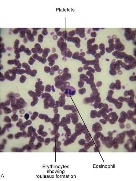
FIGURE 5.40: A. Cat blood with an eosinophil, platelets, and erythrocytes showing rouleaux formation.
B. Dog blood with erythrocytes and a nucleated red blood cell.
Continued
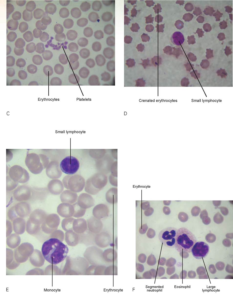
FIGURE 5.40, cont'd: C. Dog blood with erythrocytes and platelets. D. Blood showing crenated erythrocytes and a small lymphocyte. E. Dog blood with a monocyte and a small lymphocyte. F Dog blood with (from left to right) a segmented neutrophil, an eosinophil, and a large lymphocyte.
Continued
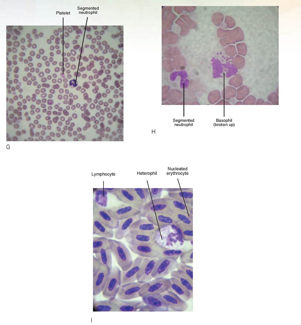
FIGURE 5.40, cont'd: G. Cat blood showing erythrocytes, platelets, and a segmented neutrophil. H. Cat blood with a segmented neutrophil and basophil (on the feathered edge and is broken up, also granules appeared darker than eosinophilic granules on same slide). I. Bird blood with nucleated red blood cells, a lymphocyte and heterophil (Wright's stain).
except for in the cat, in which they are dull gray to lavender. Because the nuclei of the granulocytes have many forms, these cells are also called polymorphonucleated leukocytes (known as polymorphs or PMNs). However, these terms have come to be used synonymously for neutrophils.
The monocytes are the largest of the white blood cells (WBCs). The shape of their nucleus is highly variable; it can be oval, irregular, kidney-shaped, or horseshoe-shaped. The nuclear chromatin is diffuse and lacy or patchy in appearance. The cytoplasm is generally a pale gray-blue and may contain small dustlike azurophilic granules. A feature that helps distinguish this cell from the large lymphocyte is that it often contains vacuoles that give it a foamy appearance. Small lymphocytes have a large, dense, often eccentrically placed nucleus that is generally round, although it is oval in the pig, sometimes slightly indented in the cat, and may be binucleated in the ruminants. Most of the lymphocytes of carnivores, horses, and pigs are small, whereas the large type predominates in the ruminants. In the small lymphocyte, only a thin rim of blue cytoplasm with a light perinuclear halo may be seen. The large lymphocyte has a less-dense nucleus with a pale blue, more-abundant cytoplasm. The nucleus may be round, oval, or kidney-shaped. In both types of lymphocytes, nonspecific azurophilic granules may be seen in certain medical conditions.
Platelets are very small and occur singly or in clusters between the RBCs. They are pale blue and contain purple central granules. They play an important role in hemostasis, the clotting of blood.
Draw all the types of cells seen in peripheral blood and described previously in the spaces below.
Special Connective Tissue: Bone
The skeletal system will be discussed in an upcoming chapter. A brief overview of the histology of compact bone will be covered here.
Description: Bone has a hard, calcified matrix containing many collagen fibers and osteocytes that lie within lacunae (Figure 5.41). Bones are well vascularized. Bone is a dynamic tissue in that over time it can lose its strength and functional ability, and thus is continually resolved and replaced. This also occurs at a fracture site of a broken bone. Osteoclasts resorb old bone, and osteoblasts secrete new bony matrix in which they become engulfed (similar to that of a chondroblast) forming lacunae with osteocytes within.
Location found: The bones of the skeletal system.
Function: Its functions are to: (1) provide the body form and structure, (2) provide protection, (3) provide a site of mineral storage, (4) act as the site of blood formation (for oxygen transport and immune function), and (5) enable the body’s leverage and mobility.
Slide: compact bone
Observe the multiple circles of bone throughout the tissue; these are called osteons, and all of these together make up the Haversian system of bone. At the center of each osteon is a central canal,
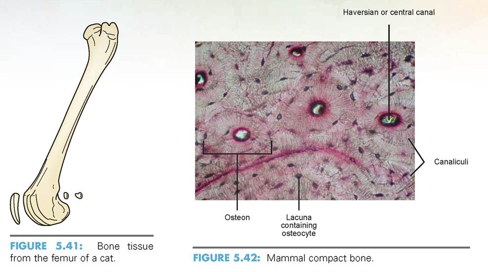
which contains a blood vessel (Figure 5.42).The dark, linear dots of the osteon that encircles the central canal are the lacunae, which contain the osteocytes. Note the thread-like lines that radiate toward each lacuna; these are canaliculi, or tiny canals that link the lacunae together and provide nutrition for the osteocytes.
In the space provided, draw a section of compact bone and label the cells, canals, and other parts discussed previously.
EXERCISE 5.4 MUSCLE TISSUE HISTOLOGY
There are three types of muscle: skeletal, smooth, and cardiac. Muscle is a highly specialized tissue designed to contract, and it produces most types of body movement.
Skeletal Muscle
Skeletal muscle is attached to the skeleton. It is under voluntary control, and its contractions move the limbs and other external body parts.
Description: Skeletal muscle cells are long, cylindrical, and have multiple nuclei. The most prominent feature of this tissue is the transverse striations across each cell. Therefore, skeletal muscle is classified as striated muscle (Figure 5.43).
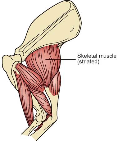
FIGURE 5.43: Skeletal (striated) muscle attached to bone.
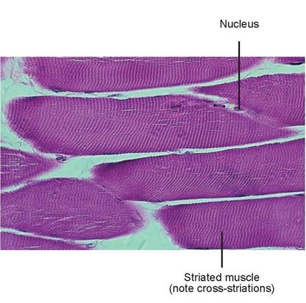
FIGURE 5.44: Longitudinal section of striated muscle cells.
Location found: Skeletal muscle can be found attached to bone and tendon, and in some areas, to skin.
Function: It allows for voluntary movement and locomotion, facial expression, and skin movement.
Slide: skeletal muscle
Note the long, red muscle cells with cross striations. On the outside of these cells, notice the numerous nuclei (Figure 5.44).
In the space provided, draw a section of skeletal muscle and label the parts of the cells.

Cardiac Muscle
Cardiac muscle is involuntary and makes up the heart’s myocardium (or heart muscle).
Description: Cardiac muscle cells are also striated, are generally uninucleated, and may be branched. They are separated from one another by specialized junctions called intercalated discs (Figure 5.45).
Location found: Located exclusively in the heart.
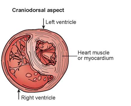
FIGURE 5.45: Cross section of myocardium of the heart.
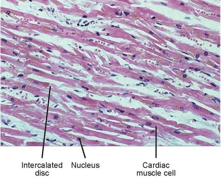
FIGURE 5.46: Longitudinal section of cardiac muscle cells.
Nucleus of smooth muscle cell
Function: Cardiac muscle contracts the atria and ventricles, moving blood into, through, and out of the heart.
Slide: cardiac muscle
The striations on cardiac muscle cells are not as noticeable as they are on skeletal muscle (Figure 5.46). Instead they appear as multiple, fine-lined, cross-striations in each cell. Note that the cells are separated on each end by dark, heavy lines, which are the intercalated discs. Notice also that some of the cells branch. The nuclei are large in relation to the cells and cannot be found in every cell because of the way the slide sample was sectioned.
In the space provided, draw a section of cardiac muscle and label the parts of the cells.

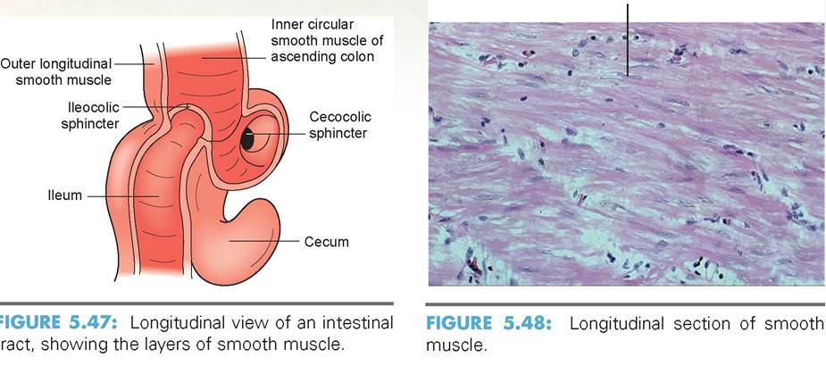
Smooth Muscle
Smooth muscle is involuntary and is found in organs that must contract to move fluids. It can also act as a sphincter muscle to control the diameter of vessels, airways, or the openings between tubular organs.
Description: Smooth muscle cells are unstriated, have spindle-shaped cells with a central nucleus, and are arranged closely to form sheets of tissue (Figure 5.47).
Location found: Smooth muscle is found in the walls of tubular organs, around bronchi, in the walls of arteries and veins, and in the uterus, and it forms the precapillary sphincter muscles.
Function: The functions of smooth muscle are to propel fluids and substances (such as foodstuffs or urine) along internal tubular passageways (such as the intestines). Smooth muscle also controls the amount of fluid or air entering or leaving certain areas of the body.
Slide: smooth muscle or intestinal tract
Smooth muscle cells are long and thin and tapered at both ends. They course in the same directions but are not perfectly parallel in configuration (Figure 5.48). The nuclei are located within the cells and sometimes appear rippled if the cell is contracting.
In the space provided, draw a section of smooth muscle and label the parts of the cells.

EXERCISE 5.5 NERVOUS TISSUE HISTOLOGY
Nervous Tissue
The brain and spinal cord possess billions of neurons, or nerve cells (Figure 5.49). You already looked at a nerve cell in Chapter 3, Exercise 3.2.
Description: Each neuron has a nerve cell body complete with a nucleus, and many short, thin processes called dendrites come off of it. The dendrites collect electrochemical signals from other nerves and route them through the cell body and down a long fiber, called an axon, to the next nerve cell, organ, or muscle.
Location found: In addition to being found in the brain and spinal cord, neurons are found in nerves throughout the body.
Function: The nerve cells’ function is to transmit information by electrochemical signals from sensory receptors to the brain and from the brain to the effectors (muscles, organs, and glands) that control their activity.
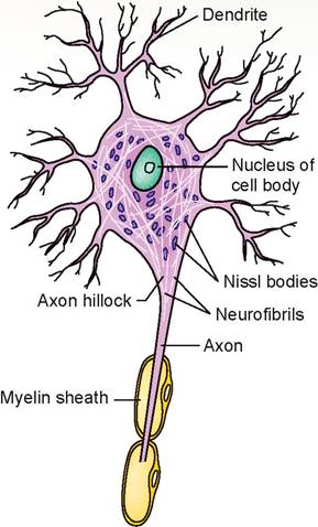
FIGURE 5.49: Nerve cells.
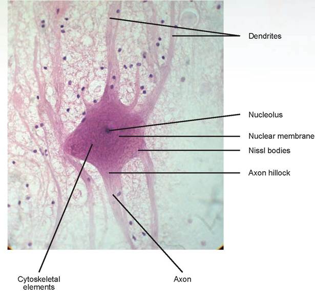
FIGURE 5.50: Mammalian motor neuron.
Slide: nerve cells (neurons), brain
Find the following structures (these were described previously in Chapter 3, Exercise 3.2): nerve cell body, nucleus, nucleoli, axon, axon hillock, dendrite, Nissl bodies, Cytoskeletal elements (Figure 5.50).
In the space provided, draw a section of nervous tissue and label the parts of the cells.

Clinical Significance
The clinically significant part of this chapter is an exercise in performing a differential white blood cell count. Because certain disease processes can be characterized by their pattern of leukocyte increase or decrease in peripheral blood, it is necessary to obtain this information as quickly as possible. Performing an in-house total white blood cell count and differential can be very helpful in obtaining a rapid diagnosis.
EXERCISE 5.6 DIFFERENTIAL WHITE BLOOD CELL COUNT
Take out your blood smear slide again and count the first 100 recognizable leukocytes using a five- punch differential cell counter to keep track. A bell should ring on the counter when 100 total cells have been counted. (Keep track on paper if you do not have a cell counter.) Fill in your results below:

This count is the relative cell count. Differential leukocyte counts should always be expressed as total cell counts rather than percentage counts (which is what the relative cell count is). The relative cell count should be just a means to obtain a total cell count; it has little interpretive value.
Assume the total white blood cell (leukocyte) count is 12,000 cells∕μl (this count may be obtained by performing manually a total white blood cell count using a hemocytometer or by an electronic blood cell counter). Now multiply each of the previous values by the total leukocyte count. This will yield the total cell count for each type of leukocyte, called the absolute cell count.
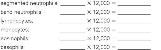
Assuming this is a dog (even if your slide is a human blood smear), the normal range of neutrophils (segs and bands combined) is 3,000 to 12,000 cells∕μl, lymphocytes is 1,000 to 5,000 cells∕μl, monocytes is 150 to 1,350 cells∕μl, and eosinophils is 100 to 1,250 cells∕μl. The normal range of basophils is not listed because they are rare in peripheral blood. Which, if any, of these levels are out of the normal range?
Summary
This chapter has been a general study of microscopic anatomy and should form the basis for the study of the organ systems we will encounter in later chapters. You learned the basic structure of epithelial tissues, their functions, and where they are located. We covered connective tissue, and you found that the fibers and matrix predominates over the cellular components in this tissue. You learned the difference between embryonal, proper, and special connective tissue. We also reviewed the structure and function of nerve cells. The final exercise was a practical application of the knowledge gained from this study and a technique most technicians should put to great use in the future.
REVIEW QUESTIONS
1. Name the four primary tissue types.
2. What do all epithelial tissue have in common as a support structure?
3. Name the areas a free surface might face.
4. Name the five characteristics of epithelial tissue.
5. What is the difference between simple and stratified epithelia?
6. What are the shapes of squamous, cuboidal, columnar, and transitional epithelial cells?
7. Describe the configuration of pseudostratified epithelial cells relative to the free surface and basement membrane.
8. What is the difference between exocrine and endocrine glands?
9. Where might you find each of the following epithelial cells: simple squamous, simple cuboidal, pseudostratified columnar, simple columnar, stratified squamous, stratified cuboidal, stratified columnar, transitional?
10. What is contained in a goblet cell?
11. A striated border is made up of what structures?
12. What is the difference between keratinized and non-keratinized stratified squamous epithelium?
13. Name the three components of all connective tissue.
14. Name the three major types of connective tissue.
15. Name the three types of fibers found in connective tissue proper.
16. What is mesenchyme?
17. Name four cell types found in areolar connective tissue.
18. True or False: There is no such thing as loose regular connective tissue.
19. Where are each of the different types of proper connective tissue found?
20. Name the three types of cartilage.
21. What types of cells are found in cartilage?
22. Where are the different types of cartilage found?
23. Name the different types of normal white blood cells found in blood. Which ones are called granulocytes, and which are called agranulocytes?
24. Name the two types of bone.
25. What is the name of the mature type of bone cell, and in what structures are they found?
26. Name the three types of muscle.
27. Which muscle types are striated?
28. Which muscle type(s) is/are voluntary, and which is/are involuntary?
29. What is the difference between a dendrite and an axon?
30. Distinguish between a relative cell count and an absolute cell count.