The Integumentary System
OBJECTIVES
• name and histologically identify the layers of the epidermis and dermis from prepared slides
• identify the cutaneous glands and hair from prepared slides
• understand the factors determining skin color
• identify the structure and parts of the equine foot; note which parts are epidermal derivatives and which are dermal derivatives
• understand the structure of a horn as compared to an antler; identify the parts and note which are epidermal derivatives and which are dermal derivatives
• understand the structure of the claws, pads, and nose of a dog
• know the anatomical structure of a cow’s mammary gland, including the names of all the ducts, lobes, lobules, and alveoli
MATERIALS
• three-dimensional model of the skin (if available)
• three-dimensional model of the equine foot (if available)
• model of the cow’s mammary gland
• longitudinal and cross-sectional displays of horn and antler
• prepared slides of bovine skin, hairy mammal skin, sweat gland, and human heavily pigmented skin
• compound light microscope
• immersion oil
• magnifying glass
• colored pencils
• prepared specimen of an equine hoof
• live dog(s), some with unpigmented nails, and others with pigmented nails
• live cat
• live horse
Introduction
The integument includes the skin and its derivatives, which are the accessory organs of the skin.
The skin consists of the epidermis and dermis. The dermis is attached to the underlying hypodermis, or subcutis (subcutaneous tissues), which is composed mostly of adipose tissue and is attached to muscle and bone underneath. The accessory organs of the skin include the sweat, sebaceous, and mammary glands, as well as the hair or feather follicles, claws, hooves, beak, and scales.86
EXERCISE 6.1 HISTOLOGY OF THE INTEGUMENTARY SYSTEM
As recommended in the previous chapter, when viewing slides of tissues, first go to low power, focus, and orient yourself on the slide (i.e., find the free surface and the deeper structures).
Then change to high power to see the cellular detail needed to understand the morphology of the tissue.For the following types of tissue, obtain the recommended slide(s), locate all the structures on the slide you are instructed to find and that are labeled in the diagrams and photomicrographs shown in the figures throughout the chapter. Then, draw and label what you see in the spaces provided. The things you will be instructed to find will include the cell, its nucleus and cytoplasm, the basement membrane, connective tissue fibers, and ground substance. The most important structures to be identified are listed in colored bold print. Subsequent slides may also contain the same structures or other related structures, which may be listed in bold print or italics; they are of no less importance and therefore should also be identified.
Epidermis
Slides recommended: Those labeled bovine skin or human skin, scalp.
Description: You may want to begin by reviewing the section of the previous chapter covering stratified squamous epithelium. The epidermis viewed in the previous chapter on pages 49-50 was nonkeratinized. However, the epidermis of thick skin (as seen here) is keratinized to assist in its protective function. Hence, the most abundant type of cells in this tissue are the keratinocytes (Figure 6.1), which are a special type of squamous cell with the ability to produce keratin. This is a tough, fibrous, waterproof protein that imparts to the skin its strength and resiliency.
Slide: skin
The layers of the epidermis, from deep to superficial, are the stratum basale, stratum spinosum, stratum granulosum, stratum lucidum, and stratum corneum (Figure 6.2).
Locate the bottom layer of the epidermis, or the basal cell layer, next to the red, fibrous dermis. This is the stratum basale; it consists of only one layer of keratinocytes, which are constantly undergoing mitosis and producing millions of new cells daily. This layer’s alternative name is the stratum germinativum.
This stratum is attached to the basement membrane. Up to 25% of the cells in this layer are melanocytes, which produce the skin pigment melanin. The darker the skin, the more melanocytes are present to produce a greater amount of melanin. Other cells located at this level are the Merkel cells, which are associated with sensory nerve endings extending from the dermis. The Merkel cell and nerve ending combine to form a Merkel disc, or a touch receptor.The stratum spinosum is a relatively thick layer of cells just superficial to the stratum basale. These keratinocytes appear stippled, an appearance that is a result of processing for histological examination: The cells shrink, but their desmosomes hold tight. Contained within these cells are thick, web-like bundles of intracellular tonofilaments of prekeratin protein. These cells are also undergoing mitosis but at a slower rate than the stratum basale. A cell called a Langerhans’ cell is found within this layer, and it functions as a macrophage. Only these two layers of the epidermis receive adequate nourishment via diffusion from the dermis. As the cells multiply, the daughter cells are pushed upward and away from their source of nutrition, and they gradually die.
The next layer of cells,just above the stratum spinosum, is the stratum granulosum, named for the abundant number of granules its keratinocytes contain. These cells are more flattened in appearance than those of the deeper layers. The granules within them are of two types: (1) lamellated granules, which contain a waterproofing glycolipid that is excreted into the extracellular space; and (2) keratohyaline granules, which combine with the tonofilaments in the more superficial layers to form keratin fibrils within the cells. At the upper border of this level, the cells begin to die.
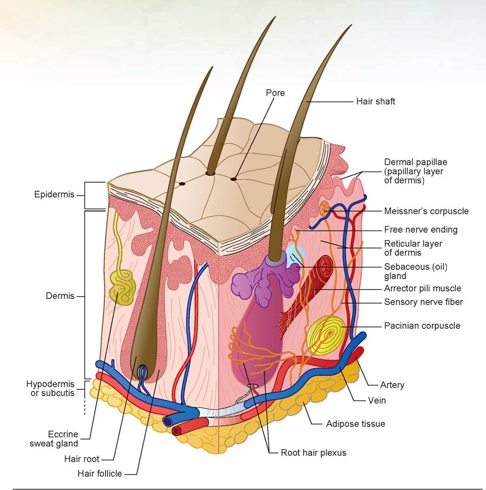
FIGURE 6.1: The structure of skin.
The thinnest layer is the next layer, called the stratum lucidum.
It is a clear or translucent band of dead keratinocytes with indistinct boundaries and keratin fibrils located within. It is present in areas of thick skin, but not thin skin.The outermost layer of the epidermis is called the stratum corneum, which consists of 20 to 30 cell layers and accounts for much of the epidermal thickness. These cells are dead, and their flattened, scale-like remnants are fully keratinized. These cells are constantly shed and replaced by division of the deeper cells (Figure 6.3).
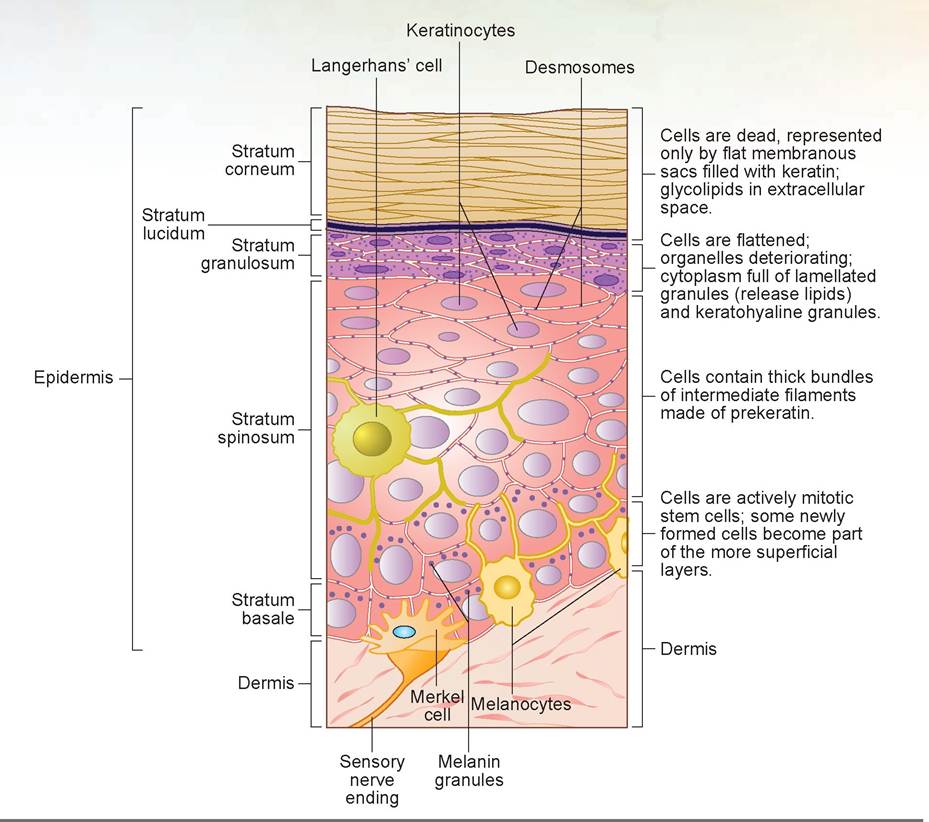
FIGURE 6.2: The layers of the epidermis, including the various cell types.
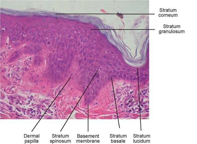
FIGURE 6.3: Mammal skin.
Dermis
Slides recommended: Those used in the previous activity, labeled bovine skin or human skin, scalp.
Description: Below the epidermis is the dermis. It is composed of two layers: (1) the papillary layer and (2) the reticular layer. The papillary layer is made of areolar (also known as loose irregular) connective tissue, and the reticular layer of dense irregular connective tissue. The reticular layer accounts for 80% of the total thickness of the dermis. Like the epidermis, the dermis varies in thickness depending on its location within the body. For example, a dog’s foot pads are much thicker than an eyelid.
Slide: mammal skin
The papillary layer is the most superficial of the two dermal layers. Its name is derived from the word papilla, which means small, nipple-shaped projection. This layer contains multiple papillary projections, called dermal papillae, which rise into the epidermis and form a wave-like pattern between the epidermis and dermis (Figure 6.3). The shape of this interface helps ensure the connection between these two structures.
Within the papilla are looping blood vessels that provide nourishment to the cells of the epidermis, as mentioned previously. These vessels also help remove waste products and assist with temperature control of the body. Also, within this layer are nerve endings for pain and touch, called Meissner’s corpuscles (Figure 6.1), and receptors sensitive to temperature changes.The boundary between the two layers of the dermis is indistinct because the collagen fibers from the papillary layer blend with the bundles of collagen fibers of the reticular layer. (You may want to review the material on deep irregular connective tissue in the previous chapter on pages 57-58). Note that in the reticular layer these layers of heavy, wavy bundles (or cords) of collagen fi bers tend to run parallel to each other (see Figure 6.4B). The orientation of these fibers is in the direction of the stress placed on them. It also consists of many small arteries and veins, sweat glands (in species containing them), and sebaceous glands. These vessels allow the skin to play a major role in temperature regulation of the body. When the body’s temperature is high, the arterioles dilate, and the capillary network
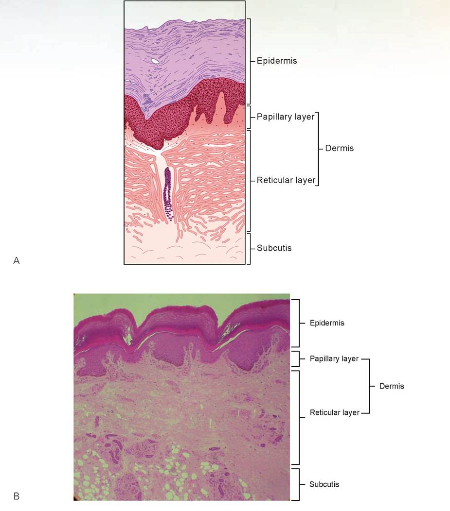
FIGURE 6.4: A. Epidermis and dermis of a digital pad. B. Primate plantar pad.
becomes engorged with the heated blood to allow the heat to radiate from the skin’s surface. If the environment is cool and the body’s heat must be conserved, the arterioles constrict so that blood bypasses the dermal capillary networks.
Both layers of the dermis, papillary and reticular, are heavily invested with collagenic fibers and elastic fibers, which give the skin its exceptional elasticity in youth. As the body ages, the number of elastic fibers decreases and the subcutaneous layer loses fat; this leads to wrinkling and inelasticity of the skin and can be observed most easily in primates and humans.
Hypodermis, or Subcutis
Slides recommended: Those used previously, labeled bovine skin or human skin, scalp.
Description: The hypodermis, or subcutis, is the thick layer of subcutaneous areolar connective tissue that is rich in adipose tissue and lies deep to the dermis (Figure 6.5). You may want to review the section of the previous chapter on pages 61-63 on adipose tissue. Blood and lymphatic vessels and nerves are also found in this region. At the dermis-subcutis border is a special type of touch receptor, called a Pacinian corpuscle, which is sensitive to heavy pressure. The boundary between the dermis and subcutis is blurred because the fibers of both merge with one another. This layer is important because it permits the skin to move freely over the underlying muscle and bone without putting tension on the skin, which might result in tearing. The hypodermis also acts to help insulate the body from environmental temperature variations.
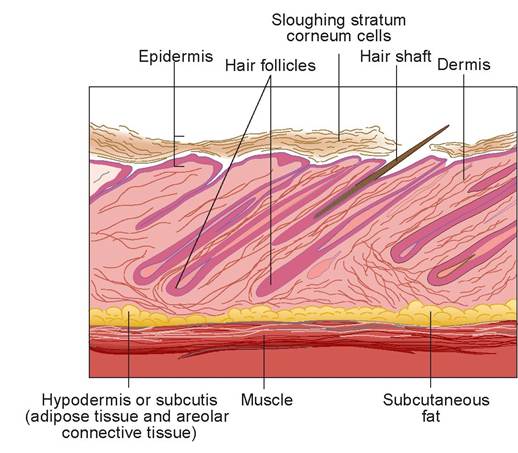
FIGURE 6.5: Skin: epidermis, dermis, and hypodermis (subcutis), including hair follicles and hair shafts.
Slide: skin
Look beneath the dermis to view the subcutaneous fat and the structures within.
In the space below, using the slide of the hypodermis, draw and label the layers, cells, and other structures observed.
Accessory Structures of the Integument: Hair
Slides recommended: Those labeled hairy mammal skin.
Description: Most skin covered with hair or fur consists of three epidermal layers: the stratum basale, stratum spinosum, and stratum corneum. However, there are regions in furry mammals where all five layers exist. These are places where the keratinization process has slowed and the skin is very thick. The surface of the skin of a hairy mammal is covered in scale-like folds, and the hair shaft emerges from beneath the scales and is directed away from the opening. Dogs usually have a cluster of three hair follicles per scale. A tylotrich hair (also known as a tactile hair) is a special type of hair that emerges from a knob-like elevation called a tactile elevation (or epidermal papilla). These hairs are important to animals because they assist with their perception of touch and space.
Slide: hairy mammal skin
Observe the shaft of the hair first; it is made of hard keratin and has a central medulla surrounded by a cortex, which is enveloped by the cuticle layer (Figures 6.6 and 6.7). The amount of melanin in
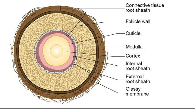
FIGURE 6.6: Cross section of a hair shaft.
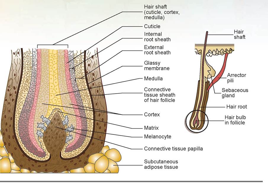
FIGURE 6.7: Longitudinal section of a hair follicle.
the cortex determines whether the hair is brown, red, yellow, or black. As pigment is lost from the cortex, which occurs in old age, the hair becomes progressively grayer. If it loses pigment entirely, the medulla fills with air, and the hair becomes white.
Next, observe the hair follicles and try to find one cut directly through the middle longitudinally. The portion of the hair enclosed within the follicle is the root. The hair bulb is a collection of well- nourished, germinal epithelial cells at the basal end of the follicle. As the daughter cells are pushed farther away from this growing region, they die and become keratinized. Thus, the bulk of the hair (i.e., the shaft) is composed of hard keratin and is no longer living material. The follicle contains both epidermal and dermal cells.
Look closely at the hair bulb and find the hair’s connective tissue papilla, which is a continuation of the connective tissue sheath that surrounds and anchors the hair in the dermis. Proceeding from the exterior inward, the next layer is a thin, clear layer called the glassy membrane. The first layer of cells in the follicle proper is the external root sheath. Beneath this is the internal root sheath. Everything inside of this becomes part of the hair shaft as it grows out, but these cell layers are not distinct in the follicular area. A thin area just inside the internal root sheath is the cuticle, the area directly in the center is the medulla, and the cells in between, which are the most abundant part of the hair root and shaft, compose the cortex. The cells of the bulb, located around the connective tissue papilla, are called the matrix. This is also where melanocytes are found. These add the pigment melanin to the cortex.
The part of the root where cells begin to lose their cellular appearance is called the keratogenous zone. Above this area,just peripheral to the internal root sheath, you should find the pale-stained, glandular epithelial cells.
These are sebaceous gland cells (modified squamous epithelial cells). This type of gland is known as a holocrine gland (an exocrine-type gland). Holocrine refers to a gland in which the whole cell is liberated during secretion (Figure 6.8). The cell ruptures and emits a white, semi-liquid mixture called sebum, the oil that lubricates the hair shaft. In the sheep, this product ultimately becomes lanolin.
Other types of exocrine glands named for their secretory process are merocrine glands and apocrine glands. In merocrine glands, the secretory product passes through the cell walls without any loss of cytoplasm; examples are sweat glands. Apocrine glands lose a slight amount of cytoplasm and/or cell membrane during secretion, although the whole cell is not lost in the process; examples include the prostate gland.
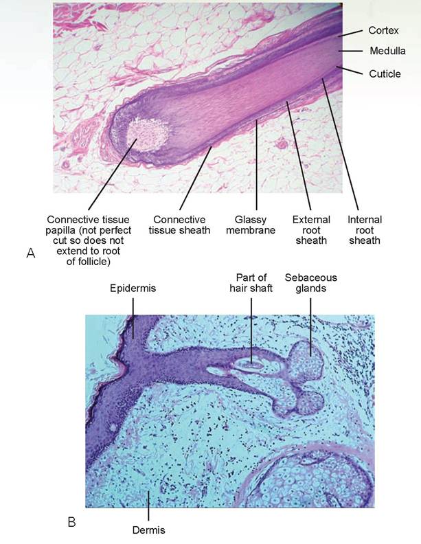
FIGURE 6.8: A. Hair follicle. B. Partial hair shaft with sebaceous glands.
In the space below, using the hairy mammal slide, draw the follicle and hair shaft and label the layers, cells, and other structures observed.
EXERCISE 6.2 THE INTEGUMENTARY SYSTEM MODEL
Locate all the items found on the slides in the previous exercise on the plastic model of the integument. The model also should have an example of the arrector pili muscle. This is composed of small bands of smooth muscle cells that connect each hair follicle to the papillary layer of the dermis. When these muscle cells contract, which is usually during cold, fright, or bluffing behavior (e.g., the animal is making itself appear larger), the slanted hair follicle is pulled upright, which dimples the skin surface. In areas of small, fine hairs, this dimpling appears as goose bumps, but in heavily furred areas it will not be seen. Contraction of the arrector pili muscles also exerts pressure on the sebaceous glands surrounding the follicles, causing a small amount of sebum to be released.
Note some of the other glands present on the model. There are the sweat glands, also known as sudoriferous glands. These are found over the entire body of most domestic species, including the dog, pig, horse, cow, and sheep. However, only the horse produces a profuse amount of sweat. There are two types of sweat glands, apocrine and eccrine. The eccrine sweat gland consists of a simple, coiled tube located in the dermis or subcutis and connected to the surface via a long duct. In dogs, this type of sweat gland is found only in the deep layers of fat and connective tissue of the foot pads. Apocrine sweat glands have a coiled, excretory portion found deep in the dermis or subcutis, and they empty into the hair follicles rather than onto the skin’s surface.
EXERCISE 6.3 PAW, PADS, CLAWS, AND NOSE OF THE DOG AND CAT
Pigmented Skin
There are numerous regions in the dog, including the skin, mucous membranes, and claws, that are darkly pigmented. Pigmentation is determined by the relative amounts of two pigments, melanin and carotene, in the skin and accessory structures. Another factor that may affect the color of the skin is the degree of oxygenation of the blood, which can give a reddish tint to areas of white skin. This may be especially noticeable in areas of inflammation.
Melanin is produced by melanocytes and becomes incorporated into the cells of the stratum basale and the deeper layers of the stratum spinosum. Because these cell layers also form the germinal layers of claws (and hooves in large animals), the amount of melanin deposited determines the color of these structures as well. The release of the melanin granules is controlled by the release of melanocytestimulating hormone, which is controlled by the pituitary gland. Carotene is a yellow-orange pigment present primarily in the stratum corneum and in the adipose tissue of the hypodermis.
Obtain a live dog and observe the tongue and other areas inside the mouth for black pigmentation. Which breeds are likely to have this pigmentation? Then, look at the top of the nose and the claws to see if they are pigmented.
The Nose of a Dog
Using a magnifying glass, observe the surface of the nose and pads of the dog. The top of the nose in dogs, cats, pigs, and sheep is called the planum nasale. In the cow and horse, this area is called the muzzle and technically is named the planum nasolabiale. Note that it is composed of many polygonal plates packed together, and that it may be further divided by grooves into polygonal plaques (Figure 6.9). The nose is usually black or dark brown and is composed of three layers of epidermis: the stratum basale, stratum spinosum, and stratum corneum. Surprisingly, considering the heavy use of the nose, stratum corneum is composed of only four to eight layers of cells. The nasal epidermis and dermis interdigitate in a manner similar to other areas of the skin; they form prominent dermal papillae deep into the epidermal cells. In the dog, the planum nasale contains no glands. However, in the sheep, pig, and cow, tubular glands are found.
Foot Pads of a Dog or Cat
Now observe the pads on a dog’s or cat’s paws. The pads adjacent to the claws are called the digital pads; the large central pad is called the metacarpal pad (or metatarsal pad in the rear feet), and the
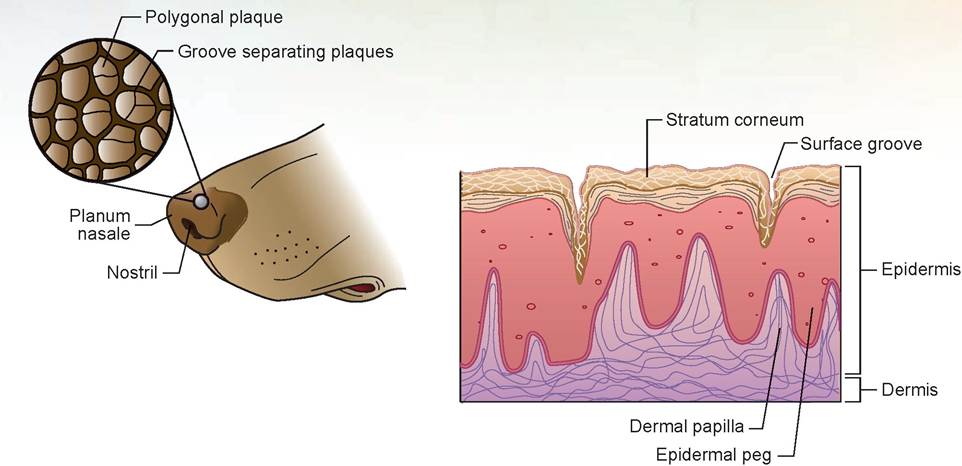
FIGURE 6.9: The nose of a dog and cross section of the epidermis and dermis.
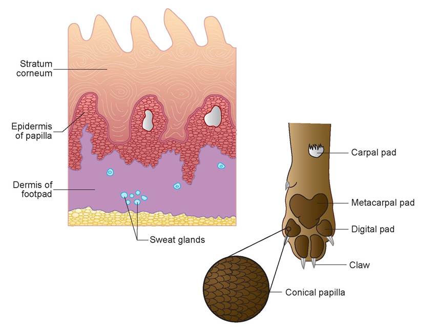
FIGURE 6.10: The pads of a dog's feet and cross section of a drawing of the epidermis and dermis.
small pad on the caudal surface of the leg (located a short distance proximal from the metacarpal or metatarsal pad) is called the carpal pad or tarsal pad, depending on its location on front or hind feet (Figure 6.10). The digital, metacarpal, and metatarsal pads are the weight-bearing pads in the dog and cat. Using a magnifying glass, observe the many minute conical papillae covering the pads. Sometimes the central areas of the pads are worn smooth from walking on hard, rough surfaces. When this happens, the central papillae are rounded or flattened. All five layers of the epidermis are present, and the
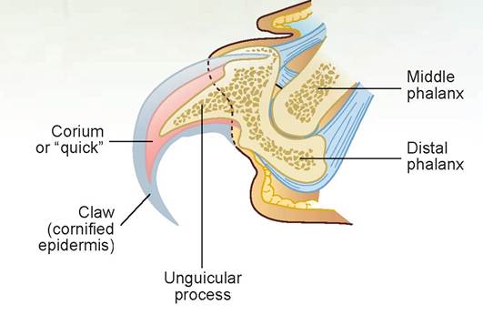
FIGURE 6.11: The claw of a cat.
stratum corneum is the thickest. There are multiple dermal papillae at the epidermal-dermal interface as well. Deep to the dermis is a thick layer of subcutaneous adipose tissue. This insulating fat, plus the thick, tough keratinized stratum corneum, forms an effective protective barrier against abrasion and thermal changes. This enables the animal to walk on rough, hot, or cold surfaces. The interior of the pads is composed of exocrine sweat glands and lamellar corpuscles. The ducts pass through the dermis to the stratum basale. The secretions of these glands are then expelled onto the surface of the pad.
The Claw of a Dog and Cat
Now look at the claws and compare a non-pigmented claw of a dog to a pigmented one. Then, look at the claw of a cat and compare it to the dog and to the diagram in Figure 6.11. In both animals, the claw is attached to the toe at the coronary band (or bed). This composes the germinal, epidermal cell matrix that gives rise to the horny wall (or unguis). Beneath the wall is the sensitive corium, which is modified dermis, otherwise known as the quick of the claw. This is the area that bleeds if you accidentally cut the claw off too short during a nail trim. The corium surrounds a bony process attached to the distal phalanx called the unguicular process.
EXERCISE 6.4 ASSOCIATED STRUCTURES OF THE INTEGUMENT
IN LARGE ANIMALS
Chestnuts and Ergots
The chestnut and ergot are dark, horny structures found on the legs of members of the equine family. Chestnuts are brown to tan or grayish, depending on the hair color of the animal. They are found on the medial side of the legs at the level of the carpus or knee (note that this is not the true knee, which is the stifle joint of the hind leg) and the tarsus or hock (Figure 6.12). The ergot is the horny tissue found buried in the long hairs below the fetlock joint.
Chestnuts are thought to be vestiges of the carpal pads (front leg) and tarsal pads (hind leg) of the first digit; and similarly, the ergots are vestiges of these pads of the second and fourth digits. Remnants of the fifth digit do not exist. As the horse progressed through its evolutionary development, the multitoed species progressively lost their digits as their need for speed increased. Now the equine species supports all of its weight on the third digit. Using the live horse, find the chestnuts and ergots.
Horns and Antlers
The horns of cattle, sheep, goats, and similar species are formed over the horn process, or os cornua, which is an outgrowth of bony tissue from the frontal bone. Horns are covered by a thin layer of modified dermis called the corium of the horn. The corium at the base of the horn where itjoins the skin is thickest; this area is the germinal area. The horn itself is made of highly keratinized stratum corneum
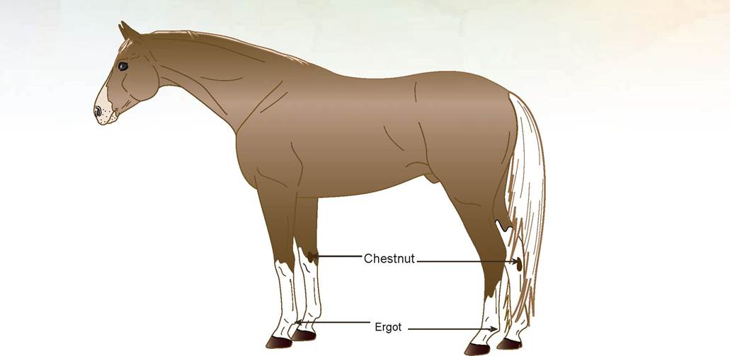
FIGURE 6.12: Location of the chestnuts and ergots of a horse.
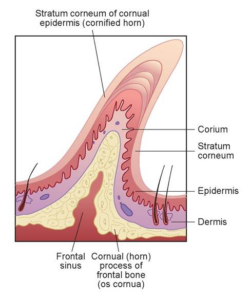
FIGURE 6.13: A horn.
of the epidermis and is constructed of many tubules of horn tissue that extend from the base of the horn to the tip. The medullary cavity of the os cornua is continuous with the frontal sinus within the frontal bone. A soft type of horn, called the epikeras, covers the surface of the horn at the base and extends for a variable distance toward the horn’s apex.
Seasonal variations in the animal’s nutritional state are reflected as variations of the horn’s growth, which resembles the rings of a tree. The age of the animal may be estimated by counting the rings in the horn. Horns occur in both males and females and are not sex-linked characteristics. Observe the cross section and longitudinal section of the display of a horn, and compare it to the diagram in Figure 6.13.
Antlers are bone developed by a process called endochondral ossification (bone growth on a cartilaginous matrix). Nutrition for this developmental process comes via the antler’s velvet. The velvet is modified skin and contains both epidermis and dermis. The antler’s progression to full size coincides with the aging and death of the velvet tissue, which the animal rubs off to expose the bony antler. Antlers undergo cyclic growth, maturation, and shedding annually and are associated with the breeding habits of the species. Antlers in the deer and elk are sex-linked; only the males of the species have them. Observe the cross section and longitudinal section of the display of antler.
The Foot of the Horse
The foot of the horse is defined as the hoof and all the structures contained within it. The horse walks on its third digit, which is equivalent to the middle finger in a human. The main bones inside of the hoof are the distal phalanx (coffin bone), the distal tip of the middle phalanx (short pastern bone), and the distal sesamoid bone (navicular bone).
Observe the plastic model and the prepared specimen of the horse’s foot, and identify the parts listed as follows. Use the diagrams to aid in this identification. After doing this, find these structures on the foot of a live horse.
1. hoof wall: The wall is the portion of the hoof visible when the horse is standing up. On the outside it is divided arbitrarily into a toe region in front, medial, and lateral quarters on the sides, and the medial and lateral heels. Internally, it is divided into three layers: the stratum externum (outer layer—the periople and a thin layer of horny scales that gives the outer surface of the wall below the periople its smooth, glossy appearance), the stratum medium (middle layer—forms the bulk of the wall), and stratum internum (inner layer—consists of the horny lamina). The wall is constructed of parallel tubular horn cemented together by intertubular horn, all of which is highly keratinized material, making it hard and durable.
Why would the hoof be constructed of thousands of small tubules of horny material? Take a piece of paper and try to get it to stand without support. Now take that same piece of paper and roll it into a coiled cylinder; it should stand on its own. The tubular structure is stronger. The stratum internum connects to the corium deep within the hoof by interdigitating with the primary and secondary lamina of the laminar corium. This forms a solid connection between these two layers (Figure 6.14).
Note the rippled laminar surface of the stratum internum on the model and specimen of the inside of the hoof wall. This layer is also called the insensitive lamina, or horny lamina. The coronary band (coronet) produces the hoof wall in an area just inside where the hair meets the wall. This is actually the coronary corium, which contains multiple papillae with the germinal cells that produce the tubular wall. This is why the hoof wall grows downward and needs to be trimmed if it does not wear off. The coronary groove is the convex surface on the top edge of the wall that connects to the coronary corium. See Figures 6.14 through 6.17 for examples of these structures.
2. periople: The periople is a thin layer of tubular horn that covers the top of the wall for a short distance distally from where the hair meets the wall. It turns a milky white color when the hoof is soaked in water. The periopole is produced by the perioplic band, a small area of perioplic corium, which is just above and concentric with the coronary band.
Note that structures 3 through 9 can be viewed from the bottom of the hoof.
3. angle of the wall: This area is where the walls turn inward at the heel, forming the bars.
4. bars: Bars are the extensions of the wall toward the apex of the frog.
5. sole: This is the horny bottom of the hoof between the wall, the bars, and the frog.
6. frog: The frog is a cornified tissue that forms a point in the middle of the bottom of the foot. It is split into two halves at the heel by the central sulcus and is more elastic than the sole. The frog, with its underlying digital cushion, aids in the absorption of concussion as the foot impacts the surface of the ground. As the frog strikes the ground, both the digital cushion and the frog are compressed between the distal phalanx and the ground. As they become thinner and wider, pressure is placed on the bars, the collateral cartilages, and the wall. The blood within the corium has a hydraulically shock-absorbing effect on the hoof wall.
The compression on the frog, digital cushion, and blood within the hoof causes the blood to be forced out of the vascular corium. This effectively pumps it into the veins of the legs and upward against gravity. Because the leg veins of horses do not have valves, movement of the horse’s legs and
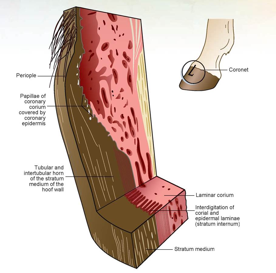
FIGURE 6.14: Longitudinal and cross section of the hoof wall, showing the primary and secondary lamina.
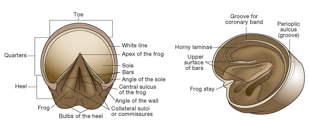
FIGURE 6.15: The bottom and inside of the hoof.
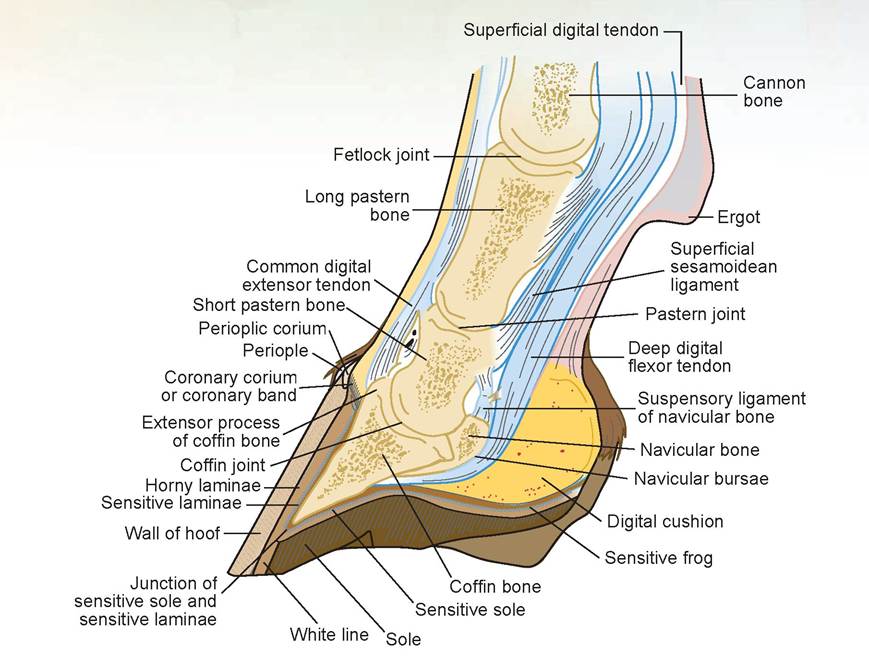
FIGURE 6.16: Sagittal section of the foot of a horse.
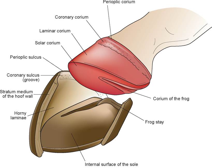
FIGURE 6.17: Dissected view of the hoof and lamina of a horse.
resulting compression within the foot keeps the blood flowing up the legs and prevents edema, or the fluid portion of blood filling the extracellular spaces.
7. commissures: Two commissures, one on each side of the frog, separate the frog from the bars. They are also known as the collateral sulci.
8. bulbs of the heel: These are paired, rounded projections—one medial, one lateral—located proxi- mocaudally from the caudal aspect of the hoof proper.
9. white line: This is the continuation of the stratum internum to the bottom of the hoof, where the sole and the hoof wall meet.
10. corium: The corium, like other coria previously mentioned, is modified dermis. Like the dermis of the skin, which contains the blood vessels and nerves and provides nutrition to the epidermis, the corium also is well vascularized and innervated and supplies nutrition to the wall. The corium is not considered part of the hoof proper, but it is part of the foot. The corium can be subdivided into different regions as follows.
The laminar corium interweaves with the stratum internum and lies between the wall and the third phalanx. It consists of primary and secondary laminae (Figure 6.18) and supplies nutrition to the stratum internum. Although the secondary lamina appears striated, it is actually made up of adjacent microvillous-appearing structures interdigitating with the stratum internum.
The perioplic corium lies in the perioplic groove (located above the coronary groove), contains the germinal cells, and supplies nutrition to the periople.
The coronary corium lies in the coronary groove, contains the germinal cells for the wall, and supplies nutrition to the stratum externum and medium.
The sole corium lies superior to the sole and supplies it with nutrition.
The frog corium lies superior to the frog and supplies it with nutrition.
Mammary Gland of a Cow
Compare the structures listed below to the model of a cow’s mammary gland. If a model is not available, use only the diagram in Figure 6.19.
The mammary gland of a cow is the ideal structure to use as an example of a compound exocrine gland because it contains all of the anatomical designated ducts, lobes, and lobules. Many other glands are named using the same terminology but may lack one or more structures. A mammary gland is a compound, alveolar, exocrine gland. It is multicellular, and its secretions are merocrine and serous in nature. It is actually classified as a modified sweat gland that produces milk. See Figure 6.19 for the anatomy of the cow’s mammary gland.
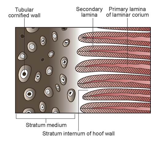
FIGURE 6.18: Microscopic appearance of hoof wall showing the primary and secondary lamina of the horse's foot.
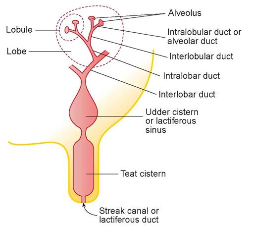
FIGURE 6.19: Anatomy of the bovine mammary gland.
Comparative Anatomy of Mammary Glands
1. Cow: As pictured in Figure 6.19, there are technically four mammary glands in the cow. However, it is conventional to describe the gland as having four quarters, each with one teat.
2. Mare: The female horse has a smaller lactiferous sinus at the base of each teat, which opens to the exterior via two or three lactiferous ducts. There are two mammary glands, one on each side of the median line in the prepubic region.
3. Ewe and doe: The mammary glands of the female sheep and goat are similar to the cow’s in structure, but they have only two mammary glands.
4. Sow: A female pig’s mammary glands contain a small lactiferous sinus at the base of each teat, and each teat has two lactiferous ducts. There are usually 10 to 12 glands or 5 to 6 pairs.
5. Bitch and queen: The mammary glands of the dog and cat contain a small lactiferous sinus at the base of each teat. The teats are short and contain 6 to 12 lactiferous ducts. There are usually 10 mammary glands or 5 pairs.
Milk Production
The hormones estrogen and progesterone cause the mammary glands to enlarge during pregnancy. Shortly before birth these hormone levels decrease, and prolactin is released, stimulating lactogenesis. Maintenance of milk production is dependent on the secretion of prolactin, growth hormone, adrenocorticotropic hormone, and thyroid-stimulating hormone. Prolactin release is also stimulated by the suckling of the offspring.
Milk Let-Down
Suckling also causes milk let-down; it stimulates nerves that send a message to the posterior pituitary gland to release oxytocin into the bloodstream. This in turn causes a contraction of the myoepithelial cells surrounding the alveoli and smaller ducts to cause milk ejection (let-down).
Clinical Significance
Because the skin is the largest organ of the animal’s body and covers the animal’s exterior, it is subjected to many types of injurious agents. Dermatology is the study of the skin and its associated diseases. The skin must protect the animal from becoming overhydrated during immersion in water, from becoming dehydrated during exposure to heat or sun, and from a variety of bacterial, viral, fungal, and parasitic agents.
When an animal becomes infested with the mite Demodex canis, this condition is known as demodec- tic mange. These mites have the ability to burrow into the skin along the hair shafts and to reside around the follicle. This parasite is present in the hair follicles of almost all dogs and in some cats. Their presence indicates a form of immunity called premunition, which is seen in some parasitic conditions and depends on the continued presence of the parasite in or on the host. In other words, having a few mites on the dog stimulates its immune system and prevents an overwhelming infestation. We occasionally see dogs that have generalized demodectic mange all over the body, and often there is an associated pyoderma with it. Many of these dogs have some form of immune deficiency. Treatment is aimed at getting rid of the mites and the secondary bacterial complications. Often these are young dogs, and it is hoped that as the animal matures, its immune system will also mature and prevent future infestations and secondary infections.
------------------------------------------- he profession of veterinary medicine is a job, and like all jobs there are things we like,
X and things we do not. Personally, I enjoy surgery. The saying “a chance to cut, a chance (j∕ to cure” really does define the psyche of the surgeon, and I think I have this affliction. If I had the opportunity to do it all over again, I would become a board-certified veterinary surgeon — either that or a dermatologist. Dermatologists never lose their patients; they never die and they never become completely cured.
The day Mel Parker burst into my hospital carrying a profusely bleeding Irish Setter was the day I had the opportunity to be both. And after looking at this dog, I wanted to be neither.
At first I thought it was Mel's own Setter, which struck me with fear.
“Take him to the treatment room,” I directed, bypassing the exam room.
“What happened, Mel? Where did you get this dog?” I asked as I immediately started to assess the problems presented and stem the flow of blood.
“I don't know where he came from. All of a sudden he was just there, right in front of my truck. I was on top of him before I could hit the brakes. You know what my truck looks like, raised up. I went right over him and stopped I don't know how far down the road—40 feet or so, I guess. I looked back in my rear view mirror, thinking I would see the dog lying there, but it was gone. I jumped out to look and heard this crying underneath my truck. Oh God! It was horrible. The dog's collar got caught on the underside of my truck, and I had drug it the entire distance. I got bit twice trying to get the dog loose.” Mel showed me the wounds on his hand.
The dog was a male, and he was not being aggressive right now. Nevertheless, we put a muzzle on him to be safe. It is hard to treat an injured animal if the veterinarian or technician is bleeding as badly as the dog. It was the dog's collar getting caught that had saved his head from injury, but his right front leg and hip suffered the rest of the damage. The bone was exposed from his elbow down to his carpus; the pavement had ground it right down to the marrow cavity. The outer muscles were in shreds and bleeding profusely.
Mel was a contractor and an old friend; in fact, we had worked together for awhile. When my hospital was being built and I was between jobs as a relief veterinarian, I pounded nails, carried lumber, and helped him build a house or two. It was just like being back on the farm. I knew Mel, and I knew he would do anything he could to help this dog recover.
After administering some hefty doses of pain medication, which had the beneficial effect of also tranquilizing the dog, we were able to stop the bleeding and clean up his wounds. His rear legs had many abrasions and a few cuts that needed suturing, but nothing of the magnitude of the damage to the front leg. We administered fluids and other anti-shock treatment to stabilize him, and within a few hours he was doing quite well. The problem, of course, was that there was not enough skin left to cover the open wound, and it was so large that sliding skin grafts would be of no help.
Fortunately, the dog's owner was located within a couple of hours. After some discussion between Mel and the owner, I got permission to do surgery. The owner was not exactly rich; and in fact, you would be stretching it to call him financially stable. So I think Mel, out of the goodness of his heart, paid for part of the bill, even though technically he did not have to. The dog was running loose in a city with a leash law.
I decided a full-thickness skin graft was our best hope. I took a strip of skin from the dog's back, removed all the subcutaneous fat down to the bottom layer of the dermis, and sewed it over the open wound.
For skin grafts to survive, they (like all tissues) must receive blood to nourish the cells and supply oxygen for metabolism. This is the critical factor that determines whether the graft will take. A sliding skin graft, in which a section of skin is slid from one area to another, retains its vascular supply. Sometimes surgeons will use a tube graft, which involves making a tubular stalk of skin containing the blood vessels, spreading the end out, and sewing it in place. When that section of skin develops a new blood supply in its new location, the tubular connection is severed and removed. This type of graft can be used in areas of the body that are not connected but are close to each other, such as from the animal's side to an area around the knee.
If these two grafts are not possible, either a full-thickness or a split-thickness graft can be used. For a split-thickness graft, a dermatome knife is used to cut a strip of skin down to the level of the dermis to prepare it for transplant. Because I did not have a dermatome and so much tissue was missing, a full-thickness graft was my only choice in this situation. Removing the subcutis so the underlying vessels could attach directly to the dermis was the critical step in the surgical procedure.
I had marked the skin with a surgical marking pen so I knew which direction the hair grew. This way I could orient the skin strip so the hair grew down the leg, not up. After suturing the skin graft in place, my technician and I put a thick cotton bandage, called a Robert-Jones bandage, on the leg temporarily to stabilize it and apply compression without cutting off circulation. After a few days, we switched to another type of splint to support both the elbow and carpal joints. Fortunately, the damage was to the side of the leg and not to the cranial surface, which might have damaged the radial nerve.
As it turned out, about 60% of the graft took, and there were no large areas that did not survive, just smaller patchy areas. Those formed scab-like crusts, and new skin from the periphery filled them in. The dog walked with a limp for about a year, and then one day the limp mysteriously went away.
All in all, we were quite lucky: Mel for not getting hurt any worse than he did, the owner because Mel helped him pay for his dog's surgery, the dog because he could have just as easily been killed, and finally me because it was the first and only time I have ever done this surgery. I'm one for one!
Summary
The integumentary system is the animal's first line of defense when the various parasites, bacteria, viruses, fungi, toxins, and other potentially injurious agents come in contact with the skin. This system also includes the structures that support weight, enable traction during limb progression, and enable functions such as digging and personal defense. Because so many disease conditions occur to the structures of this system, an understanding of both the structure and function of all the components is necessary. In this chapter we covered the structure and function of skin and its accessory organs. This included the horse's foot; the dog's and cat's pad, claws, and external surface of the nose; horns; and antlers.
REVIEW QUESTIONS
1. What are the two layers of the skin?
2. Give the three names of the layer of tissue deep to the skin, and name the type of tissue of which it is composed.
3. Name the accessory structures/organs of the skin.
4. Name the layers of the epidermis, from deepest to most superficial.
5. What is the purpose of a Merkel disc?
6. Where is a Langerhans’ cell found, and what is its function?
7. How do the epidermal cells receive nourishment?
8. Name the type of granules found in the stratum granulosum cells.
9. Name the two layers of the dermis, and indicate the type of tissue of which each is composed.
10. Name the touch receptor found at the dermal-subcutis border.
11. Name the three layers of the hair shaft.
12. In which layer is melanin found?
13. What is the purpose of the arrector pili muscle?
14. What is the anatomical name of sweat glands, and in what species are they found?
15. Name the different types of sweat glands.
16. What are the anatomical names for the top of the nose in a dog and the muzzle in a cow?
17. Name and locate the various pads found on a dog’s foot.
18. What is the anatomical name for the “quick” of a dog’s or cat’s claw?
19. Where are the chestnuts and ergots located? They are vestiges of what structures?
20. What is the bony process of a horn called?
21. What structure gives nutrition to the developing antler?
22. What is the stratum internum of the horse’s foot, and how does it relate to the “white line”?
23. The hoof wall receives its nutrition from what structure?
24. Name the number of mammary glands in each species listed in the book.
25. Which hormones stimulate lactogenesis and milk let-down?
More on the topic The Integumentary System:
- Structures of the integument
- Table of contents
- Akers R. Michael, Denbow D. Michael. Anatomy and Physiology of Domestic Animals. 2nd edition. — Wiley-Blackwell,2013. — 685 p., 2013
- The Endocrine System
- General anatomy
- Colville Thomas, Bassert Joanna M.. Clinical Anatomy and Physiology for Veterinary Technicians. 3rd edition. — Elsevier,2016. — 658 p., 2016
- Barger A.M., MacNeill A.L. (Eds.). Small Animal Cytologic Diagnosis: Canine and Feline Disease. CRC Press,2024. — 536 p., 2024
- FISH
- References
- HERPESVIRUS INFECTIONS IN WILD BIRDS