The Endocrine System
OBJECTIVES
• identify and name the major endocrine organs in the dissected cat and the brain of a sheep
• list the hormones produced by the hypothalamus and the pituitary gland, and the target organs of each
• understand the feedback mechanism that controls the release of these hormones
• understand the basic functions of the hormones produced by endocrine glands
MATERIALS
• cat cadaver, triple injected (order without skin attached)
• sheep brain, with pituitary gland attached
• Mayo dissecting scissors
• probe
• 1 ? 2 thumb forceps or Adson tissue forceps
• #4 scalpel handle with blade
• rubber gloves
• blood chemistry machine and serum glucose tests
• one live dog
Introduction
The endocrine system is system of glands that produce hormones, which are chemical messengers in the body.
These hormones are released directly into the blood and transported throughout the body. Whereas the nervous system is able to effect rapid changes in the body (via electrochemical impulses generated by neurons), hormones tend to produce slower changes. Although they are released into the bloodstream, specific hormones affect the biochemical activity of only one or a few specific organs. An organ that responds to a particular hormone is referred to as the hormone's target organ.The target tissue's response seems to depend on the hormone molecule's ability to bind with specific receptors (proteins) on the cells' plasma membranes or within the cells. Hormones are most often either steroid- or amino-acid-based molecules. When the hormone binds with the target organ's cells, it stimulates changes in the organ's metabolic activity. For example, thyroid-stimulating hormone causes an increase in the metabolic activity of thyroid cells, which increases production of thyroid hormones. An increase in thyroid hormones affects many cells of the body by stimulating their metabolic activity.
Some endocrine glands produce only hormones; others produce both hormones and certain cell types, such as reproductive cells. Examples of the former are the anterior pituitary, thyroid, parathyroid, and adrenal glands. Examples of the latter are the testes and ovaries. Both types of glands are derived from epithelial tissue during development, but the endocrine (or ductless) glands secrete their products into the blood or lymph. The difference between exocrine and endocrine glands was discussed in Chapter 5. You may remember that exocrine glands secrete their products onto an epithelial surface, whereas endocrine glands secrete directly into the bloodstream. The pancreas has both endocrine and exocrine glandular tissue.
Pancreatic islet cells contain endocrine tissue, which produces insulin and glucagon; the exocrine glands produce trypsin, chymotrypsin, amylase, and lipase (see Exercise 9.4 in Chapter 9).
Anatomy and Physiology of the Hypothalamus and Pituitary Gland
The pituitary gland, or hypophysis, is attached to the hypothalamus at the base of the brain by a slender stalk called the infundibulum. The pituitary gland consists of two major functional areas: the anterior pituitary, or adenohypophysis, and the posterior pituitary, or neurohypophysis. The adenohypophysis consists of three portions: the pars distalis, pars intermedia, and pars tuberalis. The neurohypophysis contains the pars nervosa and is directly connected to the hypothalamus by the infundibulum. The shape of the pituitary gland and the degree of development of its relative parts vary in domestic animals (Figure 13.1).
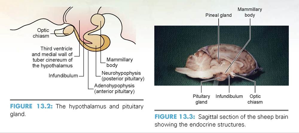
The hypothalamus is an important regulatory center for the nervous system; it regulates body temperature, thirst, hunger, sexual behavior, and such reactions as fear and rage. Despite its many functions, the hypothalamus is quite small. It consists of the optic
chiasm, infundibulum, tuber cinereum, Mammillary body, and hypophysis (Figures 13.2 and 13.3).
The hypothalamus is also a crucial regulator of the endocrine system and pituitary gland.
The hormones produced by the neurons in the ventral hypothalamus are of two types: releasing hormones, which stimulate specific cells of the anterior pituitary to produce certain hormones, and inhibitory hormones, which have an inhibitory effect and suppress production. Table 13.1 lists the hormones produced by the hypothalamus on the left, and hormones of the anterior pituitary that are affected are listed on the right.Two other hormones also are produced by the hypothalamus: antidiuretic hormone (ADH) and oxytocin. ADH is produced primarily in the supraoptic nuclei, and oxytocin primarily in the paraventricular nuclei. These are special neurosecretory neurons that allow their product to slide down an axon into the posterior pituitary gland, or neurohypophysis, for release into the blood (Figure 13.4).
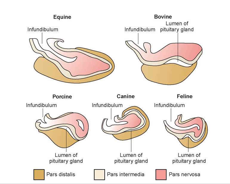
FIGURE 13.1: Comparative pituitary glands of various species.
TABLE 13.1: Hormones of the Hypothalamus and Adenohypophysis
| Releasing and inhibiting hormones | Anterior pituitary hormones affected |
| Somatotropin-releasing hormone (STH-RH) | Somatotropic hormone |
| Somatotropin releasing-inhibiting hormone (STH-RIH) | Somatotropic hormone |
| Thyrotropin-releasing hormone (TRH) | Thyroid-stimulating hormone |
| Corticotropin-releasing hormone (CRH) | Adrenocorticotropic hormone |
| Prolactin-releasing hormone (PRH) | Prolactin |
| Prolactin-inhibiting hormone (PIH) | Prolactin |
| Gonadotropin-releasing hormone (GnRH) | Follicle-stimulating hormone and luteinizing hormone |
The releasing and inhibitory hormones produced in the hypothalamus are delivered to the anterior pituitary via the hypophyseal portal system.
The capillaries in the ventral hypothalamus pick up these hormones and transfer them via the hypophyseal portal veins to the secretory cells of the adenohypophysis (see Figure 13.4).The adenohypophysis is the glandular part of the pituitary gland (adeno means “gland”), and it produces the tropic hormones. The tropic hormones (see Table 13.1) stimulate target organs (which are also endocrine glands) to secrete their hormones. The hormones of the target organ then exert their influence on other body organs and tissues. There are seven known hormones produced by the anterior pituitary:
somatotropic hormone (STH): Also known as growth hormone (GH), STH is a general metabolic hormone that plays a role in determining body growth and size. However, it also affects many tissues of the body as it regulates metabolism of proteins, carbohydrates, and lipids. It acts as an anabolic hormone in its effect on the metabolism of body proteins; in other words, it stimulates formation and buildup of
Copyright 2010 Cengage Learning. All Rights Reserved. May not be copied, scanned, or duplicated, in w
tissues, and thus is an important hormone in repair and regeneration.
thyroid-stimulating hormone (TSH): Influences growth and activity of the thyroid gland. Increasing TSH increases production of the thyroid hormones.
adrenocorticotropic hormone (ACTH): Regulates the endocrine activity of the adrenal cortex, the outer part of the adrenal gland. Increasing ACTH increases production of glucocorticoid hormones by the adrenal gland.
follicle-stimulating hormone (FSH): Stimulates follicular development in the female and spermatogenesis in the male.
luteinizing hormone (LH): Stimulates follicular rupture and formation of the corpus luteum in the female, and testosterone production in the male.
In the male, it is called interstitial cell stimulating hormone (ICSH).
prolactin: Stimulates milk production at parturition and replenishment of milk during lactation.
It may also stimulate testosterone production in males.or in part. Due to electronic rights, some third party content may be suppressed from the eBook and/or eChapter(s).
Editorial review has deemed that any suppressed content does not materially affect the overall learning experience. Cengage Learning reserves the right to remove additional content at any time if subsequent rights restrictions require it.
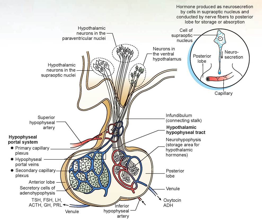
FIGURE 13.4: The hypothalamus, pituitary gland, blood flow, and neurosecretory pathways.
melanocyte-stimulating hormone (MSH): Increases skin pigmentation by stimulating dispersion of melanin granules by melanocytes. This effect is readily demonstrated in lower vertebrates, such as amphibians and fish. Other actions of this hormone are not completely understood as yet.
Two hormones are secreted into the bloodstream from the neurohypophyis:
antidiuretic hormone (ADH): Studied in the last chapter, this hormone can be considered by its action as a water resorption hormone.
oxytocin: Stimulates uterine contractions during birth and milk let-down in the lactating mother.
Physiology of the Glands of the Endocrine System detected by the hypothalamus, and secretion of the releasing factors is diminished. As levels drop, secretion of releasing factors increases. In this way, a constant level is maintained in the blood. To review, target organs regulated by the hormones of the adenohypophysis are the adrenal glands, thyroid glands, parathyroid glands, and reproductive organs.
The hormones produced by target organs have a feedback effect on the hypothalamus and pituitary gland; as they are produced, their serum levels are
Copyright 2010 Cengage Learning. All Rights Reserved. May not be copied, scanned, or duplicated, in whole or in part. Due to electronic rights, some third party content may be suppressed from the eBook and/or eChapter(s).
Adrenal Glands
The adrenal glands, like many organs, have an outer area called the cortex and a central area called the medulla. The adrenal cortex is divided into three zones:
1. The zona glomerulosa is the outer layer (under the capsule) that produces mineralocorticoids. Aldosterone constitutes 95% of the hormones produced by the zona glomerulosa and is responsible for sodium reabsorption by the kidneys.
Editorial review has deemed that any suppressed content does not materially affect the overall learning experience. Cengage Learning reserves the right to remove additional content at any time if subsequent rights restrictions require it.
2. The zona fasciculata is the middle layer and is the widest; it produces glucocorticoids.
3. The zona reticularis is the inner layer and produces a small amount of weak androgens, which are steroid hormones that have a masculinizing effect. The major androgen secreted by the adrenal gland is dehydroepiandrosterone (DHEA). This hormone has a minimal effect in males, but in females it contributes to libido.
The glucocorticoids produced are steroid-based molecules. They are cortisol (hydrocortisone), corticosterone, and cortisone. Of these three, cortisol is the most abundant and is responsible for approximately 95% of glucocorticoid activity. The metabolism of glucocorticoids is catabolic; that is, tissues are broken down to produce products needed for metabolism. The glucocorticoids’ metabolic activities include:
1. increasing gluconeogenesis in liver
2. decreasing peripheral glucose use
3. increasing proteolysis in skeletal muscle
4. increasing circulating levels of amino acids
5. increasing mobilization of free fatty acids
6. producing an anti-inflammatory effect
7. producing an anti-allergic effect
8. decreasing the immune system’s functionality
The adrenal medulla is not considered glandular tissue; instead it is modified sympathetic nervous tissue. Its products are norepinephrine, which is also released at post-ganglionic, sympathetic nerve endings, and epinephrine (adrenalin), which constitutes approximately 80% of the total secretion by this gland.
Both these hormones are sympathomimetic; that is, their effects mimic the effects of the sympathetic division of the autonomic nervous system. When released into the blood, these two chemicals:
1. stimulate an increase in heart rate and force of contraction
2. serve as vasoconstrictors that produce increased peripheral resistance
3. increase blood pressure
4. counteract the depressing action of insulin on blood sugar, thereby raising blood sugar levels
5. stimulate ACTH release
6. stimulate glucagon to initiate glycogenolysis in the liver
7. stimulate the breakdown of fats for energy
The Thyroid Gland
The thyroid gland produces the hormones triiodothyronine (T3), tetraiodo thyronine (T4), and thyrocalcitonin (TCT). Of T3 and T4, T4 is the most prevalent in farm animals, dogs, and cats. In general, T3 and T4 are the hormones that regulate (1) basal metabolic rate and oxygen use, (2) cellular metabolism, and (3) normal growth and tissue differentiation. In addition, as production of T3 and T4 rises, so do the following metabolic activities:
1. glucose absorption and utilization
2. cholesterol synthesis (but under deficiency conditions, cholesterol levels rise because of decreased elimination via the bile)
3. potentiated effects of norepinephrine
The metabolism of thyrocalcitonin will be discussed with the parathyroid glands because its metabolic activity is associated with calcium and phosphorus balance in the body.
Parathyroid Glands
The parathyroid glands are small nodules within or near the thyroid glands, usually two on each side. One is located external to and the other internal to the thyroid gland. They produce parathyroid hormone (PTH), which controls calcium levels in the blood, and thus phosphorus levels. PTH acts to increase calcium mobilization from bone, calcium absorption from the intestines, and calcium resorption from the kidneys.
A decreased PTH level causes decreased blood calcium levels, blood phosphorus levels, and urine phosphorus levels. A pathologic decrease in blood calcium levels causes tremors, muscle twitches, tetany, and convulsions. This condition could be caused by loss of calcium from the blood due to increased demand from the mammary glands in a period of increased milk production. If sufficient PTH is not available for release, calcium absorption from the intestinal tract cannot keep up with the demand. In dogs this condition is called eclampsia, or hypocalcemic tetany.
As previously mentioned, the hormone thyrocalcitonin (TCT) is produced by the thyroid gland. It keeps calcium in the bones by blocking reabsorption, and it moves calcium from blood back into the bone. TCT needs vitamin D as a coenzyme to work, and there is a relationship between PTH and TCT: If blood calcium levels are high, TCT is released; if blood calcium levels are low, PTH is released.
The Pancreas
The pancreas’s endocrine activity is not controlled by pituitary hormones, and this organ has both endocrine and exocrine activity. The exocrine portion of the pancreas secretes digestive enzymes into the duodenum. Histologically, the endocrine portion of the pancreas is composed of numerous islands of cells, called pancreatic islet cells (or islets of Langerhans), scattered throughout its tissue. The islets are composed of three types of cells: alpha cells, beta cells, and delta cells. Beta cells produce insulin; alpha cells produce glucagon; and delta cells secrete somatostatin, gastrin, and a vasoactive intestinal peptide.
Insulin has the following anabolic effects:
1. facilitates transfer of glucose into cells (hypoglycemic effect)
2. once inside a cell, causes glucose to be used more rapidly
3. enhances glycogen formation
4. increases glucokinase (which stimulates glucose to become glucose-6-phosphate)
5. increases amino acid uptake in skeletal muscle
The primary effect of insulin is to facilitate transfer of glucose into cells. However, during exercise cells do not need insulin for glucose to enter. This fact has a practical application when considering a dog with diabetes mellitus. A consistent amount of daily exercise should reduce the quantity of insulin required daily.
Glucagon has the following effects:
1. increases glyconeogenesis from glucose in the liver
2. increases gluconeogenesis from amino acids and lactic acid (via pyruvic acid) in the liver
3. increases insulin secretion
4. when given intravenously, relaxes smooth muscle
The hormones of the reproductive glands will be discussed in the next chapter. In the exercises in this chapter, the important structures are listed in colored bold print. If a structure is mentioned prior to its dissection, it is italicized. Structures discussed prior to dissection may also be in bold print for special emphasis.
EXERCISE 13.1 DISSECTION OF THE ENDOCRINE GLANDS
Most of these glands have been located at least once during previous dissections.
Procedure
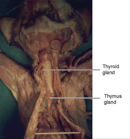
1. Locate the paired thyroid glands on either side of the cranial end of the trachea,just below the cricoid cartilage (Figure 13.5).
2. Examine the surface of the thyroid glands for two tiny parathyroid glands on each side. The external parathyroid gland is located at the cranial pole of the thyroid gland and is external to the thyroid capsule. The internal parathyroid gland is located within the thyroid capsule (is intracapsular) and lies within the parenchyma of the thyroid gland. Often these glands are not visible with the unaided eye because they are so small, or because previous dissection and the preservative have destroyed them (see Figure 13.5).
3. The thymus gland is cranial to the heart, in the ventral medial area of the thorax (see Figure 13.5). This gland was probably destroyed during your dissection of the vascular system. The hormones produced by the thymus gland are thymosin, thymic humoral factor (THF), thymic factor (TF), and thymopoietin. They promote the proliferation and maturation of T cells (a type of white blood cell), which destroy microbes and foreign substances and may retard the aging process.
4. The adrenal glands are located cranial and slightly medial to each kidney (Figure 13.6). Each gland resembles a small lymph node and has the blue phrenicoabdominal vein traversing across it.
5. The pituitary gland (hypophysis) is attached to the hypothalamus by a stalk called the infundibulum. If you remove the brain of the cat, the pituitary can be seen on the floor of the cranial cavity in the hypophyseal fossa of the skull. In the sheep brain with the meninges left intact, the rounded bulb of the pituitary gland should be easily located (see Figures 13.3 and 13.7).
6. The pineal gland may be found in either the cat brain or sheep brain above the corpora quadrigemina of the midbrain (see Figures 13.3 and 13.8).
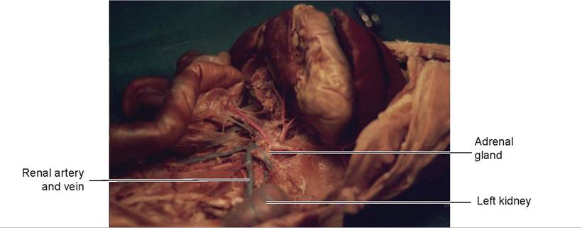
FIGURE 13.6: Left adrenal gland of a cat.
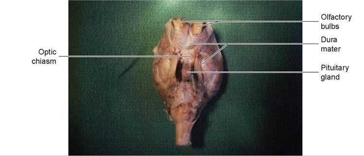
FIGURE 13.7: Ventral view of the sheep brain with dura mater and pituitary gland still attached.
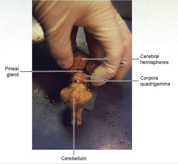
FIGURE 13.8: Caudal view of the pineal gland of a sheep.
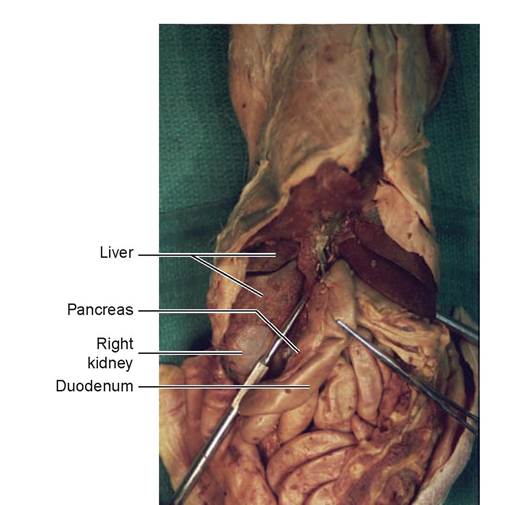
FIGURE 13.9: Pancreas of a cat.
7. The pancreas is located in the abdominal cavity, coursing adjacent to the duodenum and then curving within the mesentery medially underneath the greater omentum (Figure 13.9).
8. Locate the ovaries or testes. They will be described in more detail in the next chapter.
EXERCISE 13.2 ORAL GLUCOSE TOLERANCE TEST
This test is used to confirm the diagnosis of a deviation from normal glucose metabolism. If glucose is administered orally to a normal monogastric animal, an alteration in glucose concentration is observed over time: In phase 1, glucose absorption into the circulation exceeds excretion. As the glucose level rises, hepatic glucose output is inhibited, and secretion of insulin is stimulated. In 30 min to 1 hour, the peak blood glucose level is reached and then begins to fall. Phase 2 is the decreasing phase of the blood glucose level, and the rate of removal exceeds that of entry. During phase 3, glucose, after having returned to its original level, continues to fall to a minimal level before returning to the pre-test level.
Procedure
1. Select a live dog for the experiment and have the dog fast overnight. In the morning, take a fasting blood glucose serum level.
2. Administer glucose orally in the amount of 4 g per kilogram of body weight.
3. Take blood samples at 30- to 60-minute intervals for 3 to 4 hours.
Blood: 30 minutes: ________
1 hour: ________
1 1/2 hours: ________
2 hours: ________
2 1/2 hours: ________
3 hours: ________
3 1/2 hours: ________
4 hours: ________
4. Make a graph of your findings by plotting time on the x axis and glucose levels on the y axis.
Questions
1. At what point in the test do you know the pancreas has begun to secrete insulin?
2. Why do doctors use the term hypoglycemic effect when describing the action of insulin?
Discussion
As blood glucose rises, insulin is released. This causes cells to start absorbing glucose. When this happens, blood glucose begins to drop as glucose is removed from the serum and moved into the cells. Because glucose is removed from the blood, this process is described as having a hypoglycemic effect. A normal animal’s fasting blood glucose is less than 120 mg/dl; this value should not exceed 160 mg/dl at the end of the first hour and should return to normal by the end of the second hour. In a diabetic animal (in whom insulin is not produced or the beta cells are not responsive), blood sugar levels during the test may rise to more than 180 mg/dl and will not return to normal during the testing period. In other words, much of the glucose stays in the blood and does not enter the cells.
Clinical Significance
Hormonal problems can manifest themselves in many ways; some occur as pathological changes to the integumentary system. Two conditions in dogs cause bilaterally symmetrical alopecia, or hair loss equally on both sides of the body: hypothyroidism and hyperadrenocorticism. When testing for these diseases, you must either assay for the hormones produced by the gland, or administer a stimulating hormone and then assay for the levels of the hormones that should be produced.
For hypothyroidism, we assay for T4, usually free T4, so that results are not affected by changes in serum protein binding. Other tests that can be run are thyrotropin (TSH) stimulating test, thyrotropin-releasing hormone (TRH) stimulation test, and a TSH assay. For hyperadrenocorticism we can do an ACTH response test, a dexamethasone suppression test, or a plasma ACTH concentration. In these tests, failure to produce hormones after a signal from the stimulating hormones indicates a suppressed gland, and overproduction indicates an overactive gland.

— he day Mr. Opecia and his dog Al entered my hospital was a dark day in the history of
X veterinary medicine, at least as far as I was concerned. On that day I realized that we had
∖Jy entered a new era, one in which pharmaceuticals could be ordered from a catalog, by phone, or from an Internet site, and delivered directly to the client's doorstep. In Al's case, my doctor/ patient relationship had been abandoned and replaced by mail-order gerontology.
“Dr. Cochran, I found the greatest nutritional supplement to extend both human and animal life spans that has ever been developed,” Mr. Opecia exclaimed, puffing out his chest and smiling broadly while reaching down to pet his dog on the head. “Al and I are both on it. It is a synthetic hormone that I order from overseas through a catalog.”
Al was a 10-year-old Weimaraner with a pleasant disposition. He was not overly hyper and had a broad head and intelligent eyes. I wish I could have given the same compliment to Mr. Opecia. I stared in utter disbelief at this once-proud dog, for Al was now almost completely hairless. He had only about 12 hairs remaining in the middle of his back above the scapula. I counted them... exactly 12. Well, 14, but two came out as I counted them.
Not only was there no hair, but there had to be 10,000 comedos—tiny little blackheads—all over his body. This was the most pathetic-looking dog I had ever seen. I didn't even want to touch him. Veterinarians don't get grossed out that easily, but this was definitely gross. Al looked up at me, and somehow I think he knew what I was thinking because his entire body turned red. I had embarrassed him! Now I really felt bad. Honestly, I have never intentionally or unintentionally embarrassed a dog before; it is not in my nature.
“Mr. Opecia, he has no hair!” I exclaimed.
“Yeah, but I bought him a doggy coat so he won't get cold.”
“How do you know the supplement is working?” I asked him.
“Well, I'm on it, and I feel great,” he explained.
I glanced quickly at the top of his head. Mr. Opecia was also bald as a brick.
“So why have you brought Al in today? What's wrong with him?” I asked.
“He's got some growths around his anus I thought you should look at,” he replied.
There were three small, hard lumps in the tissue surrounding Al's anus. I did a fine-needle aspirate and found the typical large, foamy cells of a perianal adenoma.
“He's got a few benign tumors around his anus that will have to be removed,” I told Mr. Opecia. I went on to explain that these tumors were stimulated by testosterone, and that castrating Al would prevent their recurrence. I also mentioned that the tumors might be due to the hormone pills Al was on.
“No way, Doc. There's no way I'm going to do that to Al. I want to use him as a stud dog; I want to breed him!”
Al looked up at his owner and stared in apparent disbelief. “Who would have me?” he seemed to be wondering.
My concerns about the pills were correct. They must have had some potent hormones in them, because when Al stopped taking them, he regrew most of his hair. He also regrew a few more tumors, but after those were excised, they stopped returning. Eventually, Al went on to become a doggie daddy. All's well that ends well, I suppose. Oh yes, and he lived for another 8 years.
Mr. Opecia also stopped taking the pills. He is still alive and well, and he currently works as a pharmaceutical salesman. He sells injectable growth hormone, if I remember correctly.
Summary
Much of this chapter was devoted to explaining the anatomy and physiology of the hypothalamus and pituitary gland, and the target organs of pituitary hormone production. You learned that the pituitary gland varies in size and shape in the various species of domestic animals. The hormones produced by the hypothalamus were described, and the mechanisms by which they are transported to the pituitary gland were covered. You found out that the releasing hormones of the hypothalamus are transported to the anterior pituitary gland by a small portal blood system, and that oxytocin and antidiuretic hormone flow down the neurosecretory axons to the posterior pituitary. These pituitary hormones are then carried to their target organs via the bloodstream.
Each hormone causes a specific physiological effect on its target organ. You learned that the production of hormones by the target organ is controlled by a feedback mechanism to the hypothalamus, which controls output of releasing factors. Also discussed was that hormone output from the neurohypophysis is affected by certain physiological conditions, such as the effect of hydration levels on ADH release. You also learned that two hormones can have opposite physiological effects, and that homeostasis is maintained by a balance between the two hormones. For example, insulin acts to lower blood glucose, and glucagon acts to raise it; normal blood glucose levels are maintained by balanced production of these two hormones. Finally, you performed an oral glucose tolerance test to demonstrate insulin's action and its effect on serum glucose levels.
REVIEWQUESTIONS
1. What class or type of organic compounds does the endocrine system produce?
2. Define target organ.
3. Name the two parts of the pituitary gland (give both common names and anatomical names).
4. Name the structures that make up the hypothalamus.
5. What are releasing hormones, and what two types are produced?
6. Where are the products released by the neurohypophysis produced?
7. Where are the products released by the adenohypophysis produced?
8. What is a tropic hormone?
9. Describe the hypophyseal portal system.
10. Give another name for growth hormone.
11. What is the target organ for adrenocorticotropic hormone?
12. What effect does follicle-stimulating hormone cause?
13. Luteinizing hormone stimulates the production of what hormone in both males and females, and what is this hormone called in males?
14. What are the effects of the release of prolactin and oxytocin?
15. Name the zones of the adrenal cortex, and indicate what each zone produces.
16. Name the three types of glucocorticoids produced.
17. Name the eight metabolic activities of glucocorticoids.
18. What are the products of the adrenal medulla, and what metabolic activities do these compounds produce?
19. Name the products of the thyroid gland, and describe what metabolic activities these compounds produce.
20. What hormone is produced by the parathyroid gland, and what is its metabolic action?
21. Where is thyrocalcitonin produced?
22. Where are alpha, beta, and delta cells found, and what does each produce?
23. Name the metabolic activities caused by the release of insulin and glucagon.
24. Which thyroid hormone is assayed in clinical medicine?
25. Name the tests run for hyperadrenocorticism.