The Urinary System
OBJECTIVES
• name and describe the vascular supply to the kidneys
• name and identify the parts of the nephron using diagrams and models
• describe the function of the urinary system
• name and locate the major anatomical structures of the kidney using pig and cat kidneys
• understand blood pressure regulation by the kidneys
• understand the physiological principles and factors involved in an animal’s ability to concentrate urine
• measure the specific gravity of urine and understand what it means when it is elevated, fixed, and abnormally low
• understand the complexities of the physiology of micturition and the tests needed to diagnose urinary incontinence
MATERIALS
• cat cadaver, triple injected (order without skin attached)
• pig kidney, injected with red and blue latex
• necropsy knife
• Mayo dissecting scissors
• probe
• 1 ? 2 thumb forceps or Adson tissue forceps
• #4 scalpel handle with blade
• bone cutting forceps
• rubber gloves
• models of the nephron
• refractometers
• intravenous (IV) fluids and administration kit
• two live dogs
Introduction
The urinary system is one of the most important excretory pathways in the body.
The other excretory pathways are (1) the alimentary tract; (2) the biliary system, which eliminates waste into the digestive tract via the common bile duct; (3) the lungs, from which gases and chemicals are exhaled; and (4) the exocrine glands of the skin. Metabolism of nutrients produces waste products, such as carbon dioxide, nitrogenous wastes, ammonia, and detoxified organic and non-organic compounds, which must be eliminated from the body if normal function is to continue. The urinary system is primarily concerned with the removal of nitrogenous wastes from the body. In addition to their function as excretory organs, the kidneys help maintain the body's electrolyte, acid-base, and fluid balances; thus, the kidneys also act as major homeostatic organs.253
Copyright 2010 Cengage Learning. All Rights Reserved. May not be copied, scanned, or duplicated, in whole or in part. Due to electronic rights, some third party content may be suppressed from the eBook and/or eChapter(s).
Editorial review has deemed that any suppressed content does not materially affect the overall learning experience. Cengage Learning reserves the right to remove additional content at any time if subsequent rights restrictions require it.
To perform these functions, the kidneys filter the blood, taking fluid and chemicals into their functional tubular system, the nephron, where the fluid and chemicals are then processed. The nephron selectively keeps certain chemicals in the fluid, such as toxins and nitrogenous by-products; it allows needed chemicals such as glucose, sodium, and chloride to be absorbed back into the blood; and it allows excessive amounts of certain ions (such as hydrogen ions) to leave the blood via the fluid. After this process, the liquid to be excreted is urine.
The nephron also controls the amount of fluid absorbed back into the bloodstream and the amount eliminated from the body, thus helping maintain the correct amount of hydration in the system. Failure to perform these functions adequately (due to malfunctioning kidneys) will result in the animal's death.
Kidneys of various animals differ in structure and complexity. They can be monolobed, as in the pig, dog, cat, sheep, and horse, or multilobed, as in the ox and fowl. Some animals have complex duct systems, called the calyxes, leading from the renal papilla to the renal pelvis; others have no duct system and the urine flows directly into the renal pelvis. Figure 12.1 shows the kidneys of various domestic animals.
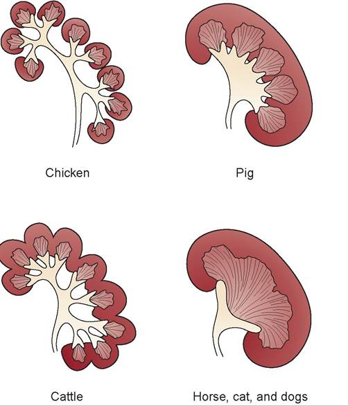
FIGURE 12.1: Structure of kidneys of various domestic animals.
The Vascular Supply to the Kidneys
The renal artery enters the kidney and branches into interlobar arteries that course between the lobes of the kidneys.
These lobes comprise separate areas of the medulla (Figure 12.2). This is easily seen in the complex kidneys of the pig and ruminants, but in the monolobed kidneys of the cat, dog, and horse, the division between the medullary areas is not distinct; the interlobar arteries course within the medulla rather than between the lobes. At the division between the medulla and the cortex, the interlobar arteries branch in an arcing pattern and are called arcuate arteries. The arcuate arteries send multiple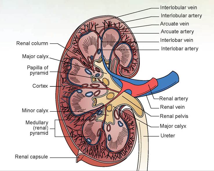
FIGURE 12.2: Frontal section of a pig kidney.
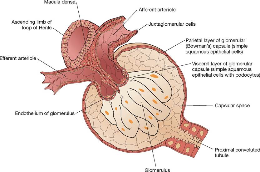
FIGURE 12.3: Renal corpuscle and the juxtaglomerular apparatus.
smaller interlobular arteries linearly through the cortex toward the periphery of the kidney.
At multiple sites along the interlobular arteries, microscopic afferent arterioles branch. Each terminates in a tufted ball of capillaries called a glomerulus (Figure 12.4). From each glomerulus, an efferent arteriole emerges that quickly branches to surround the tubules of the nephron and forms the peritubular capillary network. The part of this network that sends vessels adjacent to and parallel to the Loop of Henle is called the vasa rectae (straight vessels) and is divided into arteriae rectae, which take the blood down next to the loop, and venae rectae, which return blood to the interlobular veins. The venous system has veins that course with the corresponding arteries. From the interlobular veins, the blood travels through the
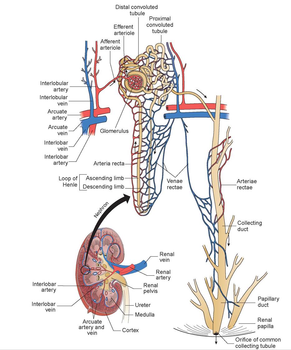
FIGURE 12.4: The nephron and its blood supply (all animals).
arcuate veins into the interlobar veins, which converge to form the renal vein and leave the kidney.
The Nephron: Anatomy and Physiology
As mentioned previously, the nephron (tubular system of the kidney) selectively absorbs its contents into the blood or excretes them via the urine.
The nephron starts as a blind sac; its visceral layer virtually shrink-wraps around the capillaries of the glomerus, and the parietal layer forms the outer wall of the sac, which is continuous with the wall of the proximal convoluted tubule. This blind sac is called Bowman’s capsule. Note in Figure 12.3 how the simple squamous epithelium of the parietal layer of Bowman’s capsule continues as the proximal convoluted tubule where the cells become simple cuboidal in shape. Refer to Figure 12.3 to identify the glomerulus, simple squamous parietal layer, Bowman’s capsule, and the lumen of the capsule.Together, Bowman’s capsule and the glomerulus are called the renal corpuscle. The fluid entering the lumen of Bowman’s capsule is called glomerular filtrate. This fluid then proceeds into a convoluted tube proximal to the loop of Henle, called the proximal convoluted tubule. From there it goes down the descending loop of the loop of Henle and up the ascending loop to the distal convoluted tubule. Multiple distal convoluted tubules, each from a separate nephron,join in a common duct known as a collecting duct. The many collecting ducts within the kidney converge to produce larger papillary ducts, which terminate in a part of the kidney called the renal papilla. The fluid, which has become urine, flows out of the renal papilla either into a minor calyx, or directly into the renal pelvis in simple kidneys like those of the cat.
As previously mentioned, the cells of Bowman’s capsule are simple squamous epithelium. Figure 5.6 contains an example of simple cuboi- dal epithelium of the kidney. The simple cuboidal cells of the proximal convoluted tubule are unique in that they have microvilli, known as a brush border (Figure 12.5). The thin descending loop
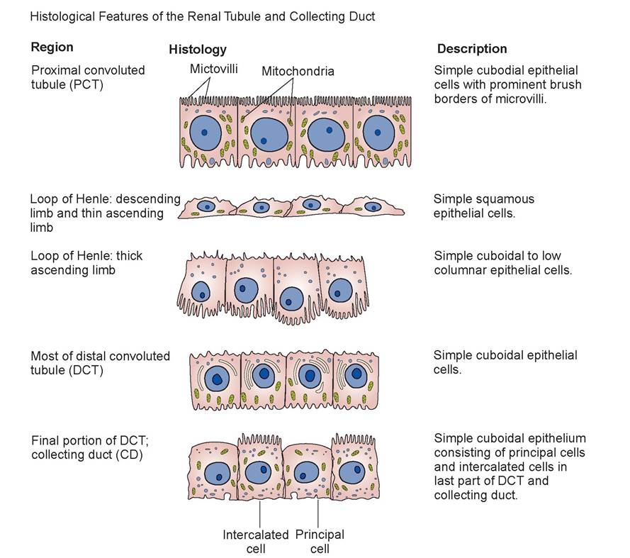
FIGURE 12.5: Histological features of the nephron's tubular cells, all species.
is simple squamous tissue again, but the thicker ascending loop is simple cuboidal or low columnar.
The distal convoluted tubule is also simple cuboidal tissue, as are cells of the collecting ducts, most of which are called principal cells, except for a few intercalated cells. The intercalated cells also are cuboidal, but they have microvilli at the apical surface and a large number of mitochondria. The principal cells have receptors for both antidiuretic hormone (ADH) and aldosterone, the two hormones that regulate water and sodium resorption. Intercalated cells play a role in the homeostasis of blood pH.The bottom of the loop of Henle is rotated such that the top of the ascending loop is adjacent to the afferent arteriole (see Figure 12.4). As the afferent arteriole approaches the glomerulus, the cells become a special type of cell, called myoepithelial cells (see Figure 12.3). These cells have some characteristics of smooth muscle and some of epithelial cells. Because they are juxtapositioned around the arteriole and glomerulus, they are called juxtaglomerular cells. A cluster of special cells that make up the wall of the ascending loop of Henle where it touches the afferent arteriole is called the macula densa (meaning “dense spot”). These are special osmoreceptors that communicate the tubular osmolality to the juxtaglomerular cells. The juxtaglomerular cells, combined with the macula densa, constitute the juxtaglomerular apparatus.
Relatively high blood pressure must be maintained to facilitate the filtering process within the glomerulus. Both the afferent and efferent arterioles are equipped with smooth muscle, so the amount of blood entering the glomerulus and the pressure within can be controlled by constricting either the afferent arteriole, the efferent arteriole, or both. The juxtaglomerular cells produce a chemical called renin (do not confuse with rennin, a digestive product), which is secreted into the blood under the following conditions: (1) when the blood pressure in the afferent arteriole falls; (2) when the sodium concentration of the blood plasma decreases;
(3) when the distal tubular osmolality decreases; or
(4) when sympathetic nerve fibers innervating the afferent arteriole are stimulated.
In the blood, renin stimulates the production of angiotensin, which acts as a vasoconstrictor to increase blood pressure. Renin also stimulates the adrenal gland to secrete aldosterone, a hormone that causes the kidney to conserve sodium ions.In the exercises in this chapter the important structures are listed in colored bold print. If a structure is mentioned prior to its dissection, it will be italicized. Structures discussed prior to dissection may also be in bold print for special emphasis.
EXERCISE 12.1 DISSECTION OF THE KIDNEY
In this exercise you will use both your cat cadaver and a pig kidney. Complete the following steps in the dissection procedure.
Procedure
1. Get out your cat cadaver. In your previous dissection of the arteries and veins you found the renal arteries coursing to the kidneys. The kidneys and ureters were found to be retroperitoneal and located along the dorsal wall. The right kidney should be relatively intact if your dissection has been careful to this point. Each kidney is surrounded by adipose tissue, called perirenal fat. The right kidney is positioned more cranial than the left (Figure12.6).
2. Remove the adipose tissue and the peritoneum that covers the ventral surface of the left and right kidneys.
3. Identify the renal artery and renal vein, which carry blood to and from the kidney, respectively (see Figures 12.6 and 12.7).
4. Find the ureters (the narrow, white tubes that drain the urine from each kidney) and trace them from the hilus (the indentation on the medial border of each kidney) to the urinary bladder (see Figures 12.6 and 12.7). The right and left ureters attach to the right and left dorsolateral aspects of the bladder, respectively, just cranial to where the urethra begins.
5. The oval urinary bladder is connected to the mid-ventral wall by a median suspensory ligament, and to the lateral walls by lateral ligaments, which contain a large amount of adipose tissue. Make a longitudinal incision in the ventral bladder wall and locate where the ureters enter on the dorsal wall. The triangular area formed by the opening of the two ureters and the opening of the urethra is called the trigonal area of the bladder. The part of the bladder that narrows, leading to the urethra, is the neck. The fundus of the bladder is the expanded part. What type of epithelial cells
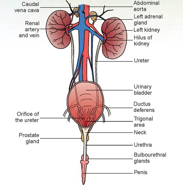
FIGURE 12.6: Urogenital system of the male cat.
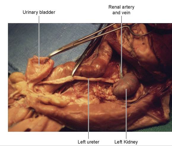
FIGURE 12.7: Kidney, ureter, and bladder of a cat.
lines the bladder and urethra? If you remember your histology, you will know they are transitional epithelial cells. The bladder’s smooth muscle layers are unique. Whereas most hollow or tubular structures contain an inner circular layer and an outer longitudinal layer of smooth muscle, the bladder has a third outer circular layer. This layer aids in contraction of the organ to expel the urine (see Figures 12.7 and 12.8).
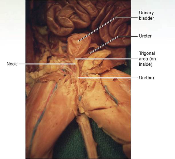
FIGURE 12.8: Urinary bladder, ureter, and urethra of a cat.
6. Locate the urethra, the duct that conducts urine from the caudal part of the bladder to the exterior. The urethra will be dissected with the reproductive system.
7. Remove the left kidney and make a longitudinal cut through it with a necropsy knife or scalpel blade, dividing it into two halves. Remove the renal capsule, a thin layer of connective tissue, from around the outside. The renal cortex is the outer, lighter-brown layer of the kidney immediately beneath the capsule. This layer contains renal corpuscles. The central layer of the kidney, the renal medulla, contains one large, dark renal pyramid (Figure 12.9). Identify the single renal papilla, called the renal crest in the cat. It is the rounded projection at the bottom of the renal pyramid of the medulla. It drains urine into the renal pelvis, which opens to the ureters.
8. Find the branches of the renal artery that lead into the kidney, the interlobar arteries within the medulla, the arcuate arteries at the division between the medulla and cortex, and the interlobular arteries within the cortex. The corresponding veins should also be identified; they can be found coursing adjacent to the arteries.
9. Obtain a pig kidney, and as you did with the cat’s kidney, make a longitudinal cut with a necropsy knife. Divide the kidney into two halves so that it resembles the one shown in Figure 12.10.
10. Locate the hilus of the pig’s kidney. All vessels enter and exit through this area. Find the renal artery, the renal vein, and the ureter exiting the renal pelvis, which is the funnel-shaped expansion of the ureter inside the renal sinus. The renal sinus is the hollow interior of the kidney; in other words, if the membrane that makes up the renal pelvis were removed, the space remaining within the kidney would be the renal sinus (see Figure 12.10).
11. The renal pelvis is joined by two (or more) large tubules, each called a major calyx. These calyxes are formed by the joining of smaller minor calyxes (see Figure 12.10). Minor calyxes do not exist in the cat.
12. Notice the darker brown areas of the renal medulla; each of these is called a renal pyramid (three- dimensionally it looks like an upside-down pyramid within the kidney). The rounded apex, or point, of the pyramid that empties into a minor calyx is the renal papilla. It is a sieve-like region that drips
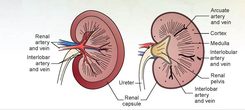
FIGURE 12.9: The cat kidney.
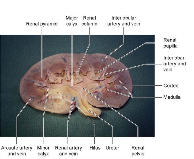
FIGURE 12.10: Frontal section of pig kidney.
urine from the papillary ducts. The striated appearance of the pyramids is caused by the numerous collecting ducts and papillary ducts, as well as the bottoms of some of the loops of Henle that extend down into the medulla. The areas between the pyramids are called the renal columns. These contain the interlobar arteries and interlobar veins (see Figure 12.10).
13. Follow the interlobar arteries and veins toward the periphery of the kidney, where they branch. Note that they arc, branching along the division between the medulla and the cortex. These are the arcuate arteries and arcuate veins, which send branches into the cortex as the interlobular arteries and interlobular veins (see Figure 12.10). The small, latex-injected vessels within the cortex are interlobular vessels.
EXERCISE 12.2 PHYSIOLOGY OF THE URINARY SYSTEM
One of the most important functions of the urinary system is maintaining the correct fluid balance in the body. The kidney’s ability to concentrate or dilute urine is dependent on its ability to resorb sodium into the interstitium and respond to the presence or absence of antidiuretic hormone (ADH) in the system. In the nephron, sodium is actively resorbed from the glomerular filtrate into the kidney’s interstitium from the proximal convoluted tubules, the ascending loop of Henle, and the distal convoluted tubules. This makes the interstitium hyperosmotic in the area of the loop and collecting ducts (which are in the medulla). The resorption of water from the collecting ducts is dependent on the permeability of the cells lining these ducts. The permeability of these cells is influenced by the secretion of ADH from the posterior pituitary gland at the base of the brain. ADH is a hormone that decreases diuresis (the production of urine), and therefore its name is based on its action to reduce the volume of urine produced: however, it might be easier for you to think of what it promotes, which is the resorption of water, making it a water resorption hormone. Under the influence of ADH, the cells become more permeable and water is resorbed from the collecting ducts (and distal convoluted tubules) into the hyperosmotic interstitium. It is then picked up by the vasa rectae and returned to the blood-vascular system.
Secretion ofADH is based on the blood’s plasma volume. If increased, the animal will have a decreased secretion of ADH; if plasma volume is decreased, there will be an increased secretion of ADH.
Procedure
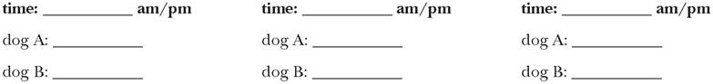
1. Locate two live healthy dogs and designate them dogs A and B for this exercise. Weigh both dogs and record your results here:
dog A:____________________________
dog B:____________________________
2. Deprive the dogs of water for six hours (or overnight) and permit them to have only food with a low moisture content. First thing in the morning, collect urine samples from both dogs by free catch or cystocentesis, and record the time and specific gravity using a refractometer.
time:________________________ am/pm
dog A:_______________________
dog B:_______________________
3. Continue to deprive dog A of water until completion of the test, but let dog B drink as much as it wants and administer 200 ml of subcutaneous fluids.
4. Weigh both dogs and record your findings.
dog A:____________________________
dog B:____________________________
For the next three hours, collect urine each hour and measure the specific gravity.
5. Graph the change in urine specific gravity over time on the x axis, and graph urine specific gravity on the y axis.
Questions
1. At the end of the test, which of the two dogs was producing the most ADH?
2. Which dog’s collecting ducts were very impermeable to water toward the end of the test?
Discussion
Withholding water and testing urine specific gravity is called the water deprivation test and is usually done by withholding water from an animal for 12 hours. Once the urine specific gravity rises above 1.030, the test is considered complete because this indicates the kidney’s ability to concentrate urine. Dog A should have had urine with a gradually increasing specific gravity, providing that its kidneys have the ability to concentrate urine. Dog B’s urine specific gravity should have risen while being deprived of water, then dropped again.
When a patient is deprived of fluids, the body’s response is to conserve water by releasing ADH; thus water is resorbed from the collecting ducts and distal convoluted tubules, which causes the urine specific gravity to rise. Administering fluids and letting the animal drink reverses this need and stops ADH secretion. When an animal’s kidneys fail and no longer respond to ADH, or when not enough nephrons survive to do thejob, the urine specific gravity falls into the fixed range of 1.008 to 1.017. In this state, urine osmolality is essentially isotonic. Therefore, the answer to question 1 is dog A, and that to question 2 is dog B.
Clinical Significance
One of the issues that continually confront veterinarians in practice is how to deal with the problem of the dog or cat that is having “accidents,” specifically inappropriate urination and soiling in the house. The first thing is to eliminate any medical and infectious causes of the problem. This traditionally involves getting a good history; knowing the amount of water the animal is consuming and whether or not it has increased; finding out if the owner has noticed blood in the urine, whether or not the accidents happen in the same place, are sporadic, or constant; and other questions such as these. A complete physical exam is necessary, including examination of the external genitalia, digital rectal exam, and (in females) a vaginal exam both digitally and with a scope. One should note the size and texture of the kidneys if possible, palpate the urinary bladder for size and tone prior to a voiding urination, check for stones, and feel the bladder wall thickness. In addition, a complete neurological exam may be necessary. The basic lab tests would need to be a urinalysis, a complete blood count, serum chemistries, electrolytes, and testing for infectious diseases. Other diagnostic tests might be radiography without contrast first, then possibly with contrast agents, such as air or air combined with a positive contrast agent. Use of ultrasound studies may be of value in lieu of using contrast radiography to find radiolucent stones or tumors. These are not the only tests that can be run; others could be cystoscopy, exploratory laparotomy, neurological testing (myelogram, MRI, or CT), and urinary system functional testing (cystometrogram, urethral pressure profile). If all these tests are negative, a behavioral problem may exist, or if it is an older dog, there may be a weakening of the bladder sphincter muscle. Noting the dog dribbling urine while sleeping may be an indication that this is the problem.
A detailed explanation of the neurophysiology of micturition is beyond the scope of this book, as it is quite involved. Micturition refers to the process of storing and periodically voiding urine. In the process of performing the above tests, the veterinarian must remember that the disorders may possibly involve urine storage, the process of urination, the urinary bladder as both a reservoir and a pump, or the urethra as the conduit. Micturition is a complex integration of the central nervous system and the sympathetic, parasympathetic, and somatic systems, with muscular activity as the result. There are many diverse causes of the problems associated with micturition because so many systems are involved. An example of this is bladder overactivity, which is due to “hyperexcitability” of the storage phase. This could be due to infection, urolithiasis, or certain drugs. The treatment is to relax the bladder. If the proper tests are not performed and the wrong treatment is implemented, the problem could worsen and cause the animal considerable distress.

⅛. elieve it or not, veterinarians are human, and they have patients they like and patients ιs—> ∕/ they dislike. I have scars from the ones I still dislike. Pointing to my hands, I can say, C c) “This scar is from Killer, and this one is from Frodo,” and so on. But some of our patients are very dear to us.
One day I heard a scratching and whining at the front door of my home. When I opened the door, one of my Golden Retriever patients was sitting on my front porch. He was lost and stopped by because he couldn't find his way home. I called his owner, and he came over and picked him up. How the dog knew where I lived still baffles me, but I will always remember that dog.
Sometimes the patients I treat are my own animals, and then of course there is no escaping emotional involvement. I used to own a beautiful yellow Lab named Sandy. On a warm, sunny summer day, while I was out playing Frisbee with my wife and son, Sandy decided to lie down in the tall grass under an apple tree and consume 2 pounds of apples. In the time it took us to walk home, his stomach was already bloating. I drove him to the local emergency animal hospital as quickly as I could. I could have taken him to the facilities at the college where I was employed, but I knew I didn't have time.
The veterinarian on duty and I tried tubing the dog to relieve the pressure, but the stomach had already twisted. I was shocked by the speed with which this had occurred; it had been no more than 15 minutes from the time I first noticed he was in distress. To relieve the pressure, we had to trocar the stomach by inserting a large-bore hypodermic needle through the skin and into the stomach to let off some gas.
Personally, I don't like operating on my own animals; I've done it, but I don't like it. In this situation there was no alternative; the emergency clinic did not allow outside veterinarians to operate in their facility—not even on their own pets. It was very distressing to me because I had lost control of the situation. Sandy survived the surgery to correct his rotated stomach, which is known as a volvulus.
Post-operatively, he started having numerous ventricular premature contractions (VPC), and I found a pneumothorax (air in the pleural cavity) to complicate his condition. How and why the spontaneous pneumothorax occurred was a mystery, both to me and to the clinician who did the surgery. I treated both; the VPCs diminished with medical treatment, and I had to insert a chest tube to correct the pneumothorax. It was almost 10 days before I could be certain the lung lobe had sealed.
Over the next month Sandy continued to improve, but he still was just not doing as well as he should have been. He started consuming large amounts of water. As soon as I noted this, I began testing his urine specific gravity. It had dropped to around 1.004, which is below the fixed level. For some reason he had developed a case of diabetes insipidus, a disease caused either by a lack of ADH from the posterior pituitary gland or by renal insensitivity to ADH. After consulting with the local internal medicine specialist, and because of Sandy's subsequent lack of response to treatment for diabetes insipidus and renal failure, I decided to put him down. He had been through enough, and after two months of daily battle, we both gave up.
I have euthanized my own animals before, but for some reason I couldn't do it this time. Fortunately for me, I have friends in the business that could come to my home and do it for me. A few times I had gone to clients' homes to put their animals down for them, and after this experience I understood just why they had been so grateful. We learn about the practice of veterinary medicine in school, but we learn about the art of veterinary medicine in practice. I still have a painting of Sandy in my home office. He lived 12 grand years, and I still miss him 15 years later!
Summary
In this chapter, you learned that the kidneys of domestic animals vary in both shape and size. Yet despite these variations in gross appearance, the functional part of the kidney, the nephron, is similar in all species. The anatomy and physiology of the nephron was studied in detail. Thus, you learned the mechanism by which the kidney is able to concentrate urine and maintain a stable hydration level within the body. By dissecting both the cat kidney and pig kidney, the comparative anatomy of the two kidneys was demonstrated. You also dissected the other parts of the urinary system and learned the parts of the bladder. Finally, you learned the mechanisms that affect the physiology of micturition.
REVIEW QUESTIONS
1. Name all of the excretory pathways of the body.
2. What is the functional unit of the kidney?
3. In which species is the kidney unilobular, and in which is it multilobular?
4. Name the arteries and arterioles of the kidney, moving from the renal artery to the glomerulus.
5. Name the parts of the nephron, moving from the glomerulus to where it empties into the renal pelvis.
6. Which is more cranial in the cat: the left kidney or the right kidney?
7. What is the function of the juxtaglomerular apparatus?
8. Under what conditions is renin secreted into the bloodstream?
9. What are the functions of angiotensin and aldosterone?
10. Name the ligaments that support the bladder.
11. Fill in the blank: The sites of entry of both ureters into the urinary bladder and the
opening to the urethra is a triangular area called the__________________________________.
12. Differentiate between the renal sinus and the renal pelvis.
13. What is the function of antidiuretic hormone?
14. Define micturition.
15. Micturition is the complex integration of what systems?