The Reproductive System
OBJECTIVES
• recognize the types of uteri in domestic animals
• understand the relationship between the endocrine system and the reproductive system, and how the endocrine hormones affect the production of reproductive hormones and cells
• understand the estrous cycle and the factors that influence its stages
• name and locate the anatomical structures of the reproductive system using diagrams and the dissected cat
• understand the production of semen, its composition, and the organs that contribute to its formation
• name and describe the layers of the uterus
• understand the types of placentation in domestic animals
• know the signs of impending parturition
• identify the various stages of the estrous cycle from vaginal cytology in the dog
MATERIALS
• cat cadaver, triple injected (order without skin attached)
• Mayo dissecting scissors
• probe
• 1 ? 2 thumb forceps or Adson tissue forceps
• #4 scalpel handle with blade
• bone cutting forceps
• rubber gloves
• compound microscope
• stained slides of the various stages of the estrous cycle of the dog
• progesterone and luteinizing hormone assay test kits
Introduction
The function of the reproductive system is to perpetuate the species.
Although it is considered a separate system, the endocrine system's hormones play a vital role in the function of the reproductive system.The target organs of endocrine hormones include the essential organs of reproduction: the gonads, which are the testes and ovaries. These produce the germ cells: spermatozoa in the male and ova in the female. During the period of female receptivity, which is hormone controlled, copulation, or mating, occurs; a haploid sperm fertilizes the haploid ovum (egg), and a diploid zygote is produced. This union occurs in the uterine tubes or oviducts.Cell division in the zygote commences quickly to prepare it for implantation in the uterine lining.
The zyzgote implants and becomes an embryo. The embryo continues to develop, and when all of the body parts are recognizable, it is called a fetus. The length of gestation, or the gestation period (the time from fertilization to birth), varies with the species. Generally, the larger the animal, the longer gestation takes. The act of giving birth is called parturition.Anatomy of the Female Reproductive Tract
The ovaries are both endocrine (hormone-producing) and cytogenic (cell-producing) organs. The medulla is the vascular center of the ovary. The cortex of the ovary is protected by a layer of dense irregular connective tissue called the tunica albuginea. This layer is surrounded by a layer of germinal epithelium. In the regions of the ovarian cortex in animals that are seasonally polyestrus (having repetitious heat cycles), various stages of the follicle may be found, such as developing and atrophying follicles, including the corpus luteum. In the development of the primary follicle, the germinal epithelium invades the stroma as a mass of cells; one cell of the mass becomes an oocyte, and the others become the follicular cells, which surround the oocyte. As the follicle matures, the oocyte enlarges and the follicular cells multiply. During follicular enlargement, a fluid-filled vesicle forms in the center. At this stage it is called a vesicular follicle, or Graafian follicle (Figure 14.1). At a certain stage of development the follicle ruptures, releasing the ovum. The follicular cells multiply to fill in the vesicle, and the structure becomes a corpus luteum (CL). It appears as a large, round, yellow-looking structure, hence the common name “yellow body.” The CL will persist if pregnancy occurs but will degenerate into a corpus albicans if pregnancy does not occur. A corpus albicans resembles a white, scar-like area.
The uterine tube, also called the tuba uterina, oviduct, Fallopian tube, or salpinx, has an open end that receives the ejected ova; this funnel-like structure adjacent to each ovary is the infundibulum.
The funnel has a fringed margin called the fimbria. The two oviducts, one for each ovary, are convoluted and extend between the ovary and the uterus. Their lining is highly folded and covered by ciliated simple columnar epithelial cells. They also contain smooth muscle. Both the cilia and the smooth muscle help move the ova and possibly the spermatozoa. As mentioned previously, the oviduct is the site of fertilization.The uterus of domestic animals consists of three parts: the neck (where the cervix is located), the body, and the horns. The uterus is a hollow structure, is lined by a mucous membrane called the endometrium, and is glandular. The endometrium’s thickness and vascularity vary with the stages of the estrous cycle. In the horse and dog, the uterus is lined by simple columnar epithelium, whereas in the pig and ruminants it is lined by stratified columnar epithelium. The uterus has intermediate smooth muscle layers called the myometrium, which consists of thick, inner, circular and thin, outer, longitudinal muscle layers. The outer serous
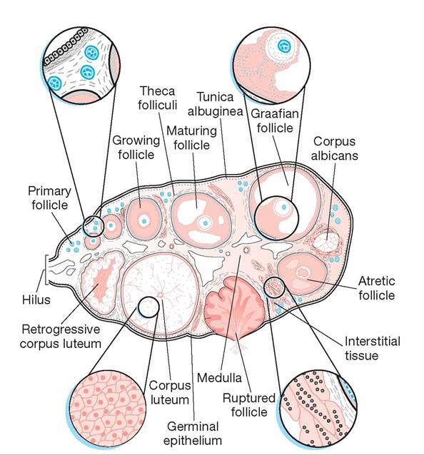
FIGURE 14.1: Ovary showing all stages of ova development and regression (all species).
layer, called the perimetrium, is visceral peritoneum and has a thin layer of connective tissue to attach it to the myometrium. The cervix is a heavy, smoothmuscle sphincter, and it is tightly closed except during estrus and parturition. There are four types of uteri in domestic animals (Figure 14.2):
1. bicornuate uterus: in the pig (sow), dog (bitch), and cat (queen)
2. bipartite uterus: in the ox (cow), sheep (ewe), and horse (mare)
3. simplex uterus: in the primate
4. duplex uterus: in the rabbit (doe)
The vagina is located within the pelvic canal and extends from the cervix cranially to the vulva cau- dally. Its mucosa is glandless, and most of the mucous secretions present there come from the mucous cells of the cervix.
The vagina is lined with stratified squamous epithelium. The muscle layer is mainly circular smooth muscle. The serosa is present only in the small cranial part that enters the abdominal cavity; the caudal part in the pelvic canal is covered by connective tissue. The fornix is an area of depression around the cervix. (The fornix is absent in the pig.)The vulva, the opening (entrance) into the genital organs, is one of the two external parts of the female genitalia; the other external part is the clitoris. The vulva consists of symmetrical, well-developed major labia, and poorly developed (also symmetrical) minor labia, which may be absent. The two labia meet dorsally at the dorsal commissure and ventrally at the ventral commissure. The vulva leads into the vestibulum, which extends cranially up to the external urethral orifice. At the ventral commissure,just deep to the external opening, is the clitoris, which is the homologue to the male penis and consists of similar parts (minus the urethra and muscles). The shaft is called a corpora clitoridis, and its size varies with the species. It ranges from 5 cm long in the mare to about 3 cm in the bitch, and it is considerably smaller in the queen. The glans clitoridis is the rounded and enlarged free end of the organ that occupies the fossa clitoridis at the ventral commissure of the vulva in female animals, and it has the same embryonic origin as the penis in the male.
The mesentery supporting the uterus is called the broad ligament. The broad ligament has three indistinct divisions:
1. mesovarium: supports the ovaries
2. mesosalpinx: supports the salpinges (singular: salpinx) or uterine tubes. (In the bitch this forms a bursa around the ovary, called the ovarian bursa.)
3. mesometrium: supports to the uterus (see Exercise 14.1, #3)
Two ligaments are found within the broad ligament. The most cranial is the suspensory ligament, which is incorporated into the mesovarium and mesosalpinx
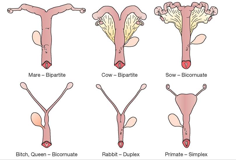
FIGURE 14.2: Shapes and types of uteri of domestic animals.
and is attached to the body walljust caudal to the kidneys.
The second is the round ligament of the uterus. This is the lateral edge of a sheet of mesentery that projects lateral and perpendicular to the main part of the mesometrium (see Exercise 14.1, #3).Anatomy of the Male Reproductive Tract
Like the ovaries, each testis (plural: testes), or testicle, is both an endocrine (hormone-producing) and a cytogenic (cell-producing) organ. The testes contain a mass of seminiferous tubules surrounded by a heavy connective tissue capsule called the tunica albuginea. The tubules are separated by fibrous septa called trabeculae, which form the support, or stroma, for the tubules. The seminiferous tubules combine to form the larger rete testis, which flow into the epididymis (via the ductuli efferentes, or efferent ducts). The interstitial cells (cells of Leydig) are located in the connective tissue between the seminiferous tubules (Figure 14.3).
The epididymis contains three parts (see Figure 14.3): The head is found on the dorsal surface of the testicle; the body is the middle part; and the tail is located on the ventral surface of the testicle (see Figure 14.3). The tubule inside is the duct of the epididymis, which is connected to the ductus deferens (vas deferens), a muscular tube that transports the spermatozoa and aids in their propulsion. The ductus deferens exits the tail of the epididymis, travels within the spermatic cord, passes through the inguinal canal, and enters the urethra just caudal to the neck of the bladder and immediately cranial to the prostate gland (if the species has this organ).
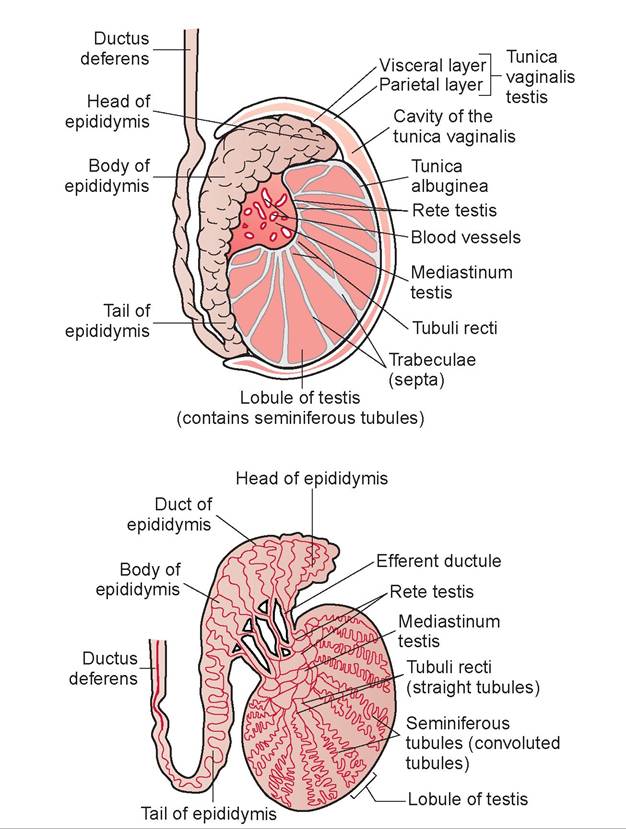
FIGURE 14.3: The testis and epididymis (all species).
The spermatic cord consists of the following:
1. ductus deferens
2. testicular artery, vein, nerve, and lymphatics
3. mesoductus deferens and mesorchium
4. tunica vaginalis visceralis (visceral vaginal tunic)
The tunica vaginalis parietalis (parietal vaginal tunic) and cremaster muscle cover the spermatic cord (Figure 14.4).
The scrotum is the sac containing the testes. It is attached to the bottom of the testicle by the scrotal ligament, which is a remnant of the gubernaculum (a ligament that pulled the testis from the abdomen, through the inguinal canal, and into the scrotum during development).
The following accessory sex glands are present in male animals, but not in all species:
prostate: Surrounds the pelvic urethra,just caudal to the bladder. The prostate is round in the dog, cat, and horse (these species have two-lobed prostates—a right and left) and more diffuse in other species. The prostate alkalinizes the semen.
vesicular glands (seminal vesicles): These paired glands lie just caudal to the neck of the bladder on both sides. Vesicular glands are hollow, pear-shaped sacs in the stallion, and large, lobulated glands in the bull, ram, and boar. These glands are absent in the dog and cat.
ampullae: These glandular enlargements at the terminal end of the ductus deferens are well developed in the stallion, bull, and ram; are small in the dog; and are absent in the boar and cat. Ampullae contribute to the seminal fluid.
bulbourethral glands (Cowper’s glands): Small, paired glands on either side of the urethra just cranial to the bony ischial arch but caudal to other glands. Bulbourethral glands are found in all domestic animals except the dog, and are very large in the boar (Figure 14.5).
The penis, which is the male organ of copulation, consists of three major components: the root, the body, and the free end surrounded by the prepuce. The root of the penis consists of the two crura and the bulbus penis. The paired crus penis is the proximal end of the corpus cavernosum penis. The corpus cavernosum penis makes up the bulk of the penis’s erectile tissue and is located dorsal to the urethra. The bulbus penis is the caudal extent of the corpus spongiosum penis. The corpus spongiosum penis is the erectile tissue surrounding the penile urethra. The urethra opens at the external urethral orifice. In the horse, sheep, and goat, a urethral process extends the urethra out from the free end of the penis. The free part of the penis starts from the attachment of the prepuce on the penis and ends as the glans penis. The glans penis is the head of the penis, which contains the corpus spongiosum glandis. The glans is a cushion that overlaps the distal end of the corpus cavernosum penis. The prepuce is the skin surrounding the free part of the penis like a muff.
The bulbus glandis is the caudal part of the penis in dogs. It swells to lock the male into the female during copulation by becoming entrapped (or tied up) within the pelvic canal. The bull, ram, and boar have a sigmoid (S-shaped) flexure of the penis (see Figure 14.5); the penis straightens during the erection process. The os penis is a grooved bone in the dog’s penis. A retractor penis muscle (made of smooth muscle) returns the penis to its normal position following an erection.
Physiology of the Female Reproductive System
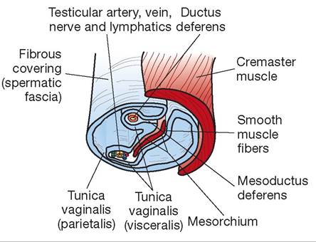
FIGURE 14.4: The spermatic cord (all species).
Follicle-stimulating hormone initiates development of the follicle. The adenohypophysis starts to liberate luteinizing hormone toward the end of follicular development, which causes the follicle to finish developing and rupture, expelling the ovum.
This process is called ovulation and is associated with estrus (heat) in most mammals. There are two types of ovulation, depending on the species. Animals that are spontaneous ovulators do not require copulation for ovulation to occur. Most species fall into this category, including the horse, ox, sheep, pig, goat, dog, primates, mouse, rat, and guinea pig. Induced ovulators, which require copulation for ovulation to occur, include the cat, rabbit, ferret, and mink.
After ovulation, the opened follicle becomes a corpus hemorrhagicum when the ruptured follicle
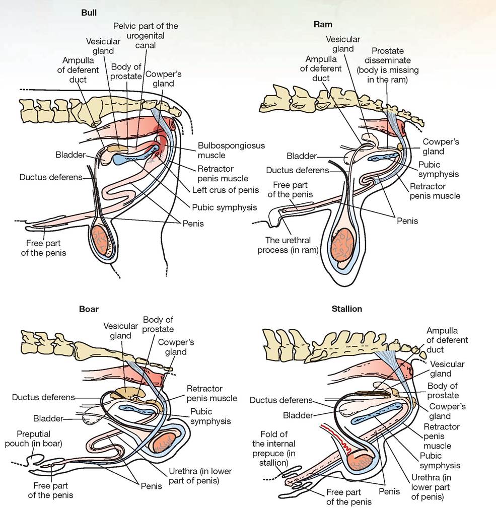
FIGURE 14.5: Comparative male reproductive systems in large animals.
forms a blood clot in the antrum area. It next develops into the corpus luteum (CL) and is influenced by luteinizing hormone to develop. The follicular cells lining the vesicle multiply to fill this open cavity. The CL persists if pregnancy occurs. Maintenance of the corpus luteum is caused by hormones produced by the placental membranes, specifically chorionic gonadotropin and prolactin, which is produced by the adenohypophysis. The CL persists throughout pregnancy in the cow, doe (goat), sow, bitch, and queen, whereas it terminates in late pregnancy in the mare and ewe. Retention of the CL in a non-pregnant animal is considered a pathological condition. The terminal stage of development is the corpus albicans. The CL degenerates to form this structure, which is a small amount of scar tissue remaining in the ovary.
There are five stages in the estrous cycle. Figure 14.6 shows the relationship between the stages of estrus and the development of the follicle and corpus luteum.
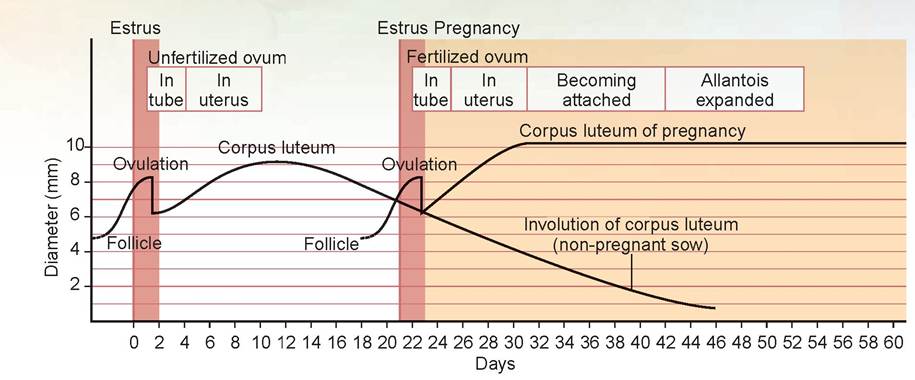
FIGURE 14.6: The estrous cycle of the sow, showing the relationship between stages of estrus and the period of follicular and corpus luteum (CL) development.
1. proestrus: The anterior pituitary gland (adenohypophysis) produces folliclestimulating hormone (FSH) and some luteinizing hormone (LH), which cause development of the follicle. The follicle starts producing estrogen, which stimulates the buildup of the walls of the vagina and uterus to prepare for copulation and pregnancy.
2. estrus: This is the period of female receptivity. Ovulation usually occurs during the end of this period in spontaneous ovula- tors. FSH levels are decreasing and LH levels are increasing.
3. metestrus: In this post-ovulatory phase, the CL starts secreting progesterone, which prevents further development of other follicles. The CL also is responsible for maintaining the uterine lining to support the fetus during pregnancy.
4. diestrus: This is the short period of quiescence in seasonally polyestrus animals. First the CL degenerates; then one or more follicles develop (depending on whether it is a litter-bearing animal) and estrus repeats.
5. anestrus: The CL degenerates during this period of quiescence between breeding seasons.
Oxytocin, as mentioned in the previous chapter, is secreted into the bloodstream by the posterior pituitary gland (neurohypophysis). Oxytocin stimulates milk let-down in the mother, and in the presence of estrogen it stimulates uterine contractions during parturition. It also stimulates the oviducts to help move spermatozoa. Prolactin from the anterior pituitary gland helps maintain the CL during pregnancy, stimulates the mammary glands to fill up with milk at parturition, and stimulates replenishment of milk (production of prolactin is stimulated via suckling).
The placenta is the membranous structure that obtains nutrients and oxygen from the mother and delivers them to the fetus. The chorion is the outermost membrane and is in contact with the maternal uterus. The amnion is the innermost membrane closest to the fetus. The amnionic sac is the sac in which the fetus is located, and it is also called the second water bag. The allantoic sac is the space between the amnion and the chorion; it is lined by the allantois (or allantoic membranes) and is called the first water bag (Figure 14.7).
The attachment of the placenta to the lining of the uterus varies according to species (Figure 14.8). The following are the types of placentation:
1. diffuse: epithelial; found in the pig and horse
2. cotyledonary: epitheliochorial; found in the sheep, goat, and cow
3. zonary: endotheliochorial; found in the dog and cat
4. discoid: hemochorial; found in primates
Figure 14.8 illustrates the types of placentation of various species; the structure of the lining of the areas of placentation also differs among species. Figure 14.9 illustrates the types of maternal and fetal tissues present in the placenta. The information in the previous list correlates to Figure 14.9.
Signs that parturition is approaching include an enlarged abdomen, enlarged mammary glands,
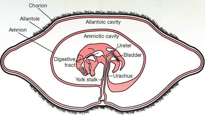
FIGURE 14.7: The fetal membranes in the dog.
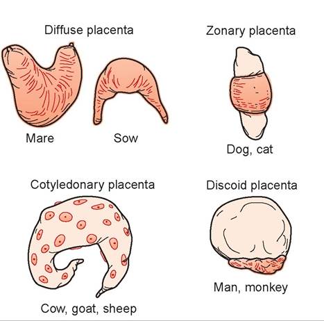
FIGURE 14.8: Types of placentation in various species.
mammary glands that fill up with colostrum, and clear mucus from the vulva. Signs in large animals include a relaxed abdominal wall, sinking in of the flanks, dropping of the belly, and sinking of the rump on both sides of the tail head. In dogs, the first stage noted is the nesting stage; the dog is restless, tears up bedding, scratches, and has no appetite. The dog’s temperature will decrease at least 2°F within 24 hours of the onset of labor. In dogs, this is called the first definitive sign of impending parturition.
Labor in dogs is characterized by intermittent straining that becomes more pronounced and continuous as delivery approaches. The fetal membranes appear as a sac filled with fluid, and the bitch will usually rupture the sac with her teeth (sometimes the sac is not seen). The first puppies generally arrive within 30 minutes after the fetal sac appears. The interval between pups may vary greatly; however, puppies usually arrive in pairs, with the mother resting 30 minutes to 2 hours between deliveries. The bitch normally licks each puppy as soon as it is born, and an experienced mother will sever the umbilical cord with her teeth. The placental membranes are delivered shortly after each puppy is born.
Placental Microscopic Structure
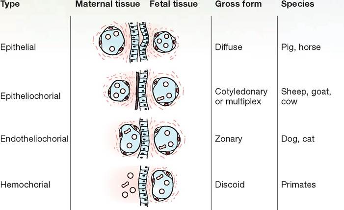
FIGURE 14.9: Layers of the placentation in various species.
Physiology of the Male Reproductive System
The main hormone produced by the testes, specifi cally, by the interstital cells (cells of Leydig), is testosterone, which is responsible for secondary sex characteristics and sex drive. Testosterone is an androgen, or anabolic steroid. Its production is stimulated by luteinizing hormone (from the anterior pituitary gland); however, in the male, the hormone is called interstitial cell stimulating hormone (ICSH). In males, follicle-stimulating hormone is the anterior pituitary hormone that stimulates spermatogenesis, or the creation of spermatozoa.
The sperm has the following parts:
1. head: This oval structure includes the nucleus containing the haploid number of chromosomes (half the chromosomes needed for an animal of the species). The head also has a cap called the acrosome, which contains enzymes to permit penetration into the ovum.
2. midpiece: This is the power plant of the sperm. Within the midpiece are numerous mitochondria that carry out the metabolism that provides adenosine triphosphate (ATP) for the sperm’s locomotion.
3. tail: This section consists of a flagellum for propulsion (Figure 14.10).
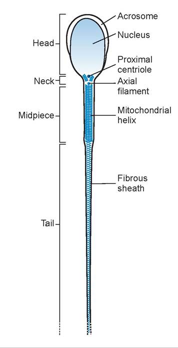
FIGURE 14.10: Parts of a spermatozoa.
The seminal fluid is produced by the accessory sex organs, and it is the medium for survival and activation of the sperm. The prostatic secretion has a specific action (in addition to those mentioned previously): It also alkalinizes the vaginal environment, preventing spermatozoa death.
EXERCISE 14.1 DISSECTION OF THE REPRODUCTIVE SYSTEM
Although you may dissect the reproductive system of only one sex, you will need to know the parts of both sexes. After dissection, trade specimens with others in the lab to study the reproductive structures in a cadaver of the opposite sex. Follow the steps in the appropriate section below, depending on the sex of your cat cadaver.
In the exercises in this chapter the important structures are listed in colored bold print. If a structure is mentioned prior to its dissection, it will be italicized. Structures discussed prior to dissection may also be in bold print for special emphasis.
Procedure
The Female Reproductive System
1. Locate the ovaries (Figure 14.11). These are small, oval structures just caudal to the caudal pole of the kidneys. They are enveloped within the mesosalpinx, which forms a bursa around each ovary called the ovarian bursa.
2. The uterine tubes (or oviducts, or Fallopian tubes) are small, highly convoluted tubes within the mesosalpinx. The dilated end is called the infundibulum of the oviduct, but it is difficult to see. The oviduct’s opening is called the ostium of the tube and is surrounded by the fimbria.
3. Trace the oviducts to their termination at the uterine horns, or cornua. The descriptive name for this type of uterus is bicornuate, or two-horned. Fertilized ova undergo multiple cell divisions as they pass through the oviducts to the uterine horns, where they implant and continue to develop into embryos and on to become fetuses. The fetuses tend to be equally spaced throughout the two horns. The broad ligament supports the uterine horns and body of the uterus. The broad ligament consists of three parts—the mesovarium, mesosalpinx, and mesometrium—but these parts are indistinguishable. The broad ligament is T-shaped; its lateral portion is a sheet of mesentery that is perpendicular to the main part of the mesometrium and anchors it to the dorsolateral wall (see Figure 14.11). The thickened, lateral edge of this lateral portion is called the round ligament of the uterus. The suspensory ligament is the thickened part of the mesovarium and mesosalpinx that connects each ovary to the dorsal body wall, just caudal to each kidney. During an ovariohysterectomy, the suspensory ligament must be broken by the veterinarian so that the ovary and ovarian artery can be elevated to the exterior and ligated.
4. The two horns come together in the middle to form the body of the uterus, which lies dorsal to the bladder and urethra (Figure 14.12).
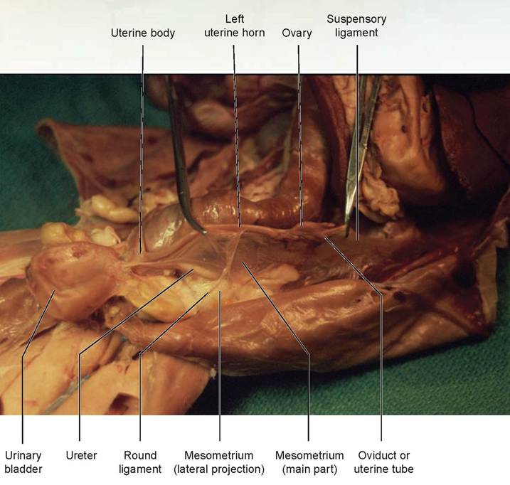
FIGURE 14.11: The broad ligament in the uterus of a cat.
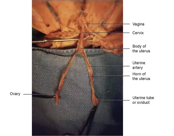
FIGURE 14.12: Parts of the female reproductive tract. The uterus has been everted and laid out on a surgical towel.
5. To dissect the remainder of the female reproductive system, it is necessary to cut through the pubic bone to expose the pelvic cavity. Using bone cutters, cut through the pelvic muscles and along the pubic bones at the pubic symphysis in the ventral midline. Be careful not to cut the urethra, which lies immediately beneath the pubic bone. Open the area further by abducting the thighs to clear the pelvic canal.
6. Locate the urethra, the tube carrying urine from the urinary bladder. Dorsal to the urethra, find the vagina, the tube leading from the body of the uterus. Note that about 2/3 of the length of the vagina, toward the uterus, is slightly thicker than the rest. This is the cervix (see Figure 14.12).
7. The vagina and urethra open into a common passage, called the vestibule of the vagina, which is the space from the external urethral orifice to the vulva.
8. Examine the external part of the vulva, which is the predominant structure of the external genitalia. The large labia majora are the lips surrounding the external genitalia. On the ventral wall of the vestibule, just deep to the opening, is the fossa clitoridis, which is difficult to identify.
9. Locate the rectum, the continuation of the descending colon, which is dorsal to the vagina.
The Male Reproductive System
1. Locate the scrotum, the sac ventral to the anus in cats. Early in fetal development, the testes are located caudal to the kidneys; they migrate before birth through the inguinal canal on either side, and into the scrotum either just before or soon after birth. They are guided by a fibrous cord, which is called the gubernaculum. The remnant of this cord is the connective tissue attaching the testis to the inside of the scrotum and is called the scrotal ligament. The scrotum is covered with skin on the outside and is lined with peritoneum on the inside. It is divided into two compartments by a median septum.
2. Open the scrotum to view and examine the testes. They are covered with tunica vaginalis parietalis (parietal vaginal tunic), an extension of the parietal peritoneum. Cut open this covering to view the testis within. The shiny surface covering the testis, epididymis, and the vessels is the tunica vaginalis visceralis (visceral vaginal tunic), an extension of the visceral peritoneum (Figures 14.13 and 14.14).
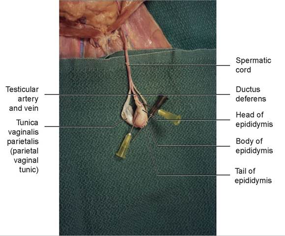
FIGURE 14.13: Testis and component parts of a cat. The lower needle is under the head of the epididymis; the upper needle (dark hubbed) is under the tail.
3. The epididymis is found tightly attached to the lateral aspect of each testis. The head of the epididymis is found at the cranial or dorsal aspect of the testis, the body of the epididymis on the side, and the tail of the epididymis at the caudal or ventral end of the testis. The natural position of the testicle varies with species; the testicle is oriented horizontally in the dog, cat, boar, and stallion, and vertically in the bull and ram (see Figure 14.5). The epididymis is a long, coiled tube that receives sperm from the testis, stores it, and delivers it to the ductus deferens during ejaculation.
4. The ductus deferens carries the sperm from the epididymis through the inguinal canal and empties it into the urethra. As the ductus deferens leaves the testis, it travels within the spermatic cord under the skin adjacent to the prepuce containing the shaft of the penis, caudally to the scrotum (one on each side), through the inguinal canal, and into the abdominal cavity. It is accompanied by the testicular artery, vein, and nerve as it courses to and through the inguinal canal. Collectively, these structures—covered with the tunica vaginalis visceralis and their serous membranous attachments, the mesoductus deferens and mesorchium—are called the spermatic cord. Trace the path of the ductus deferens from the testis through the inguinal canal; then follow it as it curves (leaving the artery and vein) and passes into the pelvic canal, where itjoins to the urethra (see Figure 14.14).
5. Locate the penis, ventral to the scrotum. The glans penis is the enlargement at the distal end of the organ. In adult, intact male cats, it possesses numerous rows of proximally directed cornified papillae known as penile spines. After copulation, these spines scrape the walls of the queen’s vagina upon withdrawal of the penis, an act that is in part responsible for inducing ovulation. The penile spines regress almost completely after castration. The penis’s opening to the outside is called the external urethral orifice. The prepuce is the skin that covers the glans penis.
6. To dissect the rest of the male reproductive system, the pelvic cavity must be exposed. Cut through the pelvic muscles and the pubic symphysis at the ventral midline. Do not cut the urethra, which lies immediately beneath the pubis. Open the area further by abducting the thighs to expose this pelvic canal.
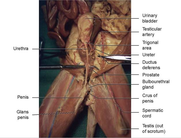
FIGURE 14.14: Ventral view of the male reproductive system of the cat.
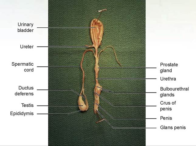
FIGURE 14.15: Dorsal view of the male reproductive tract of the cat.
7. It is now possible to view the entire urethral pathway from the urinary bladder caudally. Trace this pathway to the penis. From the distal penis working cranially, free the penis from the connective tissue and cut the crura (singular: crus) of the penis where each attaches to the ischium on both sides. Using your finger, work cranially and free the rest of the urethra from the underlying colon. Any remaining connective tissue can be cut away, but make sure it is only connective tissue. The penis and urethra should now be free and can be rotated so that their dorsal aspects can be viewed.
Immediately cranial and dorsal to the crura of the penis are the paired bulbourethral glands (Figure 14.15). Carefully dissect the ductus deferens until it reaches its point of attachment to the dorsolateral surface of the urethra. Locate the prostate gland just caudal to the attachment of the ductus deferens. The urethra can be divided into three parts: the prostatic urethra, surrounded by the prostate; the membranous urethra, between the prostate and the penis; and the penile urethra, passing through the penis.
EXERCISE 14.2 DIAGNOSIS OF BREEDING TIME IN THE FEMALE DOG
The estrous cycle of the bitch typically lasts for several weeks; however, the fertile period is short: only 48 to 72 hours. A female dog may exhibit receptive behavior, which is controlled primarily by changes in levels of the hormone estrogen, but this does not always coincide exactly with ovulation. Therefore, a combination of tests can be used to more accurately predict the best time to breed or inseminate a female dog.
The vaginal epithelium of the anestrus bitch is made up of noncornified stratified squamous epithelial cells. Under the influence of estrogen, the lining cells begin to cornify (take on keratin) in preparation for copulation. Proestrus is the time when the uterine lining, the endometrium, is developing for implantation of the embryo. The bitch will typically bleed during this stage because of red blood cell (RBC) diapedesis, or migration, into the uterine lumen. Vaginal cytology will show many uncornified squamous epithelial cells and many RBCs. Proestrus can last anywhere from four to 20 days. During
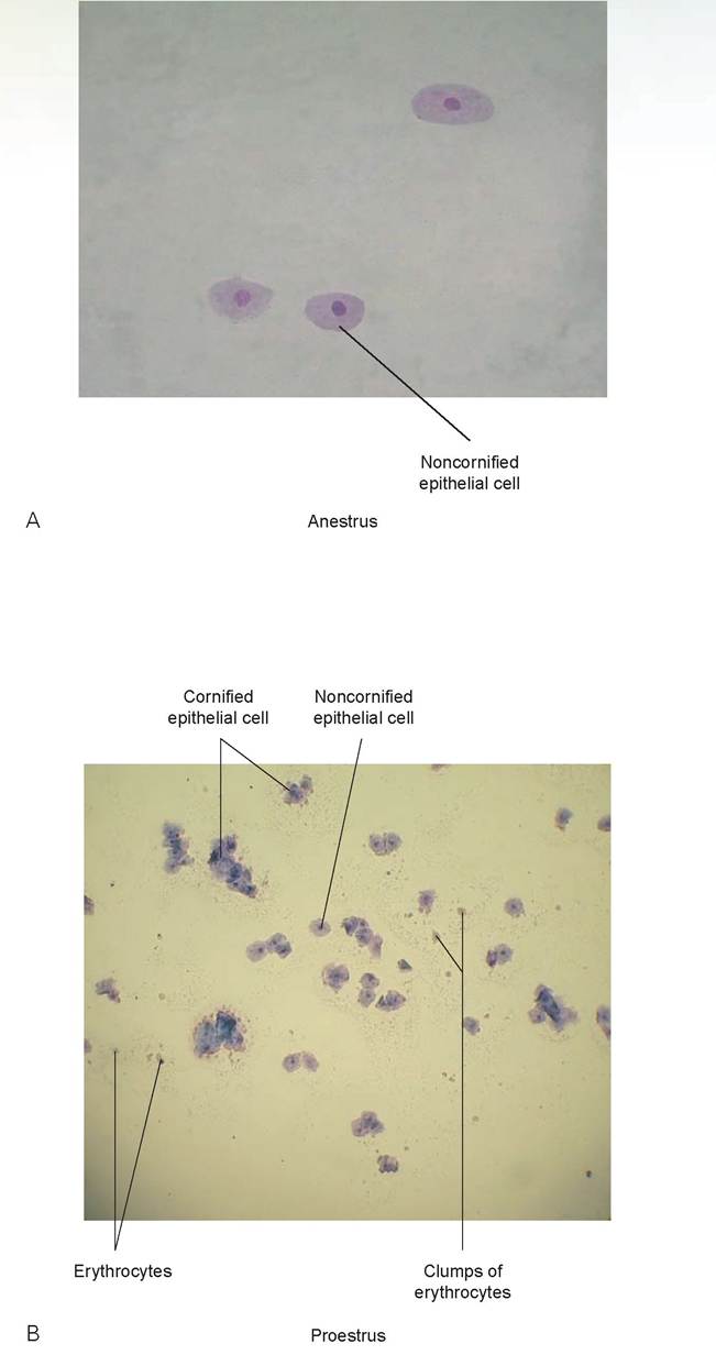
FIGURE 14.16: The stages of canine estrus as demonstrated by vaginal cytology. A. Anestrus: Note the large noncornified epithelial cells predominate. (Diestrus would look similar, but a mixture of noncornified and early cornified epithelial cells would be present.) B. Proestrus: Note the appearance of many RBCs and epithelial cells beginning cornification.
Continued
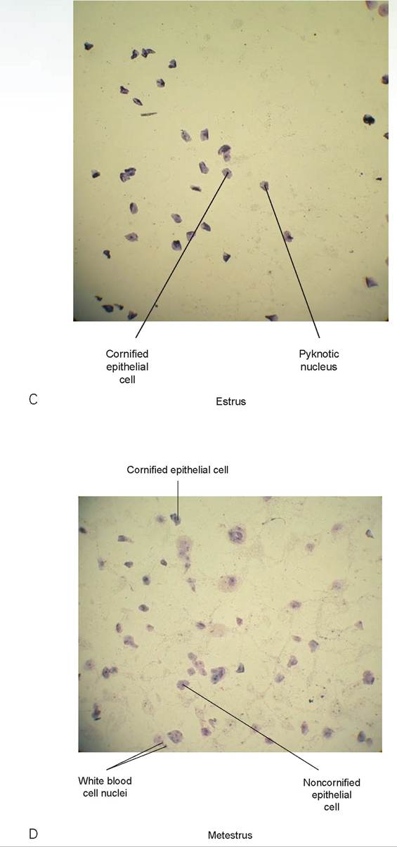
FIGURE 14.16, cont'd: The stages of canine estrus as demonstrated by vaginal cytology. C. Estrus: All cells are cornified, and nuclei are becoming increasingly pyknotic. RBCs are no longer present. D. Metestrus: Note the reemergence of noncornified epithelial cells. Some cornified epithelial cells are still present. White blood cells are usually numerous; however, on this slide there were few and it was necessary to go to high power to see them well (this photo was taken with the low power objective).
proestrus, the epithelial cells begin to cornify and gradually change from a rounded shape to a more polygonal shape with rough, angular borders, and they sometimes appear folded (Figure 14.16). The cells also stain progressively more densely. As the cells become more cornified, the nuclei begin to shrink, becoming pyknotic.
When no more RBCs are visible (occasionally RBCs will persist into estrus) and all the cells are cornified, estrus has commenced. Estrus can last anywhere from four to 13 days. The ideal time to breed is when the cells’ nuclei are completely pyknotic, but this is a subjective determination. Metestrus is characterized by the presence of white blood cells (WBCs) and new noncornified squamous epithelial cells in the smear. Many of the old cornified cells also are still present. Ovulation generally occurs 24 to 36 hours before the appearance of WBCs in the vaginal smear. Metestrus represents the luteal phase and begins when the bitch no longer accepts the male. If pregnancy does not occur, metestrus may last up to two weeks before anestrus or diestrus occurs. Diestrus is a relatively short phase of quiescence between estrus cycles in seasonally polyestrus animals.
Because of the wide variation in the length of the stages of the estrous cycle and the difficulty in predicting the exact time for ovulation, we can use serological testing to enhance the accuracy of our predictions. The following is one method of determining optimal breeding time.
1. On the first day of either noticeable blood spotting or vulvar swelling, perform vaginal cytology. Draw blood to perform a baseline progesterone assay. Continue performing vaginal cytology every other day until the vaginal epithelium is 50% cornified.
2. At this stage, start drawing blood daily to test the progesterone level. Prior to LH release, the level will remain low, generally between 0 and 1.0 ng/ml.
3. When progesterone rises to a range of 1.5 to 2.0 ng/ml, initiate daily testing of serum LH (ICG Status-LHTM, Synbiotic Corporation, San Diego, CA). The progesterone will continue to rise, but it is the initial rise in progesterone that signals the ending of the LH surge. However, there is variation from dog to dog in the time between the first rise in progesterone and the LH surge. Therefore, continue to draw blood and test for LH rise on a daily basis.
4. Ovulation will occur 2 days after the LH surge. The ova then require an additional 2 to 3 days to mature enough for implantation in the endometrium. Thus, the fertile period falls between 4 and 7 days after the LH surge, with the most-fertile days being days 5 and 6 (the day of the LH surge is counted as day 0).
5. The traditional method of counting the gestation period is 63 days from breeding. Using the LH surge offers a second method. Research has demonstrated that gestation is 65 days in length ( + /- 1 day) from the LH surge. This is true regardless of the day the bitch is bred.
Procedure
1. Using prepared slides, identify all four stages of the estrous cycle: anestrus, proestrus, estrus, and metestrus.
2. Draw blood from an anestrus dog; perform a progesterone assay.
3. Draw blood from a dog in heat (if available); perform an LH assay.
4. Record the test results.
Progesterone: ___________________________
LH:
Questions
1. Will doing these tests one time enable you to predict the optimum time for breeding?
2. If a dog becomes pregnant and progesterone is continually assayed, what will happen to progesterone levels over time?
Discussion
Because we are looking for a rise in progesterone and LH, it usually takes two or more samples to observe this. However, it is possible to draw a sample on the day of LH rise and see an increased LH compared to the predicted normal value in dogs. We could tentatively predict ovulation time by this rise in the LH level.
If a dog becomes pregnant, the corpus luteum will be maintained throughout pregnancy, and progesterone will be secreted throughout gestation. Therefore, the level of progesterone assayed will rise and level off until near the time of parturition.
Clinical Significance
Cryptorchidism is a condition in which one or both testes do not descend into the scrotal pouch during development. It is an inherited trait and is thought to be due to a single autosomal recessive gene, although recent research indicates more than one gene may be involved. Usually, testicle descent into the scrotum is expected to be complete by two months of age. Absence of palpable testes at two months is presumptive evidence of cryptorchidism. However, individual dogs have been known to have delayed descent for up to four months. Cryptorchidism is reported in all breeds, but the toy poodle, Pomeranian, Yorkshire terrier, and other toy and miniature breeds have higher incidences. Unilateral cryptorchidism is more common than bilateral (75% of cases are unilateral and 25% bilateral). The right testis is twice as often retained as the left. The prevalence in the general population of males is 1.2% in dogs and 1.7% in cats.
Cryptorchid testes are usually retained just caudal to the inguinal ring, within the inguinal ring, or inside the abdominal cavity. If the testis is caudal to or within the inguinal ring, it often can be palpated. Ultrasound is the other method of determining the position of a retained testis.
The treatment is castration of both testes. Orchiopexy, the surgical placement of a retained testis into the scrotum, is considered unethical because it masks an inherited condition. Castration is strongly recommended to owners, because of the increased incidence of tumor formation in retained testes. Owners are encouraged to castrate before the animal is four years old. Fifty-three percent of sertoli cell tumors and 36% of seminomas are found in cryptorchid testes. The risk of testicular neoplasia is approximately10 times greater in a cryptorchid testis than in normal testes.

® owner of a practice considers himself a horse doctor, and you are the newest ( I// ∖j vet on the block (or in this case, in the hospital), you get all the cow, sheep, pig, and x Zy goat work. I pulled so many calves and did so many Cesarian sections on cows that I became known as C-Section Man. In a moment of sheer lunacy I even considered sewing a big, red C on my scrub shirt. A significant proportion of my work was done on just one farm, whose owner had decided that maximum weight at weaning was better for the bottom line. Of course, any extra profit he made was being eaten up in veterinary bills. But being a pseudo-scientist and following a strict protocol for breeding, he was determined to make this work. He was doing a breeding program called a hree-breed reciprocal cross: He was trying to maximize weight at birth, milk production, and weaning weight.
I studied this during my master's degree work in genetics using Hereford, Angus, and milking Shorthorn cattle. He was using Simmental, Hereford, and Angus stock. It was the Simmental genes he was counting on to give the increase in size, but the small Angus cows and young Hereford heifers did not have the pelvic size to expel a 110-lb calf. The results were disastrous.
I was putting in the last suture on a spayed animal when the receptionist said, “Dr. C., Mr. Barkley is on the phone; another of his cows is having trouble giving birth, he says.”
“Ask him to wait a few minutes and I'll talk to him,” I told her.
Within 10 minutes, I was in my truck heading for Barkley Breeding Farm with all possible haste. I have always thought there should be a special flashing green light veterinarians can put on their trucks in an emergency situation. This would not allow the veterinarian to drive in excess of the speed limit, but it would alert people to a doctor on his way to an emergency. I guess we could let people doctors use it too.
When I arrived, they had the cow in a large stall in the barn (at least at this place I didn't have to play cowboy and rope it first). Sure enough, it was a small Hereford cow, bred to a Simmental bull. I expected to see the calf's front legs protruding from the vulva; then I could reach inside, find the head, and pull it out. In this case, there was nothing presenting at all.
“It was coming hind legs first, so we pushed it back in and tried to turn it,” Mr. Barkley told me while looking toward the ceiling of the barn.
Well, that explained why nothing was showing, I thought. I put on a plastic sleeve, lubricated it well, and pushed my arm through the vagina and up into the uterus. Being tall, thin, and lanky, I had the perfect build for this type of work. First, I located the calf's hind legs and tail, then worked my way forward along the back until I found the neck and head. Then I felt it! What the heck? I thought (or words to that effect). Why is the cow's spleen next to the calf's head? I quickly realized that they had pushed the calf so hard trying to turn it that it had ripped through the uterus and was now partly in the abdominal cavity. I wasn't sure if it was dead or alive.
“I'm going to have to do a Cesarian section. The uterine wall is torn open, and the calf's head and neck are in the cow's abdomen. I can't tell if it is still alive,” I told Mr. Barkley. The look on his face told me he knew he had screwed up.
Whenever a calf was dead in this situation, I would use a fetatome to cut it up and take it out in pieces. But in this case, it wouldn't matter. I was still going to have to open up the cow to repair the uterine wall. When I finally got the cow opened, I saw that the entire side of the uterus was torn, from its cranial tip into the pelvic canal. Most of the calf was in the abdomen, and it was dead, and had been for some time. I had to struggle to remove it because of its size and the stiffness of its muscles. I set it on the ground behind me.
“Push on its chest and see if you can get it breathing,” Mr. Barkley shouted.
“Mind you, this is only opinion,” I said. “But it's been my experience that when rigor mortis begins to set in, we rarely are able to bring them back. Sorry, Mr. Barkley, this one's dead.”
I double-sutured the uterus, using an inverting suture pattern on the second row to turn the serosa inward to ensure a good seal and prevent adhesions.
“If I were you, Mr. Barkley, because of the long tear in the uterus, I would cull this cow,” I told him. “Even though I did my best in suturing the uterus closed, I don't know if it would support another pregnancy.”
I left, thinking he would abide by my advice. A year later, I got a phone call from none other than Mr. Barkley. “Hey Doc, you know that cow you told me to cull? Well, you were wrong. It just had the nicest looking calf you'll ever see! Thanks, Doc!”
Sometimes it's nice to be wrong.
Summary
This chapter covered the anatomy of the female and male reproductive systems through examination of diagrams and by dissection of a cat. You learned the functional anatomy of both the testes and the ovaries. The processes of ovum and sperm development were included. Emphasis was placed on the comparative anatomy of the genital systems of various male domestic animals. You learned that there were different types of uteri, each with a specific type of placentation and vascular attachment to the chorion. The act of parturition in the dog was discussed in detail. In the discussion and exercise you learned how to identify the various stages of the estrous cycle through the use of vaginal cytology and a serum chemistry method to obtain more accurate predictions of the time of breeding. The anatomy and different accessory sex organs of the male were also described.
REVIEW QUESTIONS
1. The target organs, the testes and ovaries, are both endocrine and reproductive; therefore they have two types of products; what are they?
2. Define copulation.
3. Where does the union of sperm and ovum occur?
4. Define gestation period and parturition.
5. What is the mature follicle called?
6. Name the two structural stages of the life of the follicle after it has released the ovum, providing pregnancy does not occur.
7. Name the three parts of the uterus.
8. Describe the layers of the uterine wall, moving from the lumen to the exterior.
9. Name the types of uteri in domestic animals, and indicate which type occurs in which species of animal.
10. Describe the areas of the vulva.
11. What is the broad ligament, and what are the parts of its mesentery?
12. Name the three parts of the epididymis.
13. Name the parts of the spermatic cord.
14. Name the accessory sex glands in the male.
15. What are the names of the erectile tissue of the penis?
16. What is the function of the bulbus glandis?
17. Differentiate between induced ovulators and spontaneous ovulators, and indicate which types of animal fall within these two categories.
18. Name the stages of the estrous cycle.
19. Name the layers of the placenta.
20. Describe the four types of placentation, and indicate which animals fall within each of these types.
21. In the dog, what is considered the first definitive sign of impending parturition?
22. Name the parts of the sperm.
23. What is cryptorchidism?
24. Why is castration rather than an orchiopexy performed in veterinary medicine as a treatment for cryptorchidism? What may be the sequelae if a cryptorchid testis is not excised?