The Skeletal System
OBJECTIVES
• know the five functions of bone
• name and histologically identify the parts of mature compact bone from a model, diagram, or prepared slide
• identify all the bones of the cat’s skeleton, including the skull bones
• identify the major processes, depressions, and foramina in the cat’s skeleton, as specified in this chapter
• compare the bones of the skulls of the dog, cat, horse, ox, and pig for location, shape, and size
• compare the bones of the foreleg and the hind leg of the dog, cat, horse, ox, sheep, and pig for number, location, shape, and size
• identify the bones of the fowl, and recognize the analogous bones and major differences in bones in the mammal
• identify the names (anatomical and common) of the joints specified in this chapter
• locate specified ligaments, tendons, and cartilage
• understand that ionized calcium is the critical form for metabolism, and explain how it is associated with serum protein levels
MATERIALS
• three-dimensional model of mature compact bone (if available)
• prepared slides of compact and cancellous bone
• compound light microscope
• immersion oil
• colored pencils
• three-dimensional model of the equine foot (if available)
• three-dimensional model of the knee joint (human or animal)
• whole and bisected skulls of a cat
• whole skulls of a dog, horse, and cow (if available)
• articulated and disarticulated skeletons of a cat
• articulated skeleton of a dog
• articulated skeleton of a chicken
• articulated skeleton of an equine foreleg, from the carpus distally, and equine hindleg, from the hock distally
• articulated foreleg and hindleg of a sheep (Sheep legs are small, easily stored, relatively inexpensive substitutes for other large animal legs.)
• blood chemistry machine
• assay cards for calcium, albumin, and total serum protein
108
Copyright 2010 Cengage Learning.
All Rights Reserved. May not be copied, scanned, or duplicated, in whole or in part. Due to electronic rights, some third party content may be suppressed from the eBook and/or eChapter(s).Editorial review has deemed that any suppressed content does not materially affect the overall learning experience. Cengage Learning reserves the right to remove additional content at any time if subsequent rights restrictions require it.
Introduction
There are two types of bone formation: intramembranous ossification and endochondral ossification. During intramembranous ossification the osteoblasts originate from embryonal mesenchymal cells, whereas in endochondral ossification an embryonal bone is initially constructed of cartilage, which eventually is replaced by true bone. The cartilaginous bone serves as a temporary support structure and is replaced gradually so that no body part is left unsupported at any time. In both types of ossification, the first type of bone formed is woven bone; it resembles spongy bone in its appearance. This bone then goes through a process of erosion and remodeling to become either mature cancellous (spongy) bone or compact bone.
Osteoblasts are cells that actively produce bone. They are destined to become osteocytes and are so named when found in a lacuna in mature bone. Osteoclasts are cells that tear down bone so it can be rebuilt. The combined actions of the osteoclasts and osteoblasts have three important effects: (1) Bone is modeled, then remodeled in fetal life; (2) old bone is removed and replaced with new bone in adult life; and (3) bones heal by laying down a large callus of spongy bone, which is remodeled into compact bone as needed to bear weight. In addition to helping remodel bone, osteoclasts are active in the formation of bone marrow cavities and spaces, which are formed to provide sites of hematopoiesis (blood cell formation).
The skeleton has three divisions: the axial skeleton, the appendicular skeleton, and a small visceral skeleton.
The axial skeleton is composed of the bones that lie around the body's center of gravity; this includes the bones of the skull, vertebrae, hyoid apparatus, ribs, and sternum. The appendicular skeleton is composed of the bones of the limbs. The visceral skeleton consists of bones that form in soft organs (viscera). The os penis is a bone that surrounds the penile portion of the urethra in the penis of the male dog, beaver, raccoon, and walrus; the os cordis is a bone that supports the valves in the heart of cattle and sheep; and the os rostra is a bone in the nose of swine that lends support during rooting behavior.Bones have the following five functions:
1. form (shape): bones help define the shape and appearance of animals
2. protection: certain bones have a critical role in protecting internal organs, such as the skull protecting the brain and the ribs protecting the thoracic organs (heart and lungs)
3. mineral storage: bone is a primary storage site of calcium and phosphorus
4. blood formation (oxygen-carrying cells and immune function): the marrow cavities of bones are sites of blood cell formation, producing both red and white blood cells
5. leverage (mobility): the muscles and bones work together to enable movement
Types of Bones
Bones are classified according to their shape and structure as follows.
1. Long bones are proportionally longer than they are wide. Each has a central marrow cavity and a proximal and distal epiphysis. Examples of long bones include the femur and metacarpals.
2. Short bones are about as long as they are wide, and each has only one growth center. Examples of short bones are carpals and tarsals.
3. Flat bones have two plates of compact bone with spongy bone in between. This forms the trabeculae crossing from one side of the bone to the other. These bones have no marrow cavity but have small, irregular marrow spaces. Examples of flat bones include pelvic bones, skull bones, and ribs.
4. Irregular bones are all the irregularly shaped bones, such as the vertebrae and some skull bones.
Sesamoid bones are a type of irregular bone and are interposed in tendon. An example of a sesamoid bone is the patella (kneecap).5. Pneumatic bones are bones with air spaces in them. These include certain bird bones.
Projections, Processes, Depressions, and Foramina
To be able to compare bones, you should know the terminology used to describe the skeletal system. The following list of definitions is provided to aid in the study of bones and their projections, depressions, and foramina.
Articular Projections
head: The proximal end of the bone; it may have a neck attached.
condyle: A rounded projection on the distal aspect of a bone.
trochlea: A grooved, sliding surface that acts like the surface of a pulley to increase the mechanical advantage in the movement of a joint.
Non-Articular Projections
process: A prominence or projection on a bone. tuberosity: A large prominence on a bone; for example, the ischiatic tuberosity on the ischium of the pelvis.
tubercle: Large and small prominences on the humerus, known as the greater and lesser tubercles. trochanter: Large and small prominences on the femur, known as the greater and lesser trochanters. epicondyle: Roughened area on the sides of a bone, just proximal to the condyles.
spine: A pointed projection off a bone, as in the spine of the scapula.
crest: A ridge on a bone, as in the crest of the ilium.
Depressions
cavities: Generally articular spaces, where the head of a bone fits; for example, the glenoid cavity of the scapula.
notch: If non-articular, this is a groove or concavity, as in the alar notch of the atlas. If articular, it is inside the joint where the ligaments attach; for example, the trochlear notch of the ulna.
facet: An almost flat surface, as found on the carpals. fovea: A small depression on a bone, such as the fovea capitis.
fossa: A large shallow depression, as in the supraspinous fossa of the scapula.
foramen: A hole in a bone.
canal: A small, tubular hole through a bone; it is longer than a foramen.
Parts of the Long Bone (Figure 7.1)
diaphysis: The shaft of the bone. This consists of compact bone, a central medullary canal, and spongy bone at the proximal and distal ends of the shaft area.
epiphyses (singular is epiphysis): The proximal and distal ends of the bone. They have a thin outer layer of compact bone with centers composed of mostly cancellous (spongy) bone.
metaphyses (singular is metaphysis): The regions in mature bone where the diaphysis joins the epiphyses. This is where the bone visually starts to widen at the proximal and distal ends of a long bone prior to the epiphyseal plates, or growth plates. The metaphyseal region includes the epiphyseal plate, the area in which the cartilage laid down by chondroblasts is replaced by bone via ossification in a growing animal.
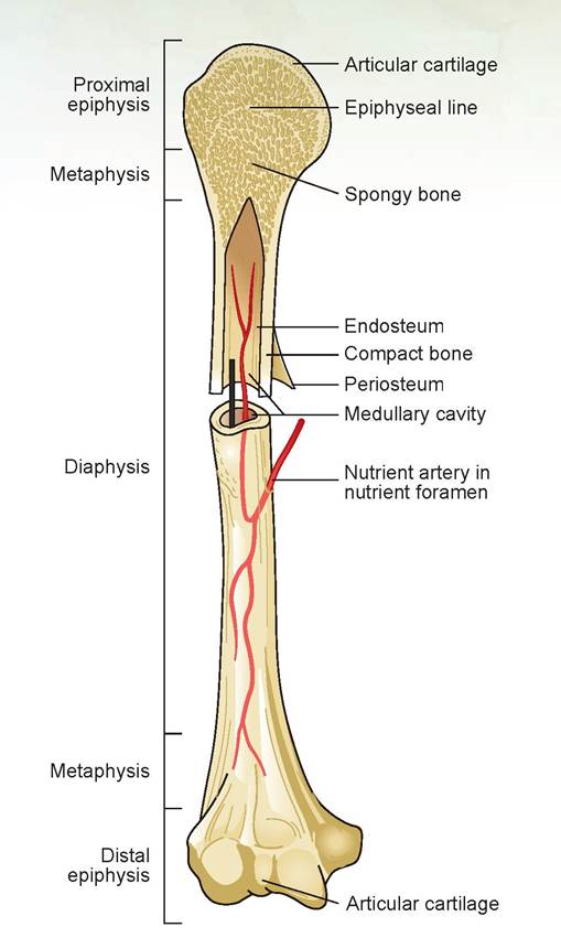
FIGURE 7.1: Parts of the long bone.
EXERCISE 7.1 HISTOLOGY OF BONE
When viewing the following recommended bone tissues, first go to low power, focus, and orient yourself on the slide. Then change to high power to view the cellular detail necessary to understand the morphology of the tissue. The high-power, dry objective lens should be sufficient for viewing these slides.
Obtain the recommended slide(s) and locate all the items on the slide you are instructed to find, including those labeled in the diagrams and photomicrographs contained in the figures throughout the chapter. Draw and label what you see on each slide in the spaces provided.
Compact Bone
Obtain a slide labeled compact bone, and use the plastic model and diagram (Figure 7.2) as a reference to locate the parts of the bone described in the following section.
Slides recommended: Those labeled compact bone.
Description: Compact bone is a cylinder of bone containing a hard, calcified matrix and many collagen fibers. The cells, or osteocytes, lie in lacunae similar to those in cartilage.
Bones are well vascularized by vessels from the bone marrow canal and the periosteum. Approximately the outer third of the blood supply comes from the periosteum, and the inner two-thirds comes from the bone marrow.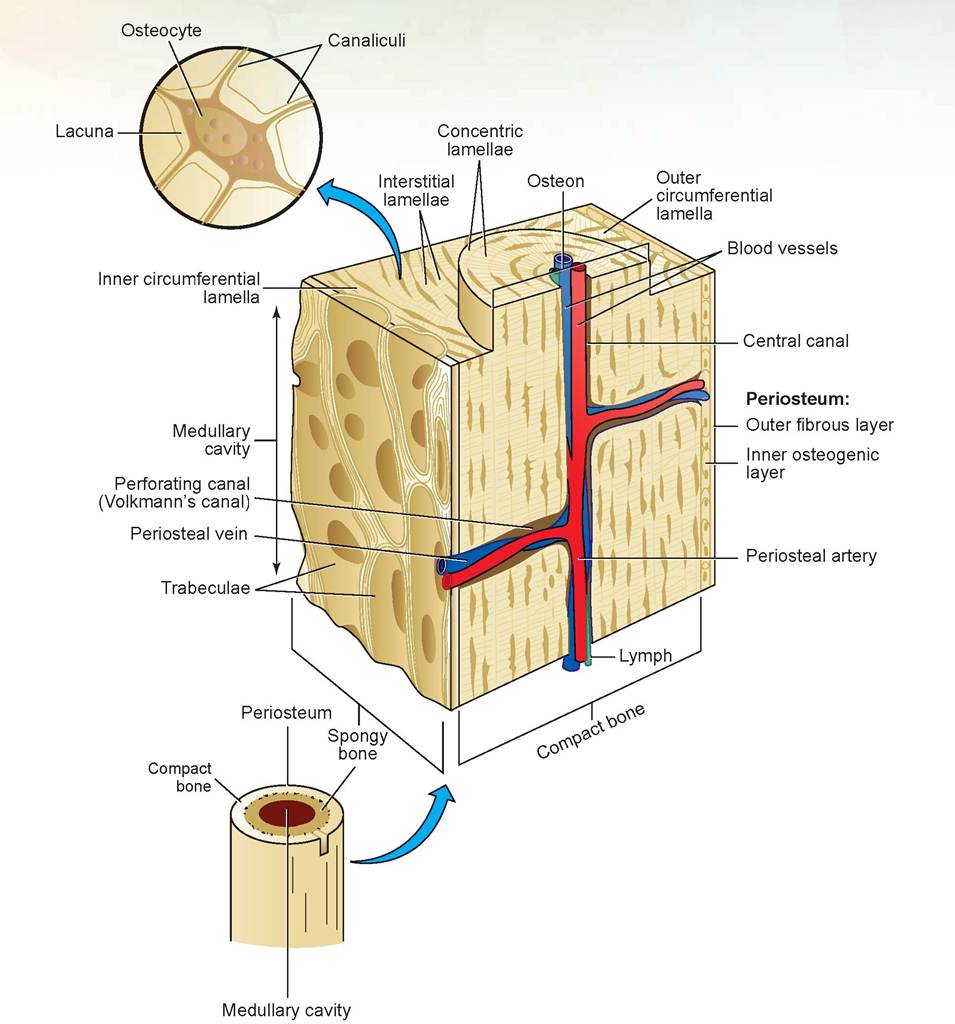
FIGURE 7.2: Diagram of cross section of compact bone.
Slide: compact bone
First, observe the dense irregular connective tissue surrounding the bone; this is called the periosteum. Because it is an important source of blood to the bone and provides nutrition to the outer third (as mentioned previously), veterinarians attempt to preserve as many periosteal attachments to the bone as possible during fracture-repair surgery. Just inside the periosteum are multiple circular layers of bone that go around the entire circumference of the bone. These are called the outer circumferential lamellae. Similarly, just inside the endosteum are the inner circumferential lamellae (Figure 7.3).
As mentioned in Chapter 5, many round cylinders of bone, called osteons, are located throughout bony tissue, and these combine to make up the Haversian system of bone (Figure 7.3). The layers of
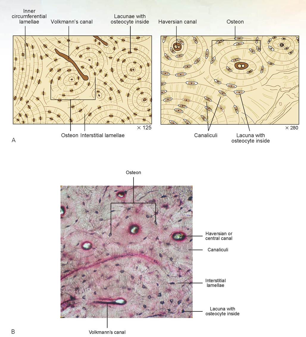
FIGURE 7.3: A & B. Cross section of compact bone.
bone between the osteons are the interstitial lamellae. At the center of each osteon is a central (or Haversian) canal that contains a blood vessel. The dark, elliptical dot-like structures are the lacunae, each containing an osteocyte. Note the thread-like lines that radiate toward each lacuna; these are canaliculi, or tiny canals, that link the lacunae together and provide nutrition for the osteocytes. The central canals of the osteons are connected by transverse canals, called Volkmann’s canals, or perforating canals. These canals also contain blood vessels that interconnect to the vessels in the central canals. These canals, together with the canaliculi, create a complex system for providing nutrition throughout the bone.
In the space provided, draw a section of compact bone and label the cells, canals, and other parts discussed previously.
Cancellous (or Spongy) Bone
Whereas compact bone contains more compact bony tissue than interosseus spaces, cancellous bone contains more interosseus spaces than bone.
Slides recommended: This type of bone is found in the epiphysis or metaphysis (just inside the epiphyseal plate), so slides of these regions can be used. Recommended slides are labeled compact and spongy bone, epiphyseal plate, or the epiphysis.
Description: Cancellous (spongy) bone is characterized by a sponge-like interconnection of bony spicules, hence the name spongy bone.
Slide: cancellous bone and/or epiphysis
On the slide, note the areas of bony matrix surrounded by white areas containing either undifferentiated cells of bone marrow or osteoblasts. Look at the area known as the Zone of ossification; you will see light purple-stained areas which have small elliptical lacunae containing osteocytes (Figure 7.4A). Note the epiphysis is similar in appearance to this layer. In the Zone of calci∣'∣catior∣ the lacunae are larger. Immediately above is the Zone of hypertrophied chondroblasts, in which they appear stacked in parallel columns. In the Zone of multiplication the lacunae and chondroblasts have a more flattened appearance. These are destined to hypertrophy and produce matrix destined to calcify in the process of ossification, thus lengthening the bone. Above this layer is the Zone of reserve cartilage, which supplies the chondroblast needed for growth. This area will eventually ossify connecting the epiphysis and metaphysis as the growth plate closes.
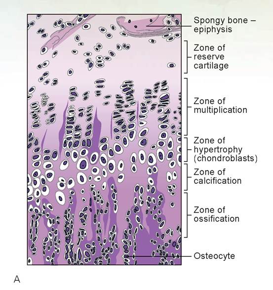
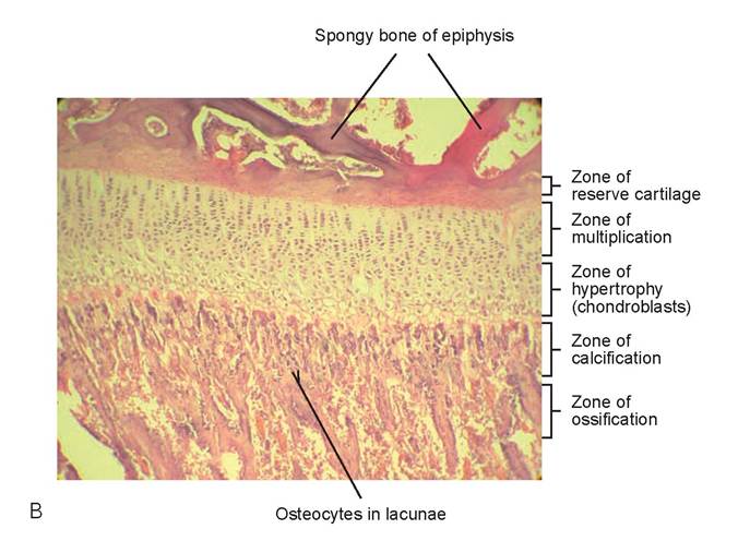
FIGURE 7.4: A & B. Cross section of an epiphyseal plate and spongy bone.
EXERCISE 7.2 THE SKELETON OF THE CAT
Locate and identify the following bones and bone parts (such as notches, foramina, protuberances, and condyles) for a cat. The names of the bones and bone parts listed here are in bold print because these are the items that are important to identify and know.
1. Identify, name, and number each of the vertebrae in the following groups of vertebrae (Figure 7.5):
a. cervical: 7 vertebrae; includes the atlas (first vertebra) and axis (second vertebra)
b. thoracic: 13 vertebrae
c. lumbar: 7 vertebrae
d. sacral (sacrum or os sacrum): 3 vertebrae (These three vertebrae are fused into one bone, which is why they are also called the sacrum or os sacrum.)
e. Caudal (coccygeal): variable number of vertebrae depending on breed of cat (You may see this abbreviated Ca+ + or Cy++.)
2. Parts of a vertebra: Using the cat skeleton, identify the parts of the vertebra listed as follows for each of the types of vertebrae listed in the previous activity (Figure 7.6).
a. vertebral arch
1. lamina
2. pedicle
b. vertebral canal
c. vertebral body
d. spinous processes
e. cranial and caudal articular processes and facets
f. lateral vertebral foramen
g. transverse process
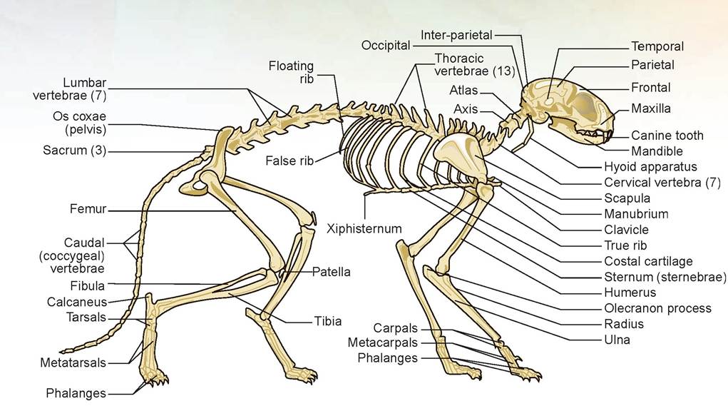
FIGURE 7.5: Side view of the skeleton of the cat.
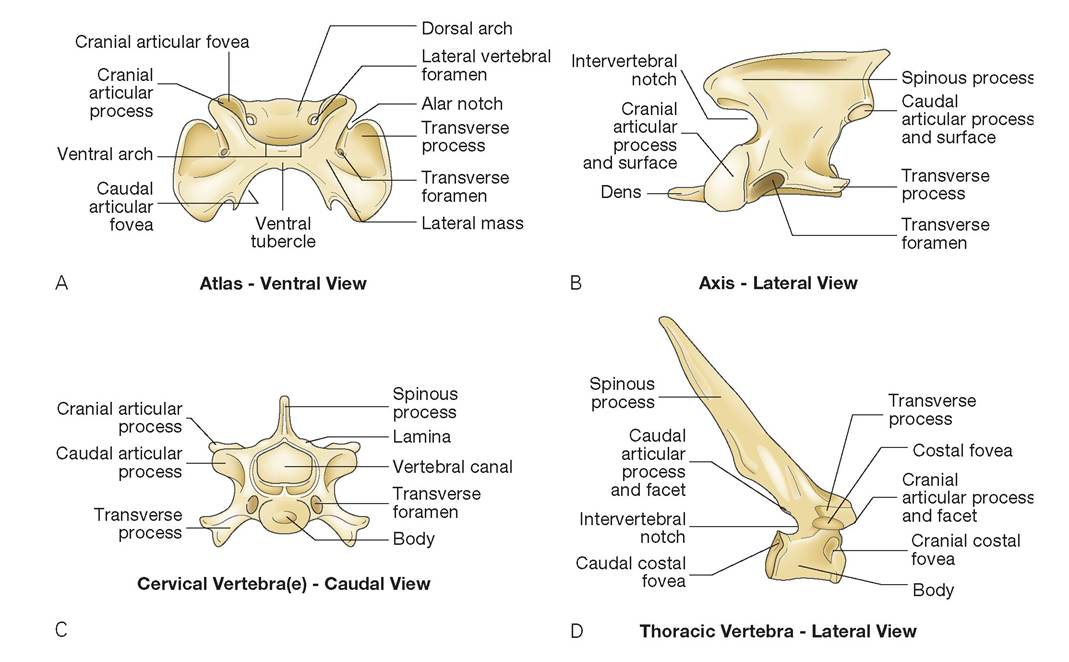
FIGURE 7.6: Vertebrae of the cat. A. Atlas, ventral view. B. Axis, left lateral view. C. Cervical vertebra(e), Caudal View. D. Thoracic vertebra, lateral view.
Continued
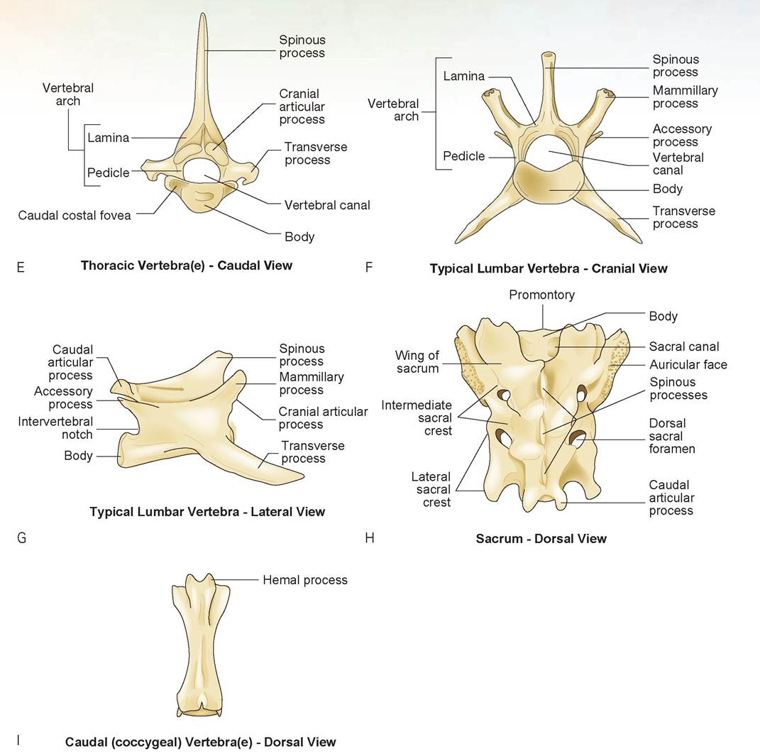
FIGURE 7.6, cont'd: Vertebrae of the cat. E. Thoracic vertebra(e), right cranial view. F. Lumbar vertebra, right cranial view. G. Lumbar vertebra, lateral view. H. Sacrum, dorsal view. I. Caudal (coccygeal) Vertebra(e) - Dorsal View.
h. transverse foramen (cervical vertebrae only)
i. costal fovea and cranial and caudal costal foveae (thoracic vertebrae only)
j. accessory process (lumbar vertebrae only)
k. atlas (C1)
1. dorsal and ventral arches
2. ventral tubercle
3. transverse processes (wings)
4. alar notch
5. lateral vertebral foramina
6. vertebral canal
7. cranial articular fovea
8. caudal articular fovea
l. axis (C2)
1. dens or odontoid process
2. cranial articular surface
3. spinous process
4. transverse foramen
m. sacrum
1. median sacral crest (the three spinous processes)
2. intermediate sacral crest
3. dorsal and ventral sacral foramina
4. promontory
5. lateral sacral crest
6. auricular face
3. Ribs, sternum, and associated structures: Using the cat skeleton, identify the parts of the ribs, sternum, and associated structures listed as follows (see Figures 7.5 and 7.7). Also, determine the number of each rib and sternebrae (i.e., the fourth rib is attached to the fourth thoracic vertebra).
a. head
b. tubercle
c. neck
d. shaft or body
e. costal cartilage
f. costochondral junction
g. true (#1-9), false (#10-12), and floating ribs (#13)
h. sternum
1. sternebrae
2. manubrium sterni
i. xiphoid process
j. xiphoid cartilage
k. intersternal cartilage
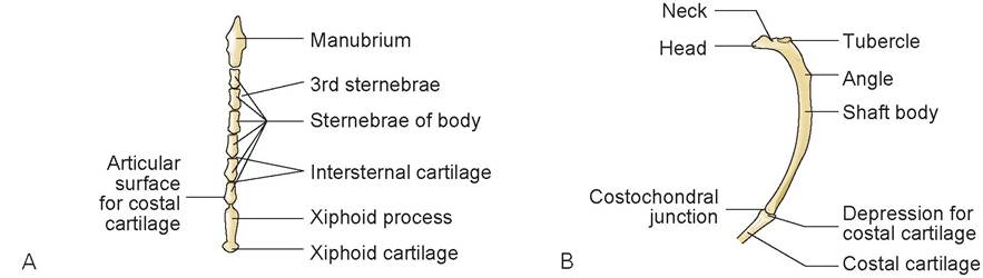
FIGURE 7.7: Sternum and ribs of the cat. A. Sternum, ventral view. B. Ribs, lateral view.
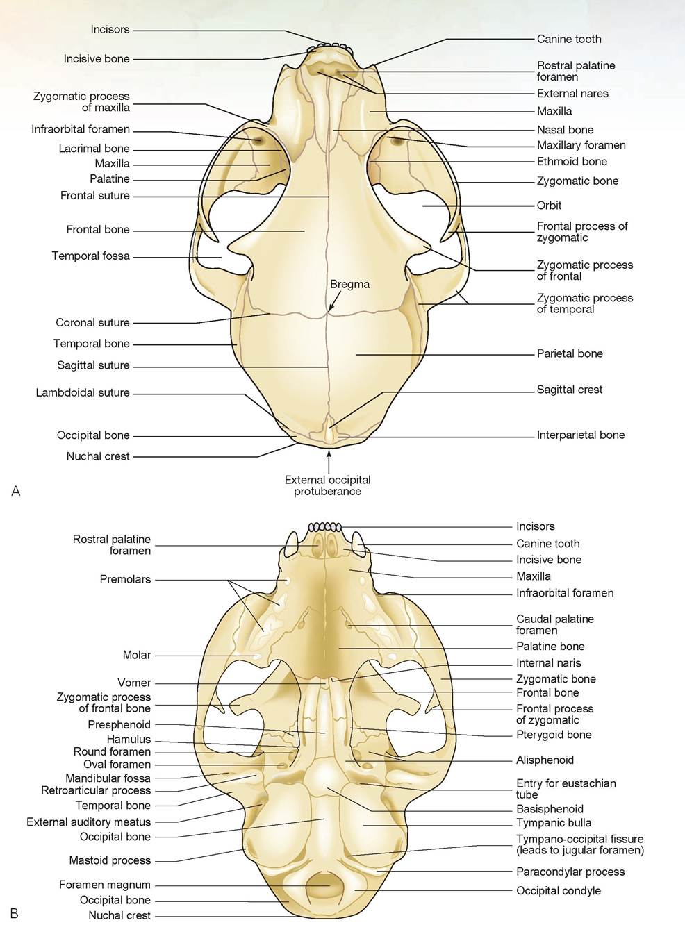
FIGURE 7.8: Skull and mandible of the cat. A. Skull, dorsal view. B. Skull, ventral view.
Continued
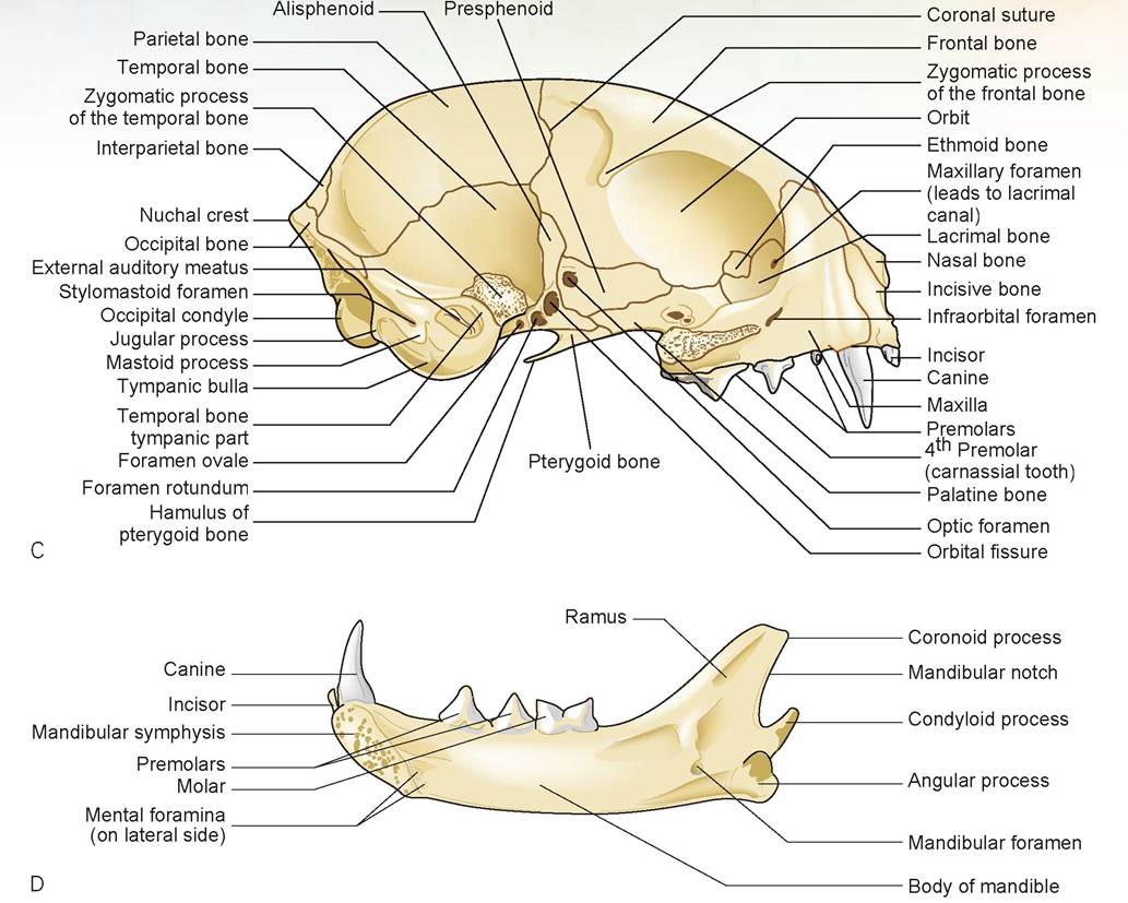
FIGURE 7.8, cont'd: Skull and mandible of the cat. C. Skull, lateral view. D. Mandible, medial view.
4. Skull: Using the whole and bisected cat skulls, identify the bones of the skull, their processes, and the foramina listed as follows (Figure 7.8). Remember, the bones of the skull are irregular bones. These bones are three-dimensional, whereas the drawings provided are two-dimensional; therefore, the same bone may be seen on two views of the skull in apparently different places. Remember that bones bend, curve, and occupy space on both the outside and inside of the skull.
a. bones of the skull
1. incisive
2. nasal
3. lacrimal
4. maxilla
5. palatine
6. frontal
7. parietal
8. temporal
9. occipital
10. tympanic bulla
11. zygomatic bone
12. interparietal
13. presphenoid
14. basisphenoid
15. pterygoid and the hamulus of the pterygoid
16. nasal concha (also known as nasal turbinate bones; bones visualized through external nares)
17. mandible
18. wings of the basisphenoid
19. ethmoid
b. processes, crests, sutures, and other protuberances
1. frontal suture
2. sagittal suture
3. coronal suture
4. nuchal crest
5. sagittal crest
6. external occipital protuberance
7. occipital condyle
8. zygomatic process of the frontal bone
9. zygomatic arch
10. frontal process of the zygomatic bone
11. zygomatic bone
12. zygomatic processes of the temporal bone and maxilla
13. bregma
14. paracondylar process
15. retroarticular process
16. mastoid process
c. parts and processes of the mandible
1. body
2. ramus
3. mandibular symphysis
4. angular process
5. condyloid process
6. coronoid process (the top of the ramus)
d. foramina and concavities of the skull
1. infraorbital foramen
2. maxillary foramen leading to the lacrimal canal
3. frontal sinus
4. maxillary sinus
5. oval foramen (or foramen ovale)
6. round foramen (or foramen rotundum)
7. orbital fissure
8. optic foramen
9. external acoustic meatus
10. jugular foramen (best seen from the inside)
11. tympano-occipital fissure leading to jugular foramen
12. foramen magnum
13. nares
14. choanae
15. orbit
16. mandibular fossa
e. foramina and depressions of the mandible
1. massenteric fossa (on lateral side)
2. mandibular foramen (on medial side caudally)
3. mental foramina (on lateral side cranially)
Note: Teeth will be covered in the chapter on the digestive system.
5. Scapula: Using the cat skeleton, identify the parts of the scapula (or shoulder blade) listed as follows (Figure 7.9).
a. coracoid process
b. glenoid cavity
c. clavicle
d. spine
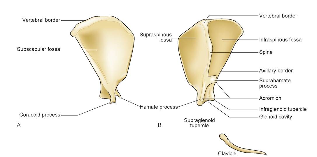
FIGURE 7.9: Scapula of the cat. A. Left medial view. B. Left lateral view.
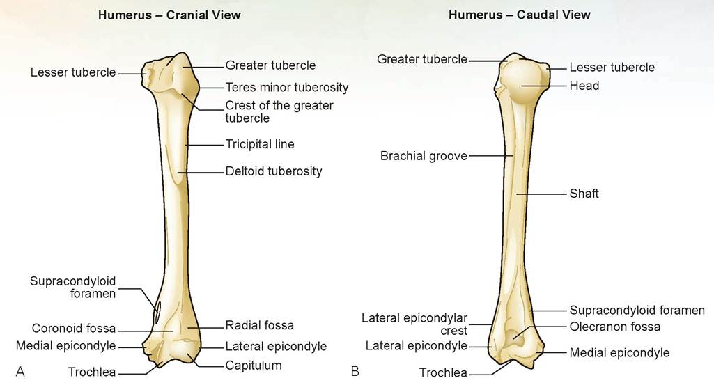
FIGURE 7.10: Humerus of the cat. A. Cranial view. B. Caudal view.
e. acromion
1. hamate process (of the acromion process)
2. suprahamate process (of the acromion process)
f. infraspinous fossa
g. supraspinous fossa
6. Humerus: Using the cat skeleton, identify the parts of the humerus listed as follows (Figure 7.10).
a. head
b. greater and lesser tubercles
c. tricipital line
d. deltoid tuberosity
e. brachial groove
f. shaft
g. supracondyloid foramen
h. medial and lateral epicondyles
i. coronoid fossa
j. radial fossa
k. trochlea
l. capitulum
7. Radius: Using the cat skeleton, identify the parts of the radius listed as follows (Figure 7.11).
a. head
b. radial tuberosity
c. shaft
d. styloid process (medial)
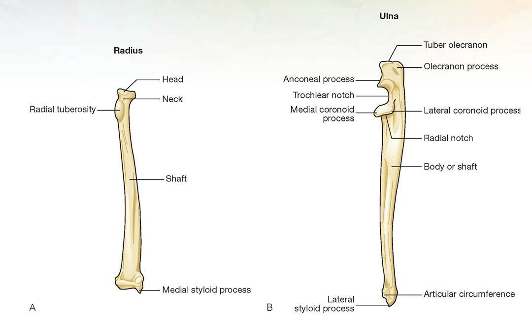
FIGURE 7.11: A. Radius. B. Ulna of the cat.
8. Ulna: Using the cat skeleton, identify the parts of the ulna listed as follows (see Figure 7.11).
a. trochlear notch
b. olecranon (Tuber olecranon) (plural: olecranoni)
c. anconeal process
d. medial and lateral coronoid processes
e. lateral styloid process
f. body of the ulna
9. Carpus, metacarpal bones, and phalanges: Using the cat skeleton, identify the parts of the carpus, metacarpal bones, and phalanges of the digits listed as follows (Figure 7.12).
a. intermedioradial carpal bone
b. ulnar carpal bone
c. accessory carpal bone (on lateral aspect of leg in all species)
d. carpal bones I to IV (medial to lateral)
e. metacarpal bones I to V
f. head (proximal end)
g. body (shaft)
h. base (distal end)
i. proximal sesamoid bones
j. proximal phalanges of digits I to V
k. middle phalanges of digits II to V (the dewclaw, digit I, has no middle phalanx)
l. distal phalanges of digits I to V
m. unguicular process of the distal phalanges
10. Os coxae (pelvis): Using the cat skeleton, identify the parts of the os coxae (pelvis) listed as follows (Figure 7.13). There are four bones of the pelvis: the ilium, ischium, pubis, and acetabular bones. The acetabular bone is in the center of the acetabulum. There is no clear distinction as to where one bone starts and another leaves off.
a. acetabulum
b. acetabular fossa (the cavity of the acetabulum)
c. acetabular notch (the center area)
d. obturator foramen
e. ilium
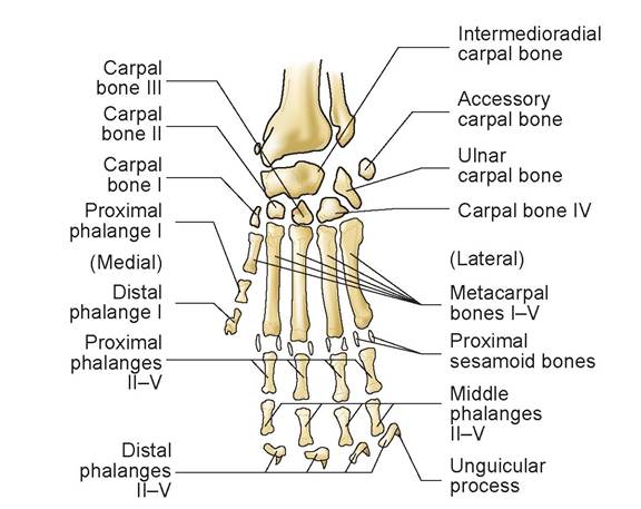
FIGURE 7.12: Carpal bones, metacarpal bones, and phalanges of the cat.
f. iliac crest
g. gluteal surface
h. tuber sacrale (very prominent and important in large animals)
i. tuber coxae (very prominent and important in large animals)
j. greater ischiatic notch
k. ischium
l. ischiatic spine
m. lesser ischiatic notch
n. ischial tuberosity (tuber ischiadicum)
o. pubis
p. symphysis pubis (or pubic symphysis)
11. Femur: Using the cat skeleton, identify the parts of the femur listed as follows (Figure 7.14).
a. head
b. neck
c. fovea capitis
d. greater trochanter
e. lesser trochanter
f. trochanteric fossa
g. intertrochanteric crest
h. shaft
i. trochlea (medial and lateral trochlear ridge and trochlear groove)
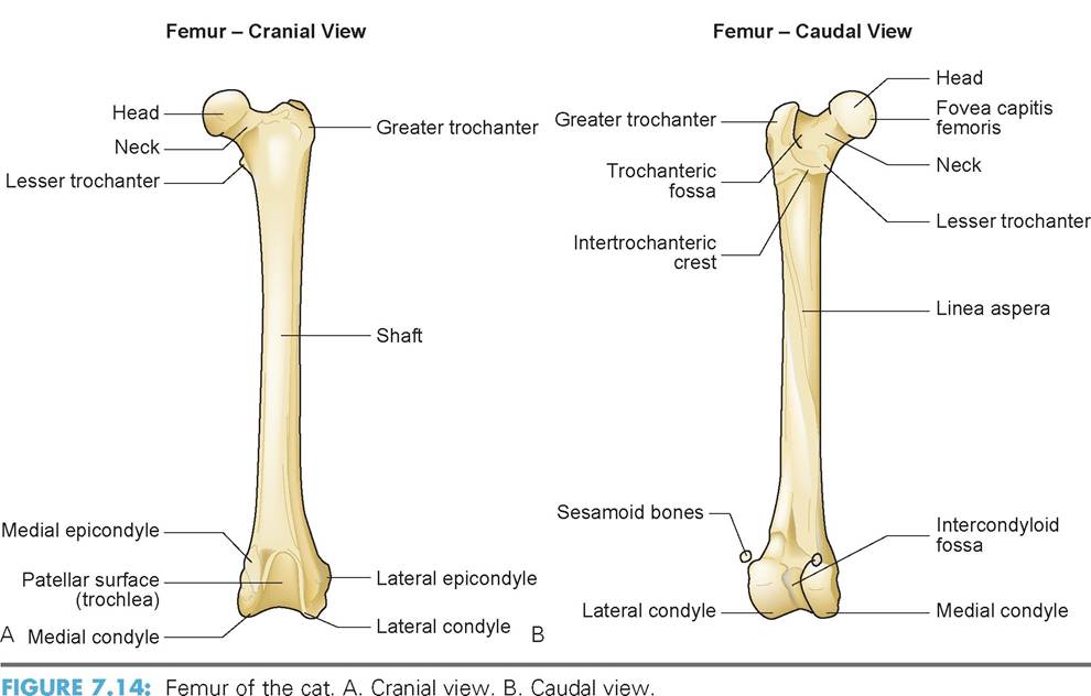
j. medial and lateral condyles
k. medial and lateral epicondyles
l. patella (or kneecap)
m. sesamoid bones
12. Tibia: Using the cat skeleton, identify the parts of the tibia listed as follows (Figure 7.15).
a. medial and lateral condyles
b. tibial tuberosity
c. shaft
d. medial malleolus
13. Fibula: Using the cat skeleton, identify the parts of the fibula listed as follows (see Figure 7.15).
a. head
b. shaft
c. lateral malleolus
14. Tarsus: Using the cat skeleton, identify the parts of the tarsus (the tarsal bones), also known as the hock, listed as follows (Figure 7.16).
a. talus (tibial tarsal bone)
b. trochlea
c. calcaneus (fibular tarsal bone)
d. tuber calcanei
e. sustentaculum tali
f. central tarsal bone
g. tarsal bones I to IV
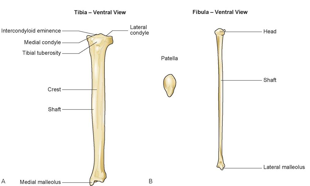
FIGURE 7.15: A. Tibia, ventral view; B. Fibula, ventral view of the cat.
15. Metatarsals and phalanges: Using the cat skeleton, identify the metatarsals and phalanges listed as follows (see Figure 7.16).
a. metatarsal bones I to V
b. body
c. proximal sesamoid bones
d. proximal phalanges of digits II to V (medial to lateral; digit I usually is missing in the cat)
e. middle phalanges of digits II to V
f. distal phalanges of digits II to V
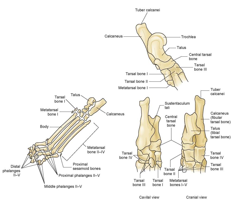
FIGURE 7.16: Tarsal bones, metatarsal bones, and phalanges of the cat.

EXERCISE 7.3 COMPARATIVE OSTEOLOGY
1. Using skulls from the dog (Figure 7.17A), horse (Figure 7.17B and C), and ox (Figure 7.17D and E), identify the following bones, and compare them to each other and to the bones in the skull of the cat (Figure 17.8).
a. maxilla
b. frontal and frontal sinus
c. parietal
d. occipital
e. temporal
f. nasal
g. lacrimal
h. zygomatic arch
i. mandible
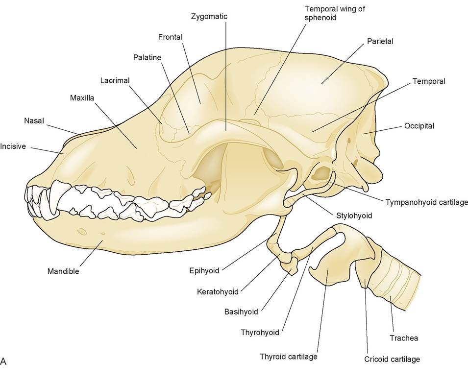
FIGURE 7.17: A. Skull of the dog.
Continued
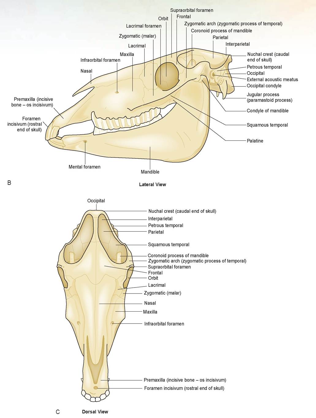
FIGURE 7.17, cont'd: B and C. Skull of the horse.
Continued
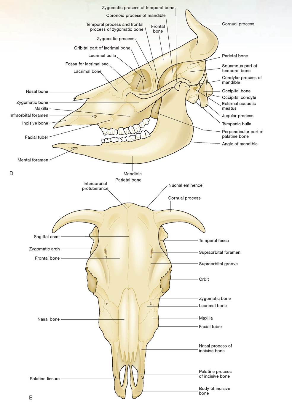
FIGURE 7.17, cont'd: D. Skull of the ox, lateral view. E. Skull of the ox, dorsal view.
2. Identify the bones listed below using the following items: (1) the front leg and hind leg of the sheep, (2) the model of the leg of a horse, and (3) the articulated skeletons of a horse and cow from the carpus and tarsus distally (Figure 7.18).
a. scapula
b. humerus
c. fused radius and ulna
d. carpal bones (The equine carpus contains the accessory, radial intermediate, and ulnar carpal bones, as well as carpal bones I to IV; see Figure 7.18B).
e. cannon bone (metacarpal [Mc] III or metatarsal [Mt] III; Mc III and IV are fused in the ruminant.)
f. splint bones (Mc or Mt II and IV in the horse)
g. proximal sesamoid bones
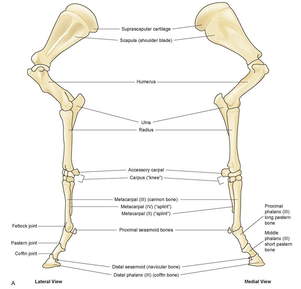
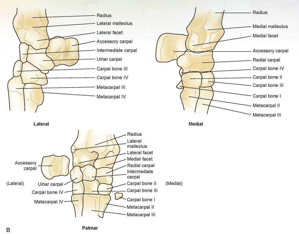
FIGURE 7.18, cont'd: B. Carpus of the horse, lateral, medial, and palmar views.
Continued
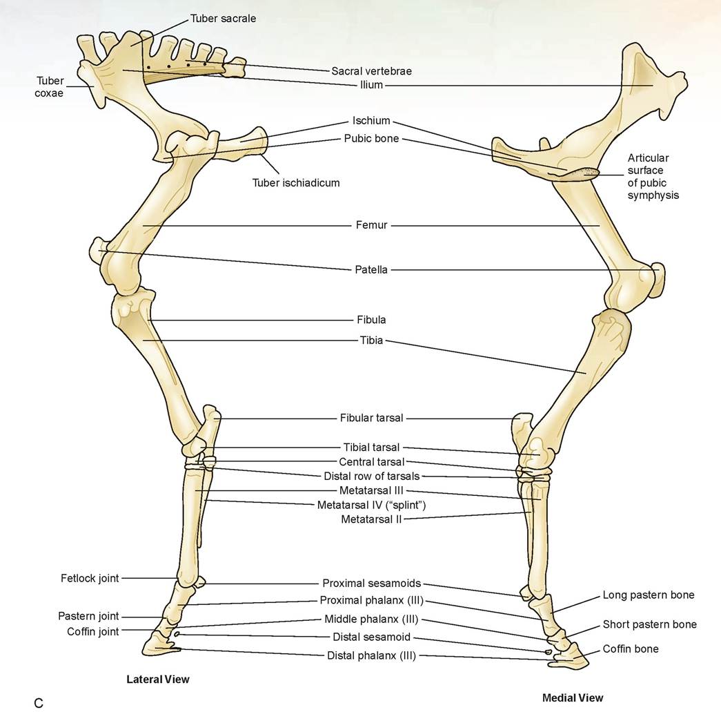
FIGURE 7.18, cont'd: C. Hind leg of the horse, lateral and medial view.
Continued
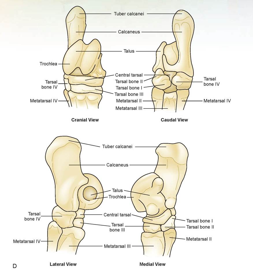
FIGURE 7.18, cont'd: D. Tarsus of the horse.
Continued
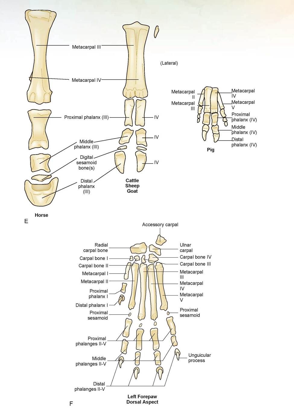
FIGURE 7.18, cont'd: E. Comparative feet of domestic animals. F Carpal bones, metacarpal bones, and phalanges of the dog, dorsal aspect of left forepaw.
h. long pastern bone (This is the proximal phalanx, digit III in the horse and digits III and IV in the ruminant.)
i. short pastern bone (This is the middle phalanx, digit III in the horse and digits III and IV in the ruminant.)
j. coffin bone (This is the distal phalanx, digit III in the horse and digits III and IV in the ruminant.)
k. navicular bone or distal sesamoid bone(s) (There is one in the horse and two in the ruminant.)
l. collateral cartilages
m. tuber sacrale
n. tuber coxae
o. tuber ischiadicum
p. os coxae and acetabulum
q. femur
r. patella
s. tibia
t. fibula
u. tarsal bones (The equine tarsus includes the talus, or tibial tarsal bone; the calcaneus, or fibular tarsal bone; the central tarsal bone; tarsal bones I and II fused; tarsal bones III and IV.
3. Using the diagram and the articulated skeleton of a chicken, locate the following bones (Figure 7.19).
a. axis
b. atlas
c. cervical vertebrae
d. thoracic vertebrae
e. synsacrum
f. caudal vertebrae
g. pygostyle
h. furcula (singular: furculum)
i. coracoid
j. humerus
k. radius and ulna
l. phalanges and digits of the wing
m. keel of the sternum
n. ribs
o. ilium
p. ischium
q. pubis
r. femur
s. patella
t. tibia and fibula
u. tarsometatarsus
v. phalanges and digits of the foot
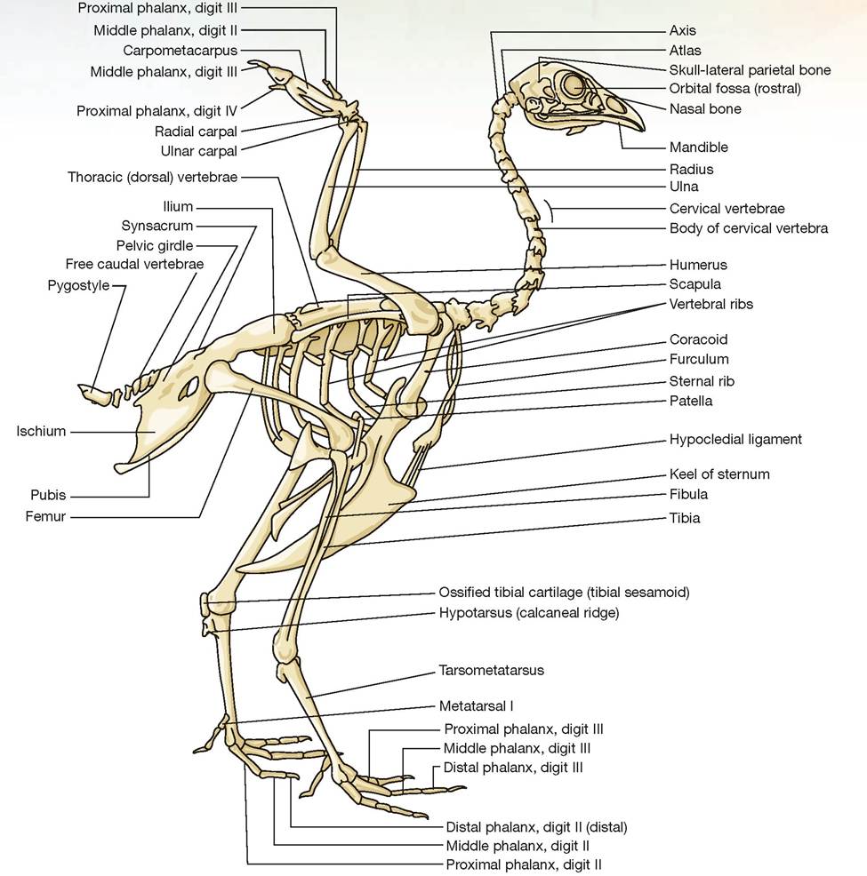
FIGURE 7.19: The skeleton of the chicken.
EXERCISE 7.4 COMPARATIVE ARTHROLOGY AND DESMOLOGY
1. Arthrology is the study ofjoints. Using the articulated skeleton, models, and Figure 7.18, find the following joints. Items are found in all species except where noted.
a. costochondral junction (where bony rib meets cartilaginous rib)
b. temporomandibular joint (T-M joint)
c. shoulder joint (scapulohumeral joint)
d. elbow joint (humeroradioulnar joint)
e. carpal joint (Found in small and large animals; composed of three joints, each with a separate synovial sac or membrane. Sometimes referred to as a knee in the equine.)
1. radiocarpal joint
2. intercarpal joint
3. carpometacarpal joint
f. fetlock joint (metacarpophalangeal joint; found in the equine)
g. pastern joint (proximal interphalangeal joint; found in the equine)
h. coffin joint (distal interphalangeal joint; found in the equine)
i. sacroiliac joint (where the sacrum attaches to the ilium of the pelvis)
j. pubic symphysis (symphysis of the two halves of the pelvis at the pubic bones)
k. hip joint (coxofemoral joint)
l. stifle joint (femorotibial joint, true knee joint)
m. tarsal joint (hock joint) The tarsus of small animals is composed of three separate joints, each with its own joint capsule. The name of each joint is based on the names of the bones from proximal to distal, e.g., the tibiotarsal joint is an articulation between the tibia and the tarsus.
1. tibiotarsal joint
2. intertarsal joint
3. tarsometatarsal joint
The tarsus of large animals is made up of four separate joints, and, as in small animals, each has its own joint capsule. As you can see, there is an extra row of tarsal bones in large animals that creates an extra joint when compared to small animals.
1. tibiotarsal joint
2. proximal intertarsal joint
3. distal intertarsal joint
4. tarsometatarsal joint
2. Desmology is the study of ligaments. Using the prepared models and diagrams, find the following ligaments.
nuchal ligament: Massive ligament that helps support the head of large animals. It runs from the nuchal crest of the skull to the spinous processes of the thoracic vertebrae. In dogs it runs from the axis to the thoracic vertebrae.
interspinous ligaments: Ligamentous bands between the spinous processes of the vertebrae.
supraspinous ligaments: Run along the top of the spinous processes from the thoracic vertebrae caudally.
transverse humeral ligament: Holds the tendon of origin of the biceps brachii muscle next to the humerus as it passes the greater tubercle medially.
sacrotuberous ligaments: Two additional supports for the pelvis. Each runs from the sacrum on either
side to the dorsal aspect of the ischiatic tuberosity (tuber ischiadicum) of the pelvis.
round ligament: Helps hold the head of the femur in the acetabulum. It runs from the acetabular bone to the fovea capitis.
patellar ligaments in the horse: Medial, middle, and lateral patellar ligaments. Horses have three patellar ligaments that run from the patella to the tibial tuberosity: one each on both the lateral and medial aspects, and a central ligament in the middle.
straight patellar ligament: In small animals, this is an extension of the quadriceps femoris tendon through and around the patella to the tibial tuberosity.
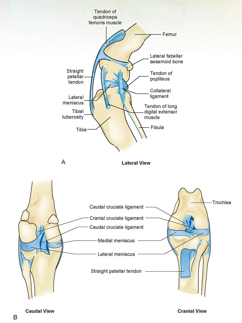
FIGURE 7.20: A. Stifle joint, lateral view of a dog. B. Stifle joint, caudal and cranial views of a dog.
collateral ligaments: These stabilize the stifle medially and laterally, coursing from the distal epicondyle of the femur to the proximal, medial, and lateral aspects of the tibia and fibula (Figure 7.20).
cruciate ligaments: These stabilize the stifle cra- nially and caudally; the cranial cruciate is lateral to the caudal cruciate within the knee joint and is attached cranially on the central head of the tibia. The caudal cruciate attaches caudally on the central head of the tibia, between the medial and lateral menisci (Figure 7.20). plantar metatarsal ligament: Mainly observed in the equine. This runs from the caudal aspect of the calcaneus to the fourth metatarsal bone (proximal aspect).
suspensory ligament: Located between the metacarpal bone (or metatarsal in the hindleg) and the deep digital flexor tendon. This attaches at the proximal end of the metacarpal (and metatarsal) and runs distally, and branches above the fetlock to attach on both sides to the proximal sesamoid bones. It then sends two symmetrical branches that pass across the fetlock to join the common extensor tendon and attach to the proximal phalanx. It is a strong support mechanism of the fetlock. In large animals with more than one digit, the suspensory ligament contains a muscle called the interosseus muscle.
Achilles tendon: This is the combined tendon of the gastrocnemius, soleus, and superficial digital flexor muscles and two additional calcanean tendons from the thigh; it attaches to the tuber calcanei.
stay apparatus: Its components are certain muscles, tendons, and ligaments which allow the horse to stand upright on the forelimbs and hindlimbs with little muscular activity. As an example, in the frontleg it includes numerous structures such as the fibrous sheet of the serratus ventralis, tendon of the biceps brachii, the extensor carpii radialis, proximal and distal check ligaments,just to name a few. The exact components of the stay apparatus in the front and hind legs is beyond the scope of this book.
check apparatus: The attachments of the superficial and deep digital flexor tendons to specific bones of the carpus; as part of the stay apparatus in the forelimbs, the attachment only of the deep digital flexor tendon on the tarsus.
reciprocal apparatus: A series of muscles and ligaments that permit the stifle and hock to both flex and extend together.
EXERCISE 7.5 PHYSIOLOGY: CALCIUM MEASUREMENTS
Approximately 50% of total serum calcium is ionized; 40% is bound to protein, especially to albumin; and 10% is complexed with anions such as citrate or phosphate. Only the ionized calcium is biologically active in bone formation, neuromuscular activity, cellular biochemical processes, and blood coagulation.
Alkalosis decreases ionized calcium levels in serum, and acidosis increases them. Ionized calcium is almost always increased in hypercalcemic conditions, and similarly, is almost always decreased in hypocalcemia (calcium. With the correction factor, the calculated results should compare well to those of the normal dog. If the reason for the decrease in serum protein is a drop in the albumin level, there should be a proportional decrease when comparing a decrease in albumin levels to a decrease in total serum protein, and the values should compare well. However, if fibrinogen or globulins are decreased and albumin stays the same, the values will vary.
Clinical Significance
The clinically significant aspects of this chapter could fill a book on orthopedic surgery. A common injury in large dogs is a rupture of the cranial cruciate ligament. In Exercise 7.4, the anatomy of the cranial cruciate ligament was discussed. The cruciate ligaments stabilize the stifle joint cranially and caudally Specifically, the cranial cruciate ligament prevents abnormal displacement of the tibia cranially from its normal articulated position with the femur.
When this ligament ruptures, a pathological movement called cranial drawer movement occurs. The name presumably results from the movement’s similarity to a desk drawer being opened, and it is the diagnostic feature of this condition. The veterinarian can assess the degree of movement of the tibia from its normal position to ascertain whether the ligament is strained or completely ruptured. Sometimes there is also a tear of the cartilage of the medial meniscus. Veterinary surgeons can open the joint capsule to observe whether this tear has occurred and trim away any remaining pieces of the torn ligament. If the remaining ligament is not removed, it may calcify and give rise to osteoarthritis in the joint. There are many corrective surgeries developed for repair of this injury.
A study conducted by Sandman and Harariand, published in the November 2001 issue of Veterinary Medicine, discussed various methods of repairing cranial cruciate ligament ruptures and the frequency with which veterinarians use the various methods. The most common was the Modified DeAngelis Technique, in which a strong, non-absorbent suture material is placed on the outside of the joint from the caudal aspect of the femur to the tibial crest and continued either through the bone or to the attachment of the straight patellar ligament, or a combination of both. This substitutes for the missing ligament, stabilizes the joint, and prevents drawer movement.

“—y' oc, Mary Thompson just called,” my receptionist told me. “She's bringing Rocky in right vC√C) away. He seems to be having difficulty breathing.”
7 ' I was just about to anesthetize my first surgery patient of the morning. I put the needle cover back on the needle and asked my technician to take the dog back to its cage. This was going to have to wait. I knew that if Mary was in a rush to get her dog here, there was something very wrong with him. Mary was a nurse anesthetist at the local hospital, and unlike some clients, she was not the type to panic unless there was good reason.
When Rocky arrived I could tell immediately he was in great distress. He was open-mouth breathing, and his gums were blue. He was cyanotic; his tissues weren't getting enough oxygen, and his mucous membrane color reflected this state.
“He seemed fine last night,” Mary told me. “Then this morning when I got up, he was like this.”
“Had he been showing any signs of problems before this, like coughing or tiring easily on exercise?”
“Yes, some coughing occasionally, but not bad. And he has been moving really slow lately, limping a bit on his front left leg, but I thought that was just age. I mean, he's a 10-year-old German shepherd! These dogs don't move fast at this age.”
It was hard to understand how this dog had become this ill so quickly without having significant clinical signs previously. The physical examination revealed two major findings. First, Rocky was in congestive heart failure, and his lungs were filled with fluid. This was causing the cyanosis. Second, I found a firm mass on the distal left humerus, just above the elbow joint. This was probably an osteosarcoma, a cancerous bone tumor.
Bone, like other tissues of mesodermal origin, forms arcomas that metastasize via the bloodstream to the lungs. Contrast this with carcinomas, which arise from tissue of ectodermal or endodermal origin (i.e., epithelial tissue) and metastasize via the lymphatics to the lungs. The usual procedure when I find a bony growth is to radiograph both the bone and the lungs. In Rocky's case, the fluid in his lungs would mask any metastatic lesions present.
I gave Rocky an intravenous (IV) injection of furosemide, a diuretic that would help remove the fluid from his lungs and from his body via the kidneys, and put him into an oxygen cage to help relieve the hypoxemia. I then discussed the prognosis and treatment options with Mary. She already knew that the prognosis for both of Rocky's conditions was grave, especially in his current state. Personally, I gave the dog little hope. Even if he did pull through his current crisis, after spending a considerable amount of money on treatment, we could find metastasis to the lungs. It was a difficult decision for Mary to make, especially with the suddenness of the onset of the disease. I thought she would put Rocky to sleep, but she told me to go ahead with my treatment plan.
Back in 1981, when this occurred, we did not have the number of good cardiac drugs that are now available. To increase the strength of the heart muscle's contractions, we put Rocky through a rapid, 48-hour digitalization with digoxin. We monitored him closely with an electrocardiograph so we would know when to back off to a maintenance dose. Rocky gradually improved over the next week. In that time period we took serial radiographs, and I sent an electrocardiogram by phone to a cardiologist with whom I conferred on the case. After one week had passed, the radiographs showed no sign of metastasis. This did not mean that it didn't exist, only that we could not see it. A lesion in the lungs must be greater than 5 mm in diameter to be visible on radiographic examination.
Ten days after entering my hospital, Rocky was stable enough for surgery. The anesthetic protocol I chose was an IV injection of a neuroleptanalgesic, a combination of a narcotic and a tranquilizer. I had a narcotic reversal agent drawn up and ready to use, just in case the dog crashed. I was able to intubate Rocky and put him on a gas anesthetic with little stress or struggle. Because this was the distal humerus, not the proximal portion, I amputated the leg at the shoulder joint rather than excising both the scapula and leg.
Having a front leg removed, especially if the dog is large, makes mobility much more difficult than does excision of a rear leg. Because about 70% of the weight is placed on the front legs, the dog must move by hopping on the remaining leg. This places great stress on the joints of that leg, and if arthritis were already present it would be difficult for the dog to move comfortably. Fortunately, Rocky's good leg was not arthritic.
Rocky went home with a strict exercise regimen, a diet low in sodium, and medications. He adapted to walking on one front leg quite well, I was told. He lived comfortably for another year and died quietly in his sleep. It is said that 5 years of survival after cancer in humans is equivalent to 6 months in dogs. Mary and I figured we had given Rocky the equivalent of an extra 10 years of life.
Summary
In this chapter we covered the comparative anatomy of the skeletal system in great detail. This system has the greatest variation in structure from one species to the next. The shape and size of the bones and joints of the limbs reflect the specific needs of mobility and functionality of each species. Learning the differences in the development of both cancellous and compact bone enables you to understand the process of bone-fracture healing in the young or adult animal. Learning the names of the bones and their processes, depressions, and foramina in the various species covered enables you to become better skilled at animal restraint, blood draws, injections, and especially radiography. All these techniques use bones and their prominences as landmarks for these tasks.
REVIEW QUESTIONS
1. Name the two types of bone formation.
2. In which of the types of bone formation do osteoblasts originate from embryonic mesenchyme?
3. Which of the types of bone formation is preceded by cartilage formation?
4. Name the two types of mature bone.
5. Osteocytes are located in what structures?
6. Name the two divisions of the skeletal system.
7. Name and define the five functions of bone.
8. Name the five types of bones according to shape and structure; then define and give an example of each.
9. Name the parts of a long bone.
10. What is the anatomical name for the growth plate in long bones?
11. Fill in the blank: Compact bone is made up of tubular bony structures
called________________.
12. The central canal of the structure in Question 11 contains what?
13. Central canals interconnect with each other by connecting canals that have what two names?
14. Name the “groups” of vertebrae in the vertebral column, and indicate how many vertebrae are in each group in the cat.
15. On which bone is the dens or odontoid process located?
16. In which group of vertebrae are the bones fused?
17. Name the area in which a bony rib becomes a cartilaginous rib.
18. What is the difference between true, false, and floating ribs, and how many of each are there in the cat?
19. The tympanic membrane is attached to what “hole” in the skull?
20. How is the acromion process different in the cat vs. the dog?
21. The dew claw is what number digit?
22. What bone has a greater tubercle, and which one has a greater trochanter?
23. What are the anatomical names for the calcaneus and the talus?
24. What is unique about the frontal bone of the ox vs. that of the dog, cat, or horse?
25. Give the correct anatomical names for the following bones and joints of the horse’s leg: cannon bone, splint bones, long pastern bone, short pastern bone, coffin bone, navicular bone, kneecaps, front leg “knee,” true knee, hip joint, hock joint, fetlock joint, pastern joint, and coffin joint.
26. What is the anatomical name for the wishbone of the fowl?
27. What ligament connects the patella to the tibial tuberosity in the dog and cat?
28. Name the ligaments of the knee, and describe how they stabilize this joint.
29. What is the difference between a tendon and a ligament?
30. What allows the horse to sleep standing up?
More on the topic The Skeletal System:
- Skeletal System
- Pelvic bones and fetal skull
- Violence and the Archaeological Record
- ANATOMIC FEATURES
- Agrawal M.. Textbook of Pediatrics. 3rd ed. — CBS Publishers,2025. — 973 p., 2025
- Violence in the Mesolithic
- BEHAVIORAL, PHYSIOLOGIC, AND ANATOMIC FEATURES
- Conceptual foundations
- 2 What Is Not in This Book?
- Tribal Religion and the Institution of Igu - the Shaman