Mixed inflammation
An inflammatory population of lymphocytes, neutrophils, and macrophages is associated with both an active (represented by suppurative inflammation) and chronic (represented by macrophages and lymphocytes) disease course.
Mixed inflammation is usually associated with chronic active hepatitis, destructive cholangiolitis, and metastatic neoplasms (Weiss et al., 2001; Stockhaus et al., 2004). As with suppurative inflammation, both infectious and noninfectious etiologies are possible. Fungi (e.g. Histoplasma spp., Blastomyces spp., Coccidioides spp.), atypical bacteria (e.g. Nocardia spp., Mycobacteria spp., Rhodococcus spp.), and parasites (e.g. Hepatozoon spp., Heterobilharzia spp.) are infectious causes, while noninfectious causes include copper-associated hepatopathy (Figures 9.29a–e; van den Ingh et al., 2006a, b). 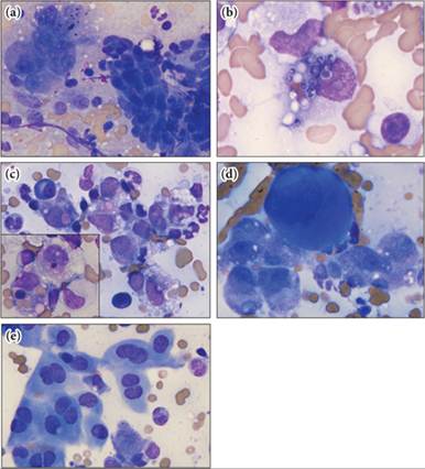
Figures 9.29a–e Feline hepatic sample. (a) Hepatocytes (left side of image) and clusters of biliary epithelial cells (right side of image) are prevalent in some areas of the slide. (b) Highly phagocytic macrophages are also present in the sample. They contain intracellular yeast that have a distinct cell wall and crescentic nuclear basophilic material within the protoplasm, consistent with Histoplasma spp. yeast. (c) Macrophages, neutrophils, lymphocytes, and plasma cells are displayed. Yeasts are found in several of these macrophages as well. The inset shows erythrophagocytic macrophages, which were a common finding. (d) Rare multinucleate giant cells are present. The deeply basophilic cytoplasm and tightly aggregated nuclei mark this cell as a megakaryocyte and denote extramedullary hematopoiesis; nRBCs and fewer myeloid precursors were also present but were not imaged. (e) Few sheets of binucleated mesothelial cells are seen and suggest reactive mesothelium (Wright–Giemsa, 1,000? magnification). The cytologic diagnosis is mixed inflammation with yeast consistent with Histoplasma spp. and extramedullary hematopoiesis.