Suppurative inflammation
A threshold of >7 neutrophils in close approximation with 200 hepatocytes found in clusters (i.e. 3.5% neutrophils) has been suggested for diagnosis of suppurative inflammation (Gardner et al., 2022).
This threshold agrees with the finding that >5% neutrophils was observed with destructive cholangitis, acute hepatitis, abscessation, and cholangiohepatitis (Figures 9.17a, b, 9.27a–d, 9.28; Weiss et al., 2001; Stockhaus et al., 2004). Inflammation can often be associated with hepatocellular atypia including variation in hepatocyte nuclear size, N:C ratio, and nucleoli (Stockhaus et al., 2004). Both infectious and noninfectious causes of suppurative inflammation have been documented. Infectious causes include viruses such as canine adenovirus-1, canine and feline herpes viruses, and feline infectious peritonitis (corona)virus; bacterial agents including Leptospira spp., Escherichia coli, Staphylococcus spp., Clostridium spp., and Brucella spp.; and protozoans, including Toxoplasma sp. (van den Ingh et al., 2006a, b). Noninfectious causes include pancreatitis and drug or toxin-induced hepatopathy and necrosis, among others (van den Ingh et al., 2006a, b). Often the underlying cause of inflammation is not found. The unifying characteristic of these is the relatively acute and active nature of the disease process. 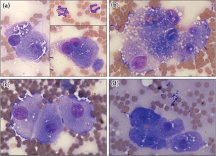
Figures 9.27a–d This dog had a remarkable septic suppurative hepatitis concurrent with hepatocellular carcinoma. (a) Degenerate neutrophils, which contain plump rod-shaped bacteria, are present (upper right panel). Cell-free bacteria are also seen (lower right panel). The hepatocytes display discrete clear vacuoles, suggestive of lipid, and dysplasia including anisocytosis, anisokaryosis, multiple nuclei, and multiple nucleoli (left and lower right panels).
(b) Two brick inclusions and two intranuclear cytoplasmic inclusions are noted in this cluster of hepatocytes. Note the prominent subterminal spore found in the bacteria in the upper right-hand corner; bacterial culture determined these to be Clostridium species. (c) The hepatocytes have variable numbers of nucleoli and distinct cell–cell junctions. Histologic evaluation diagnosed hepatocellular carcinoma. (d) Clusters of more morphologically normal hepatocytes are also present in the slide (Wright–Giemsa, 1,000? magnification).
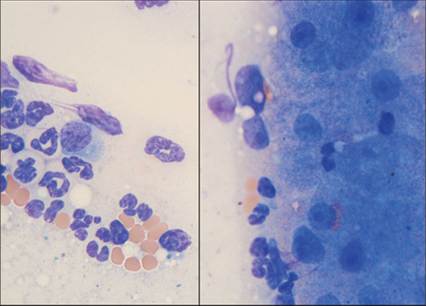
Figure 9.28 This hepatic cytology sample is from a domestic longhair cat who was sampled after poor clinical and biochemical recovery from an episode of hepatic lipidosis. The cytology has many neutrophils interspersed around the hepatic clusters (left panel). Single neutrophils are consistently observed within the hepatocyte clusters. The hepatocytes do not have lipid-type vacuolar changes (right panel). There is a suppurative process evident (Wright–Giemsa, 1,000? magnification).
More on the topic Suppurative inflammation:
- Suppurative inflammation
- Resolution
- Inflammation
- Mixed inflammation
- Cornea
- Inflammation and inflammatory lesions
- Evaluation of gallbladder fluid
- Synovial fluid
- Cases
- Bone