Muscles of the hindlimb
The hindlimb is attached to the pelvic girdle by a mobile ball and socket joint, the hip joint. Γhe sublιtm∙ bar lu∣paxial muscles of the vertebral column are extrinsic muscles attached to the vertebral column and to the pelvic girdle.
The intrinsic muscles of the hindlimb all insert on the Iiindlirnb itself or on the pelvic girdle and are described below.
Muscles of the thigh
These originate on the pelvic girdle and insert on the femur.
Ghitedls - three muscles, the superficial. middle and deep tf Iu tea Is. and the tensor fascia Iatae form the curve of the rump and are powerful extensors of the hip joint
ACllON - extend the hip joint and abduct the thigh Ilamstringflroup-form the caudal aspect of the thigh and act together to propel and extend the whole limb backwards I Hg. 4.11). I hev provide the main propulsive force of the animal and consists of three muscles:
Biceps Iemoris is the most lateral muscle in this group I Hg. 4.1 11, it originates from the pelvis and runs over the femur to the tibia and inserts on the calcaneus of the hock
ACiiON extends the hip. Ilexes the Stille and extends the hock
- Semitendinosus - runs from the pelvis and inserts on the tibia and calcaneus
action - extends the hip. Ilexes the slille and extends the hock
Semimembranosus is the most medial muscle of the hamstring group. It runs from the pelvis to the femur and tibia
ACHON - extends the hip and Ilexes the slille
(Juadrh eps Jemoris - this large muscle runs down the cranial aspect of the thigh. It consists of four parts, all of which insert at the same point on the tibial Iubcrosiiv or crest. I he Iendon of this muscle ∙Γ
contains the pah’llii. which articulates with the femur at the stille joint
action - extends the stille joint
n.Adductor muscles - lie on the medial aspect of the thigh and hold the limb close into the bodv (Fig.
4.12):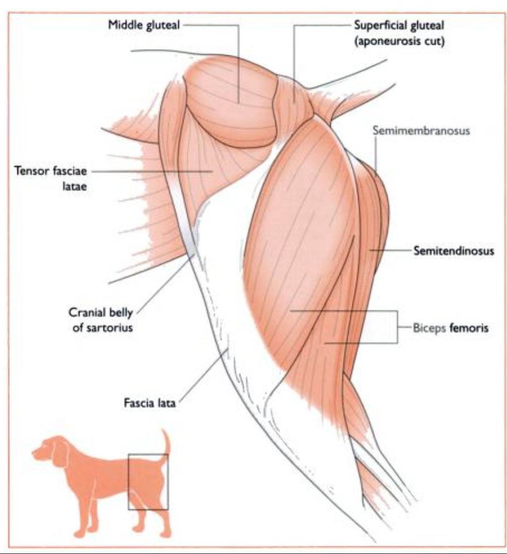
Fig. 4.11 Superficial muscles of the left lateral thigh area.

Fig. 4.12 Muscles of the med∣al aspect of the left thigh.
The tendon of the pectineus muscle is sometimes transected to relieve pressure on the hip joint of dogs with hip dysplasia. Pectineotomy does not cure the problem but may provide a degree of pain relief.
The popliteal lymph node lies within the fibres of the gastrocnemius muscle and can be palpated just caudal to the stifle joi∩L Normally it is about 2 cm long but if it is enlarged it may indicate some form of infection (or tumour) in the distal extremity.
- Pectineus - this muscle runs from the pubis to the distal
action - adducts the limb
- Sartorius - inserts on the cranial border of the tibia with the gracilis muscle
action - adducts the limb
- Gracilis - forms the caudal half of the medial Surfaceof the thigh
action adducts the limb
Muscles of the lower hindlimb
These muscles act mainly on the hock joint (Fig. 4.1 3). They include:
Uiistnx Iiemius- this muscle originates from the caudal aspect of the Iemurand inserts on the calcaneus of the hock. The tendons of this muscle contain two small sesamoid bones or fabellae that articulate with the caudal aspect of the stifle joint action - extends the hock and Ilexes stifle
Acliilles tendon - this is the large strong tendon that runs dow n the back of the leg to the point of the hock. Il includes the tendons of insertion of the gastrocnemius, biceps femoris and semitendinosus, all of which insert on the calcaneus, and also of the superficial digital flexor muscle, w hich continues over the point of the hock and dow n Io its insertion on the digits. There is a bursa at the point of insertion on the calcaneus.
Muscles of the hock and digits
As is seen in the forelimb, there are a number of muscles responsible for flexing and extending the hock and the digits:
Anterior tibialis - runs from the proximal end of the tibia to the tarsus
action - flexes the hock and rotates the paw medially
Three digital extensors - run in front of the hock and fix>1
Two ιli9itfll ∏ι,xors- run behind the fool. The Siipcrfii in! digital flexor runs from the femur to the phalanges and is one of the components of the Achilles tendon.
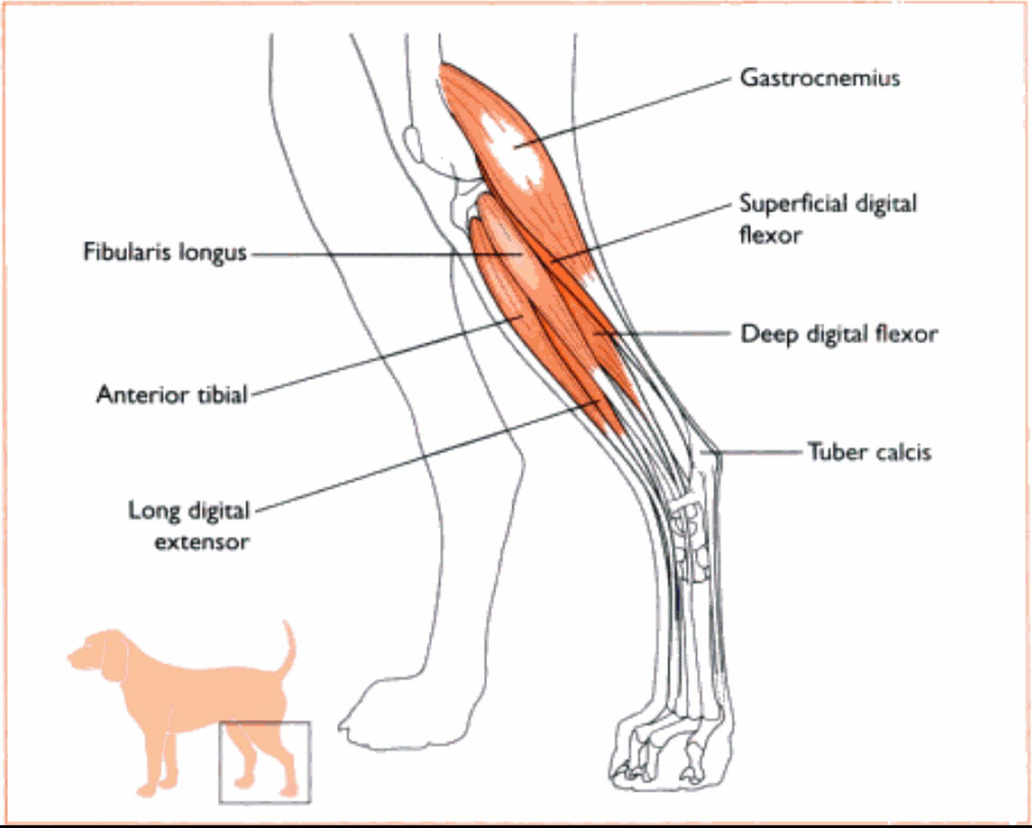
Fig. 4.13 Musdes of the tower left h∣ndlimb