MYCOPLASMAS OF BIRDS
ROBIN A.J. NICHOLAS
Mycoplasma Group, Animal Health and Veterinary Laboratories Agency (Weybridge), New Haw, Weybridge, UK
Mycoplasma infections produce significant disease in the poultry industry, particularly in battery farms, where they can rapidly disseminate through birds housed at high densities, leading to huge losses.
The ability of some, such as M. gallisepticum and M. synoviae, to spread vertically through the egg makes control particularly difficult. Even in free-range flocks, mycoplasmas can be a problem because of the close contact at roosting, although losses are generally not so high. The situation in the wild is not as clear, but generally typical mycoplasma diseases such as sinusitis, conjunctivitis and chronic respiratory disease are only seen sporadically. However, where wild species such as pheasants and partridges are intensively bred to meet the growing demand for hunting, they suffer the mycoplasma diseases seen in high-density flocks typical of the poultry industry. This intensification, seen in Europe over the last 20 years, has led to adult birds that are susceptible to a range of infectious and non-infectious diseases, and mortality rates of 5—10% or higher are commonly seen. The largest causes of death and morbidity in game birds are sinusitis and conjunctivitis (Figure 29.2), which are most often associated with mycoplasma infection. The sourcing of eggs from flocks free of M. gallisepticum has led to an improvement in this sector over recent years, although losses are still seen from time to time.Many mycoplasmas have been isolated from pheasants, including M. pullorum, M. iners and M gallinarum, but they are not considered major pathogens; their main effect seems to be in obscuring the detection of the more fastidious proven pathogens such as M. gallisepticum and M.
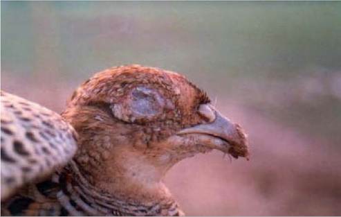
FIGURE 29.2 Pheasant showing severe eye lesions caused by Mycoplasma gallisepticum.
synoviae. However, the development of sensitive and specific molecular tests that are capable of detecting these mycoplasmas directly in clinical material, often with other species, has improved the accuracy of diagnosis.
Racing pigeons, which are often kept in overcrowded lofts throughout Europe, suffer respiratory disease. This has been linked, although not definitively, with mycoplasmas such as M. columborale, M. columbinum and M. columbinasale. In an outbreak of both ocular and respiratory disease affecting 10 and 20% of show and racing pigeons respectively, M. columborale was the main organism isolated from a cage of over 300 birds in Sicily that had been confined for over 2 months(23); a gradual improvement was seen in the birds following treatment with tylosin. These mycoplasmas have also been detected in apparently healthy feral pigeons, suggesting they may be part of the natural flora of these birds.
In 1994 a new disease called ‘house finch conjunctivitis’ spread across the eastern USA and soon became ubiquitous in the finch population. The disease caused unilateral and bilateral conjunctival and periorbital swelling, which was often complicated with nasal discharge and mucopurulent drainage, impairing both vision and respiration. Following initial reports of the disease in the house finch (Carpodacus mexicanus), American goldfinches ( Carduelis tristis), blue jays (Cyanocitta cristata) and house sparrows (Passer domesticus)(24) have also become infected, indicating a wider host range than previously thought. No such epidemic has yet been seen in Europe, although conjunctivitis is seen sporadically in a range of bird species, including birds of prey, but rarely involving M. gallisepticum. Other Mycoplasma species have been reported in wild birds, including Mycoplasma sturni, which has been associated with conjunctivitis in the European starling (Sturnus vulgaris’15'’; these European outbreaks were clinically very similar to those seen in American finches.
These observations offer further evidence that mycoplasma- associated conjunctivitis is a potentially important emerging infection affecting the welfare of both wild free- living and captive birds. This infection may have ecological effects as a consequence of altered population densities of affected species. The 60% fall in the eastern house finch population was attributed to the arrival of M. gallisepticum, probably from contact with infected domestic poultry1-26’.Mycoplasma sturni was the predominant Mycoplasma species found during a study to determine the causes of mortality in wild birds in Britain. It was detected in oropharyngeal or conjunctival swabs taken from blackbirds (Turdus merula), rooks (Corvus frugilegus’, carrion crows (Corvus corone), magpies (Pica pica) and starlings not required as given earlier. Birds that were infected with M. sturni were suffering from a range of infectious and non- infectious conditions, although none that would be considered typical mycoplasma diseases, so its significance remains unknown. More recently M. sturni was detected from some of a large population of rooks which had died, or were dying, from acute respiratory disease in the UK, although it is likely that the main cause of disease was Pasteurella multocida517(
Although reported many times in birds of prey, the role of mycoplasmas as pathogens is still unclear. Mycoplasma spp. were detected by culture and a genus-specific PCR in healthy, free- ranging raptor nestlings and birds of prey from rehabilitation centres, sampled during a routine ringing programme1-28’. These included Mycoplasma bute- onis, M. falconis and M. gypis. Unidentified mycoplasmas were also isolated from western marsh harriers (Circus aeroginosus), a Eurasian hobby (Falco subbuteo) and a barn owl ( Tyto alba). Given the lack of clinical signs or pathology, it was likely that mycoplasmas in these raptors were commensals rather than pathogens.
However, in an earlier study many of the same strains were isolated from birds of prey held at a sanctuary that showed respiratory disease, suggesting that the mycoplasma may cause or contribute to the cause of disease under the stress of captivity1-29’.The isolation and characterization of four mycoplasmas found in the upper respiratory tract of four sick Eurasian griffon vultures ( Gyps fulvus) that were housed in a Sicilian rehabilitation centre has been reported1-30’. These included Mycoplasma gallinarum, an unidentified mycoplasma highly similar to Mycoplasma glycophilum, and two unidentified mycoplasmas with similarities to Mycoplasma falconis and Mycoplasma gateae, a mycoplasma normally associated with cats. No other pathogens were detected. The significance of large numbers of as yet unidentified mycoplasmas in great white pelicans (Pelecanus onocrotalus}, a threatened European breeding species, is also unknown, but birds appeared to be healthy.
A mycoplasma closely related to M. gallisepticum was identified from the lung and liver of different captive Humboldt penguins (Spheniscus humboltdi, in a zoo in the UK(31’. It was speculated that the most likely source of this infection was ducks and other wild or semi-wild species in adjoining enclosures. Later a new species, M. sphenisci, was isolated from choanal discharge from a jackass penguin (Sphenus demersus}, which may represent a unique species found in the penguin. Interestingly, although M. sphenisci was believed to be pathogenic in the jackass penguins, king penguins (Aptenodytes patagonicus) which were close by showed no clinical signs. A similar situation arose in the UK, where M. gallispecticum and M. gallisepticum-like strains were isolated from captive Humboldt penguins exhibiting high mortality. King penguins co-housed with the Humboldts were found to have M. lipofaciens and M. spenisci but showed no clinical signs. It was thought that M. gallisepticum may be an important cause of morbidity and mortality in Humboldt penguins, an endangered species, although it is not clear how they would become infected in the wild.
It is common to isolate unidentifiable mycoplasmas from birds, and many of these will remain unidentified for many years owing to the time- consuming nature of the characterization process. Although the use of 16S rDNA sequencing has greatly improved the screening process, much biochemical, serological, morphological and genetic analysis is still required for describing new species.