NEOPLASMS
Spontaneous tumors are rare in guinea pigs under 3 years of age and uncommon even in older animals. There appear to be variations in genetic susceptibility to spontaneous neoplasia.
A thorough review of neoplastic diseases of the guinea pig was provided by Manning (1976).Cutaneous Neoplasia
Trichoepitheliomas/trichofolliculomas are the most common tumors of the skin (Fig. 5.55). These tumors contain multiple discrete epithelial structures reminiscent of hair bulbs and keratinized structures reminiscent of hair sheaths. Cutaneous papillomas, sebaceous gland adenomas, penile papillomas, lipomas, fibrosarcomas, fibromas, and carcinomas have also been described.
Mammary Neoplasia
Mammary adenocarcinomas occur in both male and female guinea pigs. The majority are interpreted to be of ductal origin. Metastases may occur to regional lymph nodes. Some are of low-grade malignancy and remain localized to the original site. Other mammary tumors
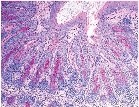
FIG. 5.55. Trichofolliculoma in the skin of an aged guinea pig. Note the aggregations of follicular structures that are typical of these tumors.
include mammary gland adenoma and malignant mixed mammary tumor.
Pseudo-odontoma: Odontoma
Pseudo-odontomas arise rarely from the elodont tooth roots of rabbits and rodents, including guinea pigs (see Rabbit Chapter 6, “Pseudo-odontoma”). They consist of disorganized epithelial and mesenchymal elements of tooth origin that are well differentiated. They are not true neoplasms, but are rather believed to be hamartomas.
Cavian Leukemia
On rare occasions, lymphoid leukemia occurs as a spontaneous disease in various inbred and noninbred strains of guinea pigs. Cases are most frequently seen in young adult animals. Leukocyte counts in the peripheral blood are relatively high, varying from 50,000 to over 200,000/ mm3.
Leukemia has been produced experimentally with transplanted cells and cell-free filtrates. Leukocytosis (up to 180,000 mm3 or greater) with a preponderance of lymphoblastic cells is the typical picture seen in blood samples. At necropsy, lymph nodes, such as cervical (Fig. 5.56), axillary, mesenteric, and inguinal, are enlarged and firm, homogeneous, and tan on the cut surface. There is marked splenomegaly and hepatomegaly. Microscopically, there is usually moderate to marked infiltration of lymphoblastic cells in the spleen, liver, bone marrow, interstitium of the lung, thymus, alimentary tract lymphoid tissue, heart, eyes, and adrenals. Guinea pig leukemia is associated with, but not necessarily caused by, an endogenous retrovirus. The primary differential diagnosis for cervical lymphadenomegaly is S. equi subsp. zooepidemicus.Respiratory Tract Neoplasia
Pulmonary tumors represented approximately 35% of reported tumors in 1 survey. The majority were benign papillary adenomas, and most were interpreted to be of bronchogenic origin. The changes were similar to those produced by certain infectious agents, and it was suggested that there may be hyperplastic and adenomatous changes in airways and alveoli in response to various stimuli, not bona fide primary pulmonary tumors. Small, white, circumscribed nodules, visible macroscopically, on microscopic examination consist of papillary structures lined by a single layer of hyperchromatic cuboidal epithelium. Primary malignant tumors of the lung are rare in guinea pigs. Nasal adenocarcinoma is another malignancy reported to occur in this species.
Reproductive Tract Neoplasia
Tumors of the reproductive tract represent approximately 25% of spontaneous tumors in this species. Of the ovarian tumors, granulosa cell tumors occur, but the majority are teratomas. A variety of tissue types may be evident in teratomas, including ciliated and mucous epithelial cells, striated muscle, and cells of ectodermal origin. These tumors should not be confused with cystic rete ovarii seen commonly in older sows.
Uterine tumors are primarily benign and of mesenchymal origin. Most are leiomyomas (Fig. 5.57) or fibromas. Rarely, malignant uterine tumors such as myxosarcomas or leiomyosarcomas have been described. Primary malignant uterine tumors consist of poorly differentiated mesenchymal cells, with extension into the peritoneal cavity. Tumors of the male reproductive system are very rare.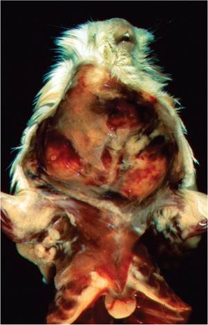
FIG. 5.56. Massively enlarged cervical lymph nodes in a guinea pig with lymphoid leukemia.
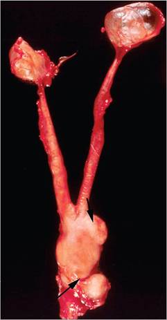
FIG. 5.57. Uterine leiomyoma (arrows) in an aged guinea pig sow with concomitant cystic rete ovarii. The leiomyoma consists of a multilobulated mass at the uterocervical junction with extension into the vaginal area.
Endocrine and Cardiovascular Neoplasia
Tumors of the endocrine system in guinea pigs include benign adrenocortical tumors and insulinomas. Neurologic signs were observed in animals with insulinomas, and Cushing's syndrome has been observed in guinea pigs with adrenocortical adenomas. Although rarely observed in laboratory guinea pigs, thyroid adenomas and carcinomas have been reported to be common among pet guinea pigs. Benign mixed tumors (myxomas) are the most commonly reported tumors of the cardiovascular system. They may include well-differentiated mesenchymal components, such as cartilage, bone, and fat. Primary myocardial tumors should not be confused with rhabdomyomatosis.
Other Neoplasms
Other neoplasms include bile duct tumors, undifferentiated carcinoma, lipomas, fibrosarcoma, and histiocytic lymphosarcoma.
BIBLIOGRAPHY FOR NEOPLASMS
Andrews, E.J. (1976) Mammary neoplasia in the guinea pig (Cavia porcellus). Cornell Veterinarian 66:82-96.
Field, K.J., Griffith, J.W., & Lang, C.M. (1989) Spontaneous reproductive tract leiomyomas in aged guinea pigs. Journal of Comparative Pathology 101:287-294.
Franks, L.M. & Chesterman, F.C. (1962) The pathology of tumours and other lesions of the guinea pig lung. British Journal of Cancer 16:696-700.
Gibbons, P.M., Garner, M.M., & Kiupel, M. (2013) Morphological and immunohistochemical characterization of spontaneous thyroid gland neoplasms in guinea pigs (Cavia porcellus). Veterinary Pathology 50:334-342.
Hong, C.C., Liu, P.I., & Poon, K.C. (1980) Naturally occurring lymphoblastic leukemia in guinea pigs. Laboratory Animal Science 30:222-226.
Jungeblut, C.W. & Opler, S.R. (1967) On the pathogenesis of cavian leukemia. American Journal of Pathology 51:1153-1160.
Kitchen, D.N., Carlton, W.W., & Bickford, A. (1975) A report of fourteen spontaneous tumors of the guinea pig. Laboratory Animal Science 25:92-102.
Manning, P.J. (1976) Neoplastic diseases. In: The Biology of the Guinea Pig (eds. J. E. Wagner & P. J. Manning), pp. 211-225. Academic Press, San Diego.
Opler, S.R. (1967) Pathology of cavian leukemia. American Journal of Pathology 51:1135-1147.
Suarez-Bonnet, A., de las Mulas, M., Millan, M.Y., Herraez, P., Rodriguez, F., & de los Monteros, A.E. (2010) Morphological and immunohistochemical characterization of spontaneous mammary gland tumors in the guinea pig (Cavia porcellus). Veterinary Pathology 47 (2): 298-305.
Williams, B. (2012) Non-infectious diseases. In: The Laboratory Rabbit, Guinea Pig, Hamster, and Other Rodents (eds. M. A. Suckow, K. A. Stevens, & R. P. Wilson), pp. 685-704. Elsievier, London.
Zwart, P., van der Hage, M.H., Mullink, W.M.A., & Cooper, J.E. (1981) Cutaneous tumors in the guinea pig. Laboratory Animals 15:375-377.