MISCELLANEOUS DISORDERS
Behavioral Diseases
Hair pulling, chewing (barbering), and trichophagia are common behavior patterns among guinea pigs in a group and can become an excessive activity once it is in vogue.
As noted under Alopecia (below), trichophagy may reflect a nutritional disorder. An extension of this behavior is ear chewing, which can result in notching or severe trauma and amputation of the ear pinnae (Fig. 5.45). Frequently, sexually mature boars fight when placed together, and severe lacerations or death may result. Very young guinea pigs in communal housing may be trampled by older animals in group stampedes.Alopecia
Alopecia may arise in guinea pigs for a variety of reasons, including endocrine dysfunction, nutrition, behavior vices, general illness, ringworm, and parasitism. Bilateral
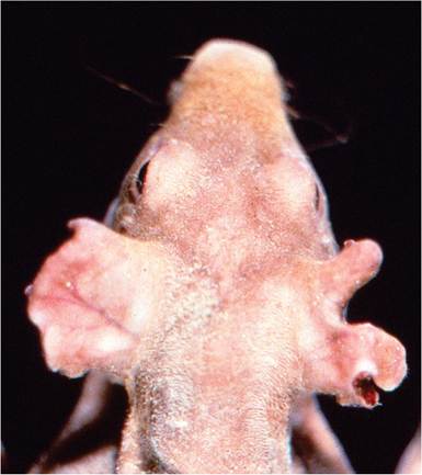
FIG. 5.45. Notching of the ear pinnae of a hairless guinea pig due to chewing by conspecifics.
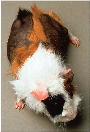
FIG. 5.46. Bilateral alopecia in an aged guinea pig sow with cystic rete ovarii. (Source: N.J. Schoemaker, University of Utrecht, Netherlands. Reproduced with permission from N.J. Schoemaker. )
alopecia commonly occurs in sows in advanced pregnancy and during lactation, particularly in older animals. In pregnant animals, hair loss may be due to reduced anabolism of maternal skin during fetal growth. The hair loss frequently occurs over the back, flanks, and rump, and the pelage will return to normal in due course in the typical case. Similar bilateral alopecia is very common in older sows with cystic rete ovarii (Fig. 5.46), and less common in guinea pigs with Cushing's syndrome related to adrenal cortical adenomas. Guinea pigs require crude fiber in their diet, which may not be met with formulated diets.
Supplementation with hay has been shown to ameliorate alopecia among breeding guinea pigs. The mechanism for alopecia in guinea pigs without hay supplementation was apparently due to trichophagia among cage mates. Barbering as a behavioral vice may also occur among conspecifics. Chronic illness due to Salmonella infection has also been shown to cause bilateral alopecia, which responded to vitamin C supplementation. Dermatitis with hair loss may occur with urine scalding and contact dermatitis. These conditions have characteristic patterns that assist with diagnosis. Acariasis, pediculosis, and dermatophytosis are also associated with alopecia.Foreign Body Pneumonia: Pneumoconiosis
Focal pulmonary lesions associated with aspirated food or bedding occur as an incidental finding, particularly in young guinea pigs. This has been observed in guinea pigs on various bedding materials, including wood products and rice straw. At necropsy, there may be foci of atelectasis or circumscribed nodules in the parenchyma of the lung, but frequently lesions are not detected on gross examination. On microscopic examination, plant fibers may be found lodged within small airways, with hetero- philic and mononuclear cell infiltration. In lesions of some duration there may be focal granulomatous bronchiolitis and/or interstitial alveolitis, with mononuclear cell infiltration and foreign body multinucleated giant cell formation. Differential diagnoses include osseous metaplasia, lesions of primary bacterial or viral origin, focal mycotic lesions (e.g., Aspergillus spp.), and granulomatous pulmonary lesions associated with the subcutaneous administration of Freund's adjuvant.
Adjuvant-Associated Pulmonary Granulomas
Pulmonary granulomas may occur in guinea pigs and other rodents or rabbits following subcutaneous injection with complete Freund's adjuvant. Microscopic changes are characterized by multifocal granulomatous inflammatory response. Posterior paresis and osteolysis was observed in guinea pigs immunized subcutaneously with Freund's adjuvant.
It was presumed that this condition resulted from inadvertent injection into epaxial muscles, with tracking of granulomatous inflammation into the spinal canal and bone. Pulmonary granulomata were also found. Differential diagnoses include perivascular lymphoid nodules, pneumoconiosis, and focal pneumonia due to infectious agents.Osteoarthritis
Laboratory-housed Dunkin-Hartley guinea pigs are prone to development of osteoarthritis involving the femorotibial joints. Lesions become apparent by 3 months of age, and progress with age. Lesions involve focal degeneration of hyaline cartilage, osteophyte formation, and synovial proliferation and fibrosis. High levels of ascorbic acid accentuated the disease, apparently through activation of TGF-beta. Wild guinea pigs have been shown to have no osteoarthritis in the knee joints, and it is unknown what the prevalence may be in the pet population.
Gastrointestinal Diseases
Chelitis
Inflammation of the oral labia and rhinarium is common in guinea pigs, and appears to be associated with acidic diets. The lesions manifest as serous excoriations, and may become secondarily infected with S. aureus or other opportunistic organisms.
Malocclusion: “Slobbers”
This condition may involve the incisors, as well as the molar and premolar teeth in guinea pigs. As elodont teeth, they grow continuously throughout life, and good opposition is required to prevent overgrowth. If the alignment is defective, maxillary teeth may overgrow labially and the mandibular teeth overgrow medially. Excessive salivation, inanition, and wasting occur in severely affected animals. Nutritional factors have
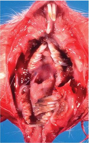
FIG. 5.47. Malocclusion and overgrowth of cheek teeth.
been implicated, and fluorosis has been associated with this condition. However, there is evidence that genetic factors play an important role in this disease. There may be a single gene involved, or more than 1 gene with incomplete penetrance.
The incidence is higher in some inbred strains. At necropsy, cheek teeth have irregular contours and sharp edges on their occlusal surfaces (Fig. 5.47). In tooth abnormalities attributed to fluorosis, lesions were characterized by impairment of dentin and enamel formation and by excessive wear. Abnormalities of this type are not evident in typical cases of malocclusion.Gastric Dilatation and Volvulus
Multiple cases of acute gastric dilatation associated with gastric volvulus have been recognized in a colony of guinea pigs, and it occurs sporadically in other facilities. Frequently, affected animals were found dead, with no previous indication of disease. Typical cases had a 180° rotation along the mesenteric axis, and stomachs are distended with fluid and gas. Death has been attributed to respiratory impairment and possibly vascular shock.
Gastric Ulcers
Gastric ulceration is common in guinea pigs, and appear to be precipitated by a number of infectious and other nonspecific stressors.
Intestinal Hemosiderosis
Accumulations of hemosiderin-laden macrophages in the lamina propria of the intestine, particularly large bowel, are a common finding in the guinea pig. There is speculation that this is due to subclinical scurvy, but
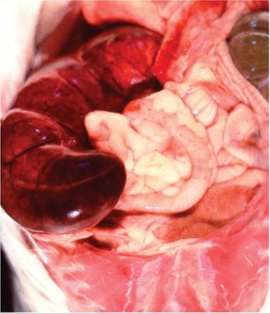
FIG. 5.48. Cecal volvulus in an adult guinea pig. The cecum is hemorrhagic and infarcted due to torsion of vascular supply.
some believe that it is due to the normally zealous iron- uptake and binding of herbivores with diets containing excess iron.
Cecal Volvulus
Deaths due to cecal torsion are occasionally observed in this species. At necropsy, the displaced organ is edematous, hemorrhagic, and distended with fluid and gas (Fig. 5.48). Cecal volvulus has been known to occur in association with impaction and may be precipitated by typhlitis of various causes.
Anorectal Impaction
Impaction of the perineal sac region with feces, sebaceous secretions, and bedding material is common in older boars, resulting in occlusion of the anus and inability to defecate.
It has been claimed to be related to muscle atony.Prolapsed Rectum
As with other species, rectal prolapse may occasionally occur in association with inflammatory conditions of the large intestine.
Focal Hepatic Necrosis
Multifocal coagulation necrosis of the liver is occasionally seen at necropsy. Affected areas tend to be subcapsular in distribution, with minimal or no inflammatory response. They are frequently interpreted to be a terminal event and may be due to hypoxic change secondary to impaired blood flow in the region. Differential diagnoses include bacterial hepatitis (e.g., Tyzzer's disease) and toxic change.
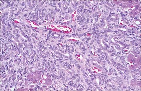
FIG. 5.49. Liver from an adult guinea pig with marked bile ductular proliferation and portal fibrosis.
Chronic Idiopathic Cholangiofibrosis
Periportal fibrosis with bile ductular proliferation is occasionally seen in adult guinea pigs as an enzootic problem in individual colonies. Lesions are characterized by hepatocyte degeneration, proliferation of cholan- gioles, and interstitial fibrosis (Fig. 5.49). The changes are suggestive of a toxin-induced change, but the etio- pathogenesis has not been resolved.
Hepatic Contusions
Capsular rupture of the liver, with hemorrhage into the peritoneal cavity, is occasionally observed at necropsy. Traumatic lesions of this type may be caused by events such as mishandling or falls. Multiple simultaneous cases of hepatic contusions should alert investigation into inappropriate restraint of struggling guinea pigs by an inexperienced handler.
Fatty Infiltration of the Pancreas
Interstitial infiltration of adipose tissue occurs in the pancreas in older guinea pigs as a normal part of the aging process. The proportion of the exocrine pancreas decreases with age, with no apparent impairment of function. Histologically, there are large areas of adipose tissue interposed between normal pancreatic tissue.
Fatty infiltration can also occur within the islets (Fig. 5.50).Urogenital Diseases
Nephrosclerosis: Chronic Renal Disease
Irregularly pitted, granular renal cortices are a common finding at necropsy, particularly in guinea pigs that are at least a year of age. It is usually an incidental finding, but lesions may be extensive enough to result in renal insufficiency. Pathogenesis has not been resolved. The renal lesions have been interpreted to be the result of a general vascular disturbance, resulting in focal areas of ischemia and fibrosis. Using immunohistochemical techniques, glomerular changes were evaluated in guinea pigs collected from several sources. Spontaneous
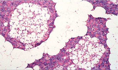
FIG. 5.50. Pancreas of an adult guinea pig with fatty infiltration of the interstitium and lipidosis of islet cells. These common changes can occur independently or together in individual animals.
deposits of IgG and complement (C3) were demonstrated along the mesangial and peripheral glomerular basement membranes. It was suggested that the antigenantibody complexes might be due to an infectious agent or endogenous tissue antigen. An accelerated disease process (and an increased incidence) has been observed in guinea pigs fed an unusually high protein diet. Mild hypertension has been recorded in affected guinea pigs.
Pathology
Multiple, granular, pitted areas may be visible on the surface of the kidney, resulting in irregular contours in severely affected animals (Fig. 5.51). On cut surface, pale linear streaks extend into the cortex, with some involvement of the medulla in advanced lesions. On microscopic examination, there is segmental to diffuse-tubular degeneration and interstitial fibrosis, with distortion and obliteration of the normal architecture (Fig. 5.52). Tubular lesions are concentrated primarily in the convoluted tubules and Henle's loop. Scattered tubules are dilated and lined with poorly differentiated, cuboidal to squamous epithelial cells. Some nephrons, interpreted to be nonfunctional, consist of tubular remnants lined by poorly differentiated cuboidal epithelium with lightly
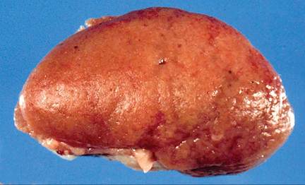
FIG. 5.51. Kidney from an aged guinea pig with chronic nephrosclerosis. The cortical surface is finely granular and pitted.
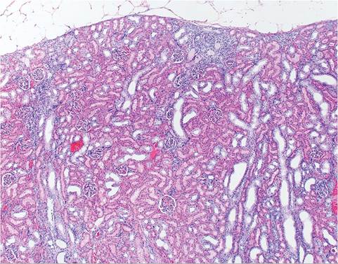
FIG. 5.52. Early nephrosclerosis in a guinea pig. Note the segmental distribution of tubular degeneration with depression of the renal capsule.
eosinophilic to amphophilic cytoplasm. Tubules are occasionally dilated and contain proteinaceous material and cellular debris. In nephrons interpreted to be fully functional, convoluted tubules are lined by hypertrophied epithelial cells with abundant, eosinophilic cytoplasm. Most glomeruli are essentially normal histologically. Occasionally, there is atrophy of individual glomeruli, with regional fibrosis. In advanced lesions, there is diffuse to segmental infiltration with fibroblasts and collagenous tissue formation. There are minimal focal aggregations of mononuclear cells consisting mainly of lymphocytes. Arterioles and arteries may have moderate medial hypertrophy, sometimes with prominent endothelial lining cells. High BUN and serum creatinine, nonregenerative anemia, and low urinary- specific gravity are clinical findings in animals with advanced nephrosclerosis.
Cystitis and Urolithiasis
Urinary tract infections occur occasionally, particularly in older sows. This may be due to the proximity of the urethral orifice to the anus in females, with fecal contaminants such as E. coli. At necropsy, changes may vary from thickening of the bladder mucosa with congestion in chronic cases to intramural and/or intraluminal hemorrhage in animals with acute cystitis. Microscopic changes seen in chronic cases are characterized by leukocytic infiltration in the lamina propria, and occasionally fibroblast proliferation. In acute cases, there may be ulceration, hemorrhage, and infiltration with heterophils. Most cases have some degree of accompanying pyelonephritis. Cystitis often occurs in concert with urolithiasis. Urinary calculi occur frequently in older sows, and less commonly in older boars. They vary from sand-like crystals to large concentric stones. They are typically composed of calcium carbonate.
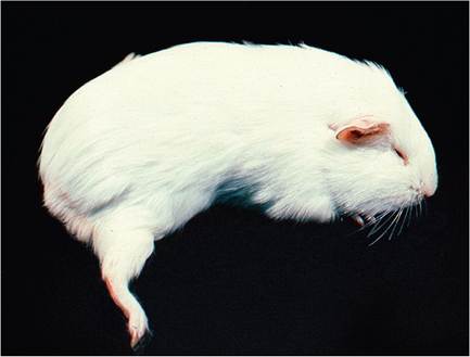
FIG. 5.53. Unilateral posterior paresis in a postpartum sow due to
obturator nerve damage during parturition.
Urolithiasis may cause obstructive uropathy, with urethral plugging in males, hydroureter, and hydronephrosis.
Male Genitourinary Diseases
In addition to obstructive uropathy and nephrosclerosis, boars are prone to accumulation of detritus in their prepuce, resulting in balanoposthitis. Older boars are prone to impaction of perineal gland secretions and other debris. Seminal vesiculitis also occasionally occurs. Guinea pigs, like other rodents, ejaculate a copulatory plug, which may cause urethral obstruction.
Disorders of Pregnancy
In addition to pregnancy toxemia, the exceptionally large fetuses at term may result in dystocia, particularly in older sows with incomplete relaxation of the pubic ligament. Obturator nerve paralysis can be a consequence of dystocia (Fig. 5.53). The large gravid uterus is prone to torsion, and ectopic pregnancies have been rarely observed in sows, which in some cases were related to uterine rupture. Stillbirth is common in guinea pigs, and the incidence may be particularly high among inbred strains, which has been shown to occur at a rate of 28.4% in strain 13 guinea pigs.
Ovarian Cysts
Cystic rete ovarii are extremely common in older sows, with 1 report documenting a prevalence of 75% in sows over 18 months of age. Bilateral ovarian involvement is very frequent (Fig. 5.54), but when unilateral, the right ovary appears to be more commonly involved. Small ovarian cysts less than 1 mm in diameter may be present on the ovaries of younger females, but they are frequently missed at necropsy. In older sows, thin-walled, fluid-filled, fluctuant cysts up to 2 cm in diameter may be present on the ovaries. Cysts may be much larger, and present clinically as abdominal distention. Smaller cysts are usually concentrated in the cephalic pole near the
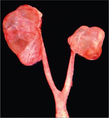
FIG. 5.54. Reproductive tract from an aged guinea pig sow, illustrating bilateral cystic rete ovarii. Large, fluid-filled cysts are on the surface of both ovaries.
hilus of the ovary. Occasionally, there is a single large cyst, with no recognizable ovarian tissue. Cysts contain clear, serous fluid. On microscopic examination, cysts are of variable size and are lined by low cuboidal to columnar epithelial cells. Solitary cilia or tufts of cilia are present on the luminal surface of some cells. Depending on the size of the cysts, there may be marked compression of the ovarian tissue, and in advanced cases only remnants of the ovary remain. Serial sections have revealed continuity between rete ovarii, follicles, and ovarian mesothelium, and the large serous cysts appear to develop from the rete ovarii.
Cystic rete ovarii have been associated with reduced reproductive performance in sows at 15 months of age and older. The most common clinical sign is bilateral symmetrical alopecia over the flank region (see “Fig. 5.46”), and there may be crusting of the skin around the nipples. Sows may manifest atypical sexual behavior. Cystic endometrial hyperplasia, mucometra, endometritis, and leiomyomas are the other changes associated with cystic rete ovarii.
Considerably less common, follicular cysts may also occur in sows. Cysts develop from preovulatory follicles, and can be differentiated from cystic rete by their lining of granulosa cells. The rarest form of ovarian cysts are paraovarian cysts, which consist of tubules arising from remnants of the mesonephric duct.
BIBLIOGRAPHY FOR MISCELLANEOUS DISORDERS
Gerold, S., Huisinga, E., Iglauer, F., Kurzawa, A., Morankic, A., & Reimers, S. (1997) Influence of feeding hay on the alopecia of breeding guinea pigs. Zentralblatt fur Veterinarmedizin A 44:341-348.
Hill, W.A., Boyd, K.L., Ober, D.P., Farrar, P.L., & Mandrell, T.D. (2006) Posterior paresis and osteolysis in guinea pigs (Cavia porcellus) secondary to Freund's adjuvant immunization. Journal of the American Veterinary Medical Association 45:53-56.
Muto, T. (1984) Spontaneous organic dust pneumoconiosis in guinea pigs. Japanese Journal of Veterinary Science 46:925-927.
Schiefer, B. & Stunzi, H. 1979. Pulmonary lesions in guinea pigs and rats after subcutaneous injection of complete Freund's adjuvant or homologous pulmonary tissue. Zentralblatt fur Veterinarme- dizin A 26:1-10.
Singh, B.R., Alam, J., & Hansda, D. (2005) Alopecia induced by salmonellosis in guinea pigs. Veterinary Record 156:516-518.
Osteoarthritis
Bendele, A.M. & Hulman, J.F. (1989) Spontaneous cartilage degeneration in guinea pigs. Arthritis and Rheumatism 31:561-565.
Bendele, A.M., White, S.L., & Hulman, J.F. (1988) Osteoarthritis in guinea pigs: histopathologic and scanning electron microscopic features. Laboratory Animal Science 39:115-121.
Jimenez, P.A., Glasson, S.S., Trubestskoy, O.V., & Haimes, H.B. (1997) Spontaneous osteoarthritis in Dunkin Hartley guinea pigs: histologic, radiologic, and biochemical changes. Laboratory Animal Science 47:598-601.
Kraus, V.B., Huebner, J.L., Stabler, T., Flahiff, C.M., Setton, L.A., Fink, C., Vilim, V., & Clark, A.G. (2004) Ascorbic acid increases the severity of spontaneous knee osteoarthritis in a guinea pig model. Arthritis and Rheumatism 50:1822-1831.
Gastrointestinal Diseases
Hard, G.C. & Atkinson, F.F.V. (1967) “Slobbers” in laboratory guinea pigs as a form of chronic fluorosis. Journal of Pathology and Bacteriology 94:95-104.
Lee, K.J., Johnson, W.D., & Lang, C.M. (1977) Acute gastric dilatation associated with gastric volvulus in the guinea pig. Laboratory Animal Science 27:685-686.
Rest, J.R., Richards, T., & Ball, S.E. (1982) Malocclusion in inbred strain-2 weanling guineapigs. Laboratory Animals 16:84-87.
Smith, M.W. (1977) Staphylococcus cheilitis in the guinea-pig. Journal of Small Animal Practice 18:47-50.
Urogenital Diseases
Alves, D.A. (2012) Pathology in Practice. Journal of the American Veterinary Medical Association 241:185-187.
Araujo, P. (1964) A case of ectopic abdominal pregnancy in guinea pig. Laboratory Animal Care 14:1-5.
Bean, A.D. (2013) Ovarian cysts in the guinea pig (Cavia porcellus). Veterinary Clinics of North America Exotic Animal Practice 16:757-776.
Doyle, R.E., Sharp, G.C., Irvin, W.S., & Berck, K. (1976) Reproductive performance and fertility testing in strain 13 and Hartley guinea pigs. Laboratory Animal Science 25:573-580.
Hawkins, M.G., Ruby, A.L., Drazenovich, T.L., & Westropp, J.L. (2009) Composition and characteristics of urinary calculi from guinea pigs. Journal of the American Veterinary Medical Association 234:214-220.
Hong, C.C. & Armstrong, M.L. (1978) Ectopic pregnancy in 2 guinea pigs. Laboratory Animals 12:243-244.
Keller, L.S.F. & Lang, C.M. (1987) Reproductive failure associated with cystic rete ovarii in guinea pigs. Veterinary Pathology 24:335-339.
Kunstyr, I. (1981) Torsion of the uterus and the stomach of guinea pigs. Zeitschrift fur Versuchstierkunde 23:67-69.
Nielsen, T.D., Holt, S., Ruelokke, M.L., & McEvoy, F.J. (2003) Ovarian cysts in guinea pigs: influence of age and reproductive status on prevalence and size. Journal of Small Animal Practice 44:257-260.
Peng, X., Griffith, J.W., & Lang, C.M. (1990) Cystitis, urolithiasis and cystic calculi in aging guinea pigs. Laboratory Animals 24:159-163.
Pliny, A. (2014) Ovarian cystic disease in guinea pigs. Veterinary Clinics of North America Exotic Animal Practice 17:69-75.
Quattropani, S.L. (1977) Serous cysts in aging guinea pig ovary: light microscopy and origin. Anatomical Record 188:351-360.
Steblay, R.W. & Rudofsky, U. (1971) Spontaneous renal lesions and glomerular deposits of IgG and complement in guinea pigs. Journal of Immunology 107:1192-1196.
Takeda, T. & Grollman, A. (1970) Spontaneously occurring renal disease in the guinea pig. American Journal of Pathology 40:103-117.
Wood, M. (1981) Cystitis in female guinea pigs. Laboratory Animal Science 15:141-143.