Nephrocalcinosis
Renal mineralization has been observed on occasion in laboratory rats, including animals on standard commercial diets. The disease has been produced by a variety of dietary manipulations, including those with a low-mag- nesium content, high-calcium content, high concentrations of phosphorus, and preparations with a low calcium-phosphorus ratio.
Lesions are characterized by the deposition of lamellar calcium phosphates in the interstitium of the corticomedullary junction, with intratubular aggregations in the same region. In advanced cases, there may be detectable manifestations of renal dysfunction, including albuminuria.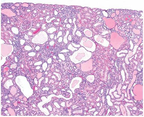
Fig. 2.64. Chronic progressive nephropathy in an aged rat. The cortex has severe tubular dilation with eosinophilic protein casts and interstitial fibrosis with mononuclear leukocyte infiltration.
Urolithiasis
Urolithiasis in laboratory rats is normally rare and usually sporadic. When present in the urinary bladder (Fig. 2.66),
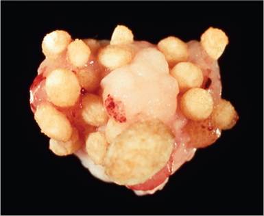
FIG. 2.66. Multiple calculi in the urinary bladder of a rat.
calculi may be associated with hemorrhagic cystitis, hematuria, and urinary obstruction. Calculi may also be located at other sites (e.g., renal pelvis, ureter, and urethra). The composition of calculi is variable. Analyses have revealed combinations such as ammonium magnesium phosphate, mixed carbonate and oxalate, and mixed carbonate and phosphate with magnesium and calcium. In male rats there are also erroneous reports of a high incidence of mucoid calculi, which are agonally excreted copulatory plugs into the urethra and bladder.