Nerves and ganglia
Nerves
Components of a neuron were described in Chapter 8. A nerve, however, is a collection of axons from many neurons and is found in the peripheral nervous system. Nerves vary in size and are surrounded by a series of connective tissue layers (Fig.
10.1). The epineurium is the outermost layer, consisting of a dense network of collagen fibers. The perineurium is the next innermost layer, and it partitions the nerve into a series of fascicles each containing a bundle of axons. The innermost layer is the endoneurium that surrounds each individual axon.Arteries and veins enter through the epineurium and branch within the perineurium. Capillaries can penetrate into the endoneurium where they nourish axons and Schwann cells of the nerves as well as the fibroblasts of the connective tissue. Therefore, a nerve consists not only of axons, but its bulk is composed of other tissues, including blood vessels, glial cells, and connective tissue.
Anatomy and Physiology of Domestic Animals, Second Edition. R. Michael Akers and D. Michael Denbow. © 2013 John Wiley & Sons, Inc. Published 2013 by John Wiley & Sons, Inc.
Classification of nerves
Since the peripheral nervous system has both sensory and motor components, nerves are classified according to their function. Nerves carrying impulses toward
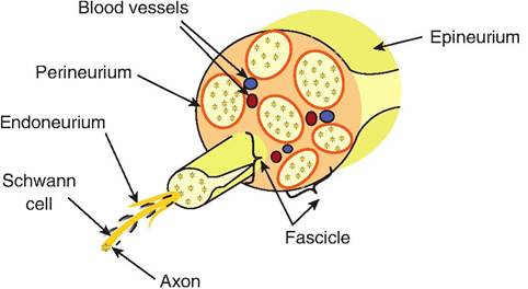
Fig. 10.1. Peripheral nerve. A peripheral nerve consists of many bundles of axons, each called a fascicle. There are three connective tissue layers surrounding various parts of the nerve. The epineurium is the outermost layer, wrapping around the entire nerve. The perineurium surrounds each fascicle, while the endoneurium surrounds each axon.
the CNS are called sensory or afferent nerves, whereas those carrying impulses away from the CNS are motor or efferent nerves.
Remember that efferent nerves carry impulses toward an effector. A nerve that carries impulses in both directions is called a mixed nerve.Peripheral nerves can function within the autonomic (visceral) nervous system or somatic nervous system. Therefore, they can be further classified as visceral afferent, visceral efferent, somatic afferent, or somatic efferent.
Spinal nerves
Nerves leaving the CNS are called either spinal nerves or cranial nerves. Cranial nerves were discussed in Chapter 8. There is a pair of spinal nerves exiting at each spinal segment. Each spinal nerve has a dorsal and ventral root that enters and exits the spinal cord, respectively (Fig. 10.2). Thus, the dorsal roots contain afferent fibers, and the ventral roots contain efferent fibers consisting of motor neurons of both the somatic and autonomic nervous system. Near the spinal cord, the two roots merge, forming a spinal nerve, which is a mixed nerve containing both afferent and efferent fibers. Each dorsal root has a swelling, called the dorsal root ganglion, situated near the spinal cord, which contains the cell bodies of the neurons running through the dorsal root. The dorsal and ventral roots pass through the intervertebral foramen located between adjacent vertebrae; the dorsal root ganglion lies between the pedicles of adjacent vertebrae.
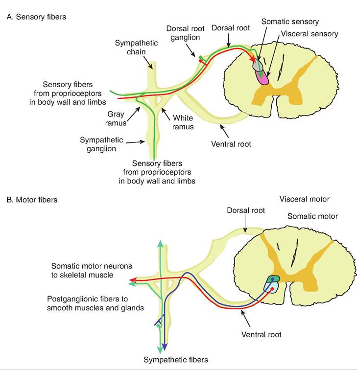
Fig. 10.2. Sp inal nerves. (A) The sensory neurons from the periphery enter the spinal cord through the dorsal root, and their cell bodies are located in the dorsal root ganglion. These fibers synapse in the dorsal gray horn of the central gray area of the spinal cord. The sensory visceral (autonomic) fibers may or may not pass through the sympathetic chain. (B) The motor fibers exit the spinal cord via the ventral root. The visceral motor fibers may enter the sympathetic chain via the white ramus.
Spinal nerves exit at every vertebra. The first spinal nerve exits superior to the first cervical vertebra while an additional spinal nerve exits inferior to each vertebra.
Therefore, cervical spinal nerves are named for the vertebrae immediately following where they exit. Since there are seven cervical vertebrae, there are eight cervical cranial nerves (Cl-C8). All other cranial nerves are named for the vertebra immediately preceding where they exit (i.e., Tl, T2). After leaving the spinal cord, both the dorsal and ventral roots merge, forming a spinal nerve. Shortly thereafter, the spinal nerve branches into the dorsal and ventral ramus. The ventral ramus carries fibers to the skeletal muscles of the body wall and limbs, as well as postganglionic fibers to smooth muscles, glands, body walls, and limbs. The dorsal ramus carries similar fibers to the back.Degeneration and regeneration of nerves
Like other cells, neurons die. It is believed that neurotrophic factors are responsible for keeping neurons alive. The presence of these factors suppresses a latent biochemical pathway present in all cells that causes the cells to commit suicide. Cells can die by a process called apoptosis, or programmed cell death, which involves four steps. The cell shrinks, the chromatin condenses, the cell fragments into apoptotic bodies, and the cellular remnants are phagocytized by macrophages or other such cells.
Using sympathetic neurons as a model, a proposed mechanism for apoptosis is as follows (Fig. 10.3). The loss of neurotrophic factors, such as nerve growth factor, decreases activity of the MAP kinase and phosphatidylinositol 3-kindase pathways, resulting in an increase in reactive oxygen species. This leads to an increase in c-jun N-terminal kinases and phosphorylation of c-jun protein. Then there is an increased expression of genes, including c-jun, cyclin Dl, and c-fos, and a decrease in RNA and protein synthesis. There is a decrease in Bcl-2 family proteins. Bcl-2 family proteins are expressed on the outer surface of mitochondria, and are bound to a molecule of Apaf-I. When damaged, Bcl-2 family proteins release Apaf-1, which activates caspases (cysteine aspartate specific prote-
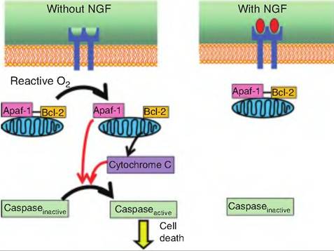
Fig.
10.3. Apoptosis. In a healthy cell, Bcl-2 is found on the outer mitochondria membrane and is bound to Apaf-1. Internal damage to the cell, such as the presence of reactive oxygen species, or lack of neurotrophic factors, such as nerve growth factor (NGF), causes Bcl-2 to release Apaf-1. A related protein called Bax also penetrates the mitochondral membranes, causing the leakage of cytochrome C into the cytoplasm. Released Apaf-1 and cytochrome C bind to inactive caspase. The resulting complex containing cytochrome C, Apaf-1, caspase 9, and ATP is called the apoptosome. Once activated, caspase 9 activates other caspases leading to digestion of structural proteins in the cytoplasm and degradation of chromosomal DNA and phagocytosis of the cell.ases). Caspases are a family of over a dozen proteins that cleave cellular proteins at aspartate residues.
Cells can also die from trauma or necrotic cell death, a process called necrosis, which is distinguishable from apoptosis. Traumatic death is characterized by an initial swelling of the cell with only modest condensation of the chromatin, and then rapid lysis of cellular membranes without endogenous programmed cell death. Necrotic cells elicit an inflammatory response that recruits macrophages that eliminate the cellular debris. In contrast, during apoptosis, individual cells are generally phagocytized prior to releasing their contents.
Ganglia
Ganglia are collections of neuron cell bodies located in the peripheral nervous system. Recall that within the CNS, a collection of cell bodies is called a nucleus. The ganglia for afferent (sensory) neurons are located in the dorsal root ganglia, discussed earlier. Somatic motor neurons do not have ganglia since these motor neuron cell bodies are located in the dorsal horn of the spinal cord. However, autonomic motor neurons are associated with ganglia since there are two consecutive nerve fibers associated with each autonomic motor pathway. These autonomic ganglia will be discussed in the section "Autonomic Nervous System."