Neuron structure
The main cell of the nervous svstem is the neuron Isee W
Ch. 2. Fig. 2.9). Neurons are responsible for the transmission of nerve impulses throughout the nervous tissue. Nerve impulses pass from one neuron to another by means of structures known as synapses.
All nerve pathways are made up of neurons and synapses. Complex nervous tissue, such as that of the brain and spinal cord, is made of neurons supported by connective tissue and neuroglial cells, whose function is to supply nutrients to and carry waste materials away from the neurons.Each neuron consists of the following parts:
A cell body containing a nucleus
Several short processes called dendrons, which are formed from many liner dendrites. These carry nerve impulses towards the cell body. A single cell body may receive as many as 6000 dendrons from other neurons
One long process called an axon. These carry nerve impulses away from the cell body. The axon leaves the cell at a point known as the axon hillock.
Nerve impulses from the dendrons are directed across the cell body towards the axon hillock and continue down the axon, reaching their final destination very rapidly. The speed of transmission along the axon is increased by the presence of a myelin sheath. Myelin is a Iipoprolcin material made by Schwann cells surrounding the axon. Its whitish appearance contributes to the colour of the more visible nerves in the bodv and to the white matter of the CNS. The mvelin sheath is interrupted al intervals of about I mm by spaces known as the nodes of Ranvier and it is through these that the axon tissue receives its nutrient and oxygen supply, ∖on-myelinated fibres are embedded within the Schwann cells IFig. 5.1) and despite their name arc actually covered in a single layer of myelin. Totally uncovered fibres are rare and may be found in areas such as the cornea of the eve.
Neurons vary in size - the diameter of the axons and dendrons may be a few micrometres and the length depends on the destination of the nerve fibre.
This may be anything from a few millimetres to more (han a metre long. Neurons also vary in shape (Fig. 5.21.Synopses
Each axon terminates in a structure called a synapse (Fig. 5. J I. Where an axon terminates on an individual muscle fibre, the synapse is referred to as a neuromuscular junction and the nerve impulse stimulates contraction of the muscle fibre. Within the button-like Prcsynaptic ending are vesicles containing chemical transmitter substances. The most common chemical is acetyl choline, but others found in the body include adrenaline, serotonin and dopamine.
As the nerve impulse travels down the axon, the vesicles drift towards the presy naptie membrane (Fig. 53) and release the transmitter into the synaptic cleft.
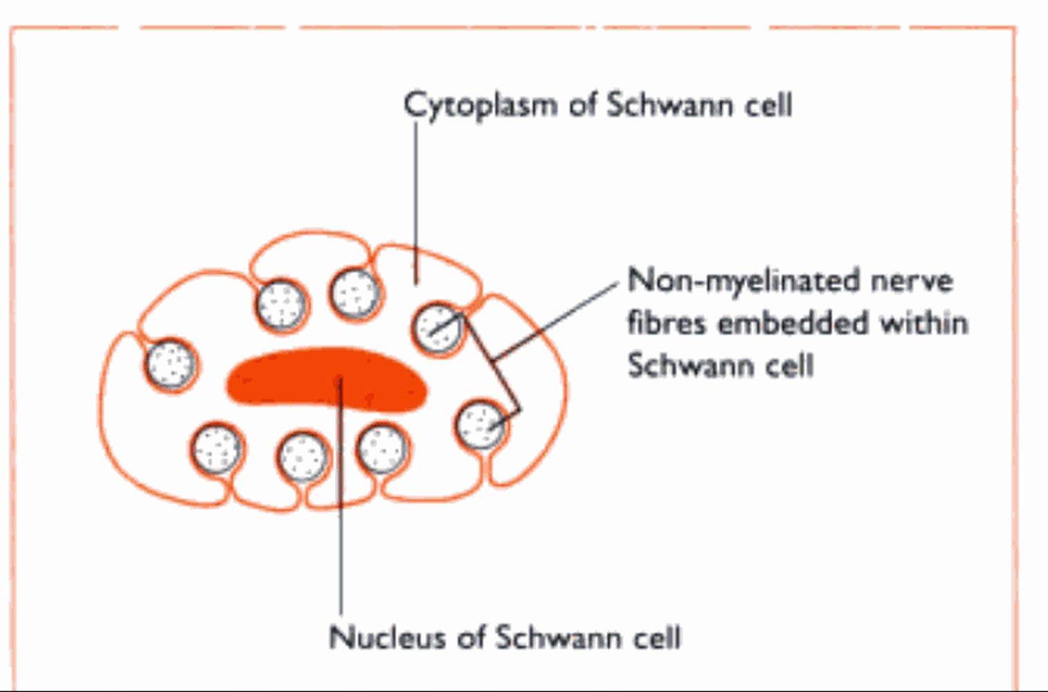
Fig. 5.1 Cross-sector through a Schwann cell showing nonmyelinated nerve fixes embedded wrth∙n the cytoplasm,
The nervous symptoms of eclampsia in the bitch and milk fever in the cow are caused by low levels of calcium in the blood, affecting the transmission of nerve impulses.
It diffuses rapidly across this gap and combines with the pastsι∣naptic membnme. making the membrane more ’excitable' and allowing the transmission of the nerve impulse to continue on down the nerve fibre. The effect is stopped by the release of cholinesterases - enzymes which destroy any acetyl choline remaining in the synaptic cleft - and the synapse returns to its resting state ready for the next nerve impulse. Effective transmission of a nerve impulse across the synapse will only occur in the presence of calcium ions.