Nonlymphoid Hematopoietic Neoplasms
The most significant spontaneous nonlymphoid hemaştopoietic neoplasm among common inbred mice is hisştiocytic sarcoma. Other forms of nonlymphoid hematopoietic tumors occur sporadically, or not at all, except under experimental circumstances or in GEMs (see Kogan 2002 for details on the MMHCC consensus classification/nomenclature for this group of tumors).
Histiocytic Sarcoma
Neoplasms of histiocytic origin are especially common in certain strains of laboratory mice, particularly in aged B6 and SJL mice. Neoplasms of this type have been produced experimentally using retroviruses or carcinoşgens in intact or thymectomized mice. Based on immuşnohistochemistry, these neoplasms arise from mononuclear phagocytic cells, such as Kupffer cells and tissue macrophages. Gross findings may include marked enlargement of the spleen, with nodular involvement of other tissues, such as liver, uterus, vagina, kidney, lung, and ovaries. In some cases, only 1 organ (e.g., uterine wall) may be involved. On microşscopic examination, there are circumscribed nodular to
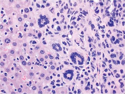
FIG. 1.128. Histiocytic sarcoma infiltrating the liver. The tumor consists of neoplastic histiocytes and multinucleated giant cells.
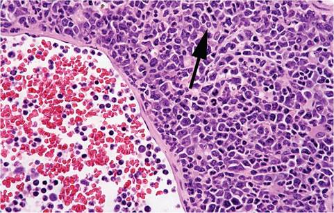
FIG. 1.129. Liver from a mouse with myeloid leukemia. Note the spectrum of differentiation, including some cells with doughnut-shaped nuclei (arrow), and the presence of neoplastic cells within the blood of the hepatic vein.
multifocal infiltrates in tissues such as liver, spleen, lymph nodes, intestine, bone marrow, female reproducştive tract, and lung. Neoplastic infiltrates consist of large histiocytic cells with irregular basophilic nuclei, fibrillar, eosinophilic cytoplasm, and indistinct cytoplasmic outşlines.
Neoplasms may vary in composition, from elonşgated fibrillar cells forming pallisading patterns to rounded cell types. Large nuclei and multinucleated giant cells are a common finding (Fig. 1.128). The neoşplastic cells tend to be particularly elongated in those arising in the uterine wall, and thus have on occasion been diagnosed as malignant Schwannomas. Erythro- phagocytosis may be associated with the neoplastic infiltrates, particularly in the liver. Occasionally tumors of this type involve a solitary lymph node or multiple ones. They consist of a prominent stromal component interspersed within a dense population of well-differenştiated lymphocytes. They may be difficult to differentişate from histiocyte-associated diffuse large B-cell lymphomas.Myeloid Leukemia
Spontaneous myeloid (granulocytic) leukemias are occaşsionally observed in some strains of older laboratory mice. They have been associated with retroviral infecştions and may be produced experimentally with chemişcal carcinogens or irradiation. The neoplastic process usually originates in the spleen, with subsequent involvement of a variety of tissues, including bone marrow, liver, lung, adrenal, and kidney. Clinically, animals are anemic and depressed, and peripheral leuşkocyte counts may approach 200,000/mm3. Mice usuşally have marked splenomegaly, with variable involvement of liver, kidneys, and other organs. There is massive infiltration of the splenic red pulp with maligşnant myeloid cells, with sparing of the splenic follicles. Diffuse infiltration of the bone marrow commonly occurs. Focal to diffuse infiltration of lung, liver, kidney, and adrenal may also occur (Fig. 1.129). Myeloid cells have large, vesicular nuclei that vary in shape with round, indented, and ring forms. Marked extramedullary myelopoiesis in the spleen and liver has been misconşstrued as myeloid leukemia by the uninformed.