Signature GEM Phenotypes: Molecular Pathology of Neoplasia
The microscopic appearance of spontaneous epithelial tumors of the mouse seldom resembles human cancers. In contrast, GEM tumors are often quite different from spontaneous mouse tumors and may more closely mimic the human condition.
It is becoming increasingly apparent that oncogenic transgenes induce “signature phenotypes” in GEMs that can be recognized among tumors derived from multiple tissue types. For example, tumors arising in a single organ, such as mammary gland, display different but consistent morphology depending upon the signal transduction pathway that is affected. These signature phenotypes cluster with genes that share common signal transduction pathways. Age, sex, immune status, strain background, and tissuespecific promoters may modify biology of GEM tumors, but seldom affect the signature phenotype. Furthermore, signature phenotypes span not only a single tissue but can be recognized in a variety of tumors arising in other organs. The field of “molecular pathology” is highly informative and needs the expertise of mouse pathologists who are aware of this evolving concept.Lymphoid Neoplasia
Tumors of lymphoid and nonlymphoid hematopoietic lineages are a major cause of morbidity and mortality among laboratory mice. The overall prevalence among all mouse strains is estimated to be 1-2%, but thymic lymphoma may occur in virtually 100% in some strains, such as AKR and C58 mice, by 12 months of age (Fig. 1.44). SCID mice also develop a high incidence of thymic lymphomas (Fig. 1.127). In contrast, BALB/c mice develop a high incidence of multicentric lymphomas (Fig. 1.45). The mouse is prone to these entities because of its inbred nature, which provides the requisite combinations of endogenous retroelements that are a
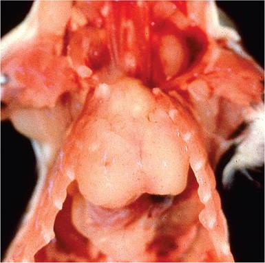
FIG.
1.127. Thymic lymphoma in an adult C.B-17-scid mouse. These neoplasms are relatively common in SCID mice.consequence of the mosaic origins of the laboratory mouse genome and selective inbreeding for tumor phenotypes (see “ Retroelements and Retrovirus Infections”). Furthermore, the mouse is unique because of the persistence and proliferative nature of the thymus into adult life, resulting in vulnerability to T-cell tumors. The mouse spleen also provides a unique milieu for evolution of neoplasia. It is a major hematopoietic organ throughout life, and it functions as the major secondary lymphoid compartment (in contrast to humans), with very active marginal zones.
The classification of mouse lymphomas has been in continuous flux but seems to be resolving with the use of immunological markers and more precise comparison with human disease. There is not much point in reviewing the various classification and nomenclature schemes that have been proposed in the past. The MMHCC consensus classification (Morse et al. 2002) generally agrees with but builds upon the WHO classification (Mohr 2001). The reader is encouraged to seek more detailed information regarding these types of neoplasms and their differential immunologic markers. Many of them rarely occur as spontaneous diseases in laboratory mice. Those that are most likely to be encountered in common laboratory mice are discussed below.
Small B-Cell LyphomaILeukemIa
These neoplasms occur sporadically in a number of strains of aged mice. They arise multisystemically, with infiltration of numerous organs, including lung and kidney, and often have a leukemic phase. They are composed of small round cells with scant cytoplasm, condensed chromatin, and a sIg+/B220+/CD19+ immunophenotype.
SplenIc Marginal Zone Lymphoma
This form of lymphoma occurs in low incidence (1-2%) among most inbred strains of mice. They may be supplanted by follicular lymphomas and have therefore been largely overlooked until recently.
They are common in NFS.N mice that are congenic for ecotropic proviruses contributed by AKR and C58 mice, and NZB mice. These lymphomas arise in the marginal zones of the splenic white pulp. Early lesions are often multicentric in the spleen, with extension from the marginal zone into the white and red pulp as the disease progresses. The spleens become enlarged, with occasional involvement of splenic lymph nodes, but disease in other tissues is generally not present. They consist of medium, uniform cells with abundant, grayish to pale eosinophilic cytoplasm, round to ovoid nuclei containing stippled or vesicular chromatin, and a sIgM+/B220+/ CD19+ immunophenotype.Follicular B-Cell Lymphoma
These lymphomas are the most common spontaneous lymphoma among many inbred strains of mice. They involve the spleen, Peyer's patches, and mesenteric lymph nodes. The tumors arise in follicles of the splenic white pulp, with a nodular appearance that can be seen as white mottling or nodules at necropsy. Microscopically, neoplastic cells are small to large, with scant cytoplasm and large, vesicular, irregularly folded to cleaved nuclei, and poorly delineated cytoplasmic boundaries. They typically are low grade, with cell populations resembling germinal center cells (large centroblasts or immunoblasts, and small centrocytes), as well as concurrent infiltration of other cell types, including significant numbers of T cells. Less than half of the cells are centroblasts or immunoblasts. Their immunophenotype is sIgM+/B220+/CD19+.
Diffuse Large B-Cell Lymphoma
These tumors are also common among inbred strains of mice and resemble follicular B-cell lymphomas. Gross enlargement of the spleen and abdominal lymph nodes is seen. They may also arise in the mediastinum with thymic enlargement. DLBCLs arise from centroblasts in the splenic white pulp. Cells are medium-sized and have scant cytoplasm with round vesicular nuclei. Nucleoli are prominent, often multiple, and typically adherent to the nuclear membrane.
Mitotic activity is high. Over half of the cells are centroblasts, and less than 10% are immunoblasts. The immunophenotype is sIgM+/ B220+/CD19+.Histiocyte-Associated Diffuse Large B-Cell Lymphoma
This form of lymphoma has a variable incidence among inbred strains of mice. It is a diffuse large B-cell lymphoma but features sheets of pink, fusiform to round, vacuolated histiocytes with large numbers of lymphocytes. Greater than half of the cells are histiocytes, which are not considered to be neoplastic. They frequently involve liver. Spleens are nodular, and lymph node involvement tends to be diffuse. It may be difficult to differentiate these neoplasms from histiocytic sarcomas because of their large populations of histiocytes.
Burkitt-Like (Lymphoblastic) Lymphoma
“Lymphoblastic” is less anthropomorphic, not associated with herpesvirus, and is unique to the mouse, but the MMHCC lists these tumors as “Burkitt-Like” as a comparative feature to human disease. These tumors arise frequently in some inbred strains of mice, especially in aged animals. Typically, there is generalized lymphadenopathy, splenomegaly, and sometimes thymic involvement. The tumors are composed of mediumsized, uniform lymphoblastic cells with round to ovoid nuclei, fine chromatin, and single or several small central nucleoli. Mitotic activity is high, and there is sometimes a “starry sky” appearance. Their immunophenotype is sIgM+/B220+/CD19+.
Plasmacytoma
This type of B-cell neoplasm is rare among most strains of laboratory mice but is notable because it can be readily induced with pristane in some strains of mice, such as BALB/c, NZB, or F1 crossed mice, or by infection with acutely transforming retroviruses. It may be common in some types of GEMs. Cells resemble plasma cells with eccentric nuclei, marginated chromatin, and moderate amounts of cytoplasm. Pristane-induced tumors arise in the peritoneum following intraperitoneal injection. Their immunophenotype is cytIg+/CD43+/CD138+.
Precursor T-Cell Lymphoblastic Lymphoma
These tumors are typically CD3+, CD4-/CD8-, TCR+, cytTdT+ thymic lymphomas that are common in some inbred strains of mice, such as AKR and C58. Some may be CD4+/CD-, CD4-/CD8+, or CD4+/CD8+. They also arise frequently in SCID mice and are commonly induced by irradiation and chemicals. Affected mice have enlarged thymuses, which may be associated with dyspnea, with variable involvement of spleen and lymph nodes. Cells are medium-sized and uniform with scant cytoplasm, round nuclei, and fine chromatin. Nucleoli are small, prominent, and multiple. Mitotic activity is high.
Small T-Cell Lymphoma
These lymphomas occur rarely in inbred mice and tend to arise in aged mice. They resemble small B-cell lymphomas and cause splenomegaly and lymphadenomegaly but not thymic involvement. Their immunophenotype is CD3+/ TCR+/CD4+ or CD8+/cytoTdT+.