Nutrients and Metabolism
Joanna M. Bassert
OUTLINE
METABOLISM, 428
Catabolic Metabolism, 429
Anabolic Metabolism, 430
Control of Metabolic Reactions, 430
Metabolic Pathways, 435
INTRODUCTION, 417 NUTRIENTS, 417 Oxygen and Water, 418 Carbohydrates, 419 Fats and Lipids, 420 Proteins, 423 Vitamins, 425 Minerals, 428
LEARNING OBJECTIVES
When you have completed this chapter you will be able to:
1.
List the six categories of nutrient.2. List and describe the three categories of carbohydrate.
3. List and describe the four categories of lipid.
4. Give the general structure of proteins.
5. Differentiate between water-soluble vitamins and fat-soluble vitamins and list their dietary sources and functions.
6. List the common macrominerals, microminerals, and trace elements found in the body.
7. Describe the processes of catabolism and anabolism.
8. List the events that occur in each stage of cellular metabolism.
9. Describe the process of glycolysis, the Krebs cycle, and the electron transport system.
10. Describe the general structure of enzymes and explain the role of enzymes in initiation and control of metabolic reactions.
VOCABULARY FUNDAMENTALS
Adenosine diphosphate (ADP) ah-dehn-o-sen dι-fohs-fat Adenosine triphosphate (ATP) ah-dehn-o-sen trι-fohs-fat Anaerobic glycolysis ahn-or-rδ-bihck glι-kohl-ih-sihs Anaerobic respiration ahn-or-rδ-bihck res-puh-ra-shuhn Biologic value bι-ah-lohj-ihck vahl-yoo
Cell metabolism sehl meh-tahb-uh-lihz-ehm
Cellular respiration sehl-u-lar res-puh-ra-shuhn Cellulose sehl-u-los
Citric acid cycle siht-rihck ah-sihd sι-kuhl
Coenzyme ko-ehn-zιm
Cofactor ko-fahck-tar
Contractile protein kohn-trahck-tehl prδ-ten
Crude protein krood prδ-ten
Cytochrome sι-to-krom
Deamination de-ahm-ihn-a-shuhn
Electron transport system e-lehck-trohn trahnz-pohrt sihs-tehm
Essential amino acid eh-sehn-shuhl ah-me-no ah-sihd
Essential fatty acid eh-sehn-shuhl faht-e ah-sihd
Essential nutrient eh-sehn-shuhl noo-tre-ehnt
Fat soluble faht sohl-yuh-buhl
Functional group fuhngk-shuh-nuhl groop
Glycolysis glι-kohl-ih-sihs
Ketone body ke-ton boh-de
Krebs cycle krehbz sι-kuhl
Lipolysis lι-pohl-ih-sihs
Macromineral mah-kro-mihn-ar-ahl
Membrane protein mehm-bran prδ-ten
Metabolic turnover meh-tah-bawl-ihck tarn-o-var
Micromineral mι-kro-mihn-ar-ahl
Mitochondrial matrix mι-to-kohn-dre-ahl ma-trihks
Monounsaturated fat mohn-o-uhn-sahch-ar-ra-tihd faht
Monounsaturated fatty acid mohn-o-uhn-sahch-ar-ra- tihd faht-e ah-sihd
Myositis mι-o-sιt-uhs
NADH N-A-D-H
Negative balance nehg-uh-tihv bahl-ehnz
Nicotinamide adenine dinucleotide (NAD) nihck-uh- tihn-ah-mιd ahd-eh-nιn dι-noo-kle-o-tιd
Nitrogen balance nι-truh-jehn bahl-ehnz
Nonessential amino acid nohn-eh-sehn-shuhl ah-me-no ah-sihd
Osmoregulator aws-mo-rehg-u-la-tar
Peptide bond pehp-tιd bohnd
Phosphorylation fohs-fohr-eh-la-shuhn
Polypeptide pohl-e-pehp-tιd
Polyunsaturated fat pohl-e-uhn-sahch-ar-ra-tihd faht
Polyunsaturated fatty acid pohl-e-uhn-sahch-ar-ra-tihd faht-e ah-sihd
Positive balance pawz-eh-tihv bahl-ehnz
Product molecule prohd-uhckt mohl-uhl-kyool
Protective protein pro-tehck-tihv pro-ten
Provitamin pro-vι-tah-mihn
Regulatory protein rehg-u-lah-tohr-e pro-ten
Saturated fatty acid sahch-ar-ra-tihd faht-e ah-sihd Short-chain fatty acid shohrt - chan faht-e ah-sihd Simple sugar sihmp-ehl shoog-ar
Starch stahrch
Storage protein stohr-ihj pro-ten
Structural protein struhck-shar-uhl pro-ten
Sugar shoog-ar
Trace element tras ehl-eh-mehnt
Transamination trahnz-ahm-ih-na-shuhn
Transport protein trahnz-pohrt pro-ten
Triacylglycerol trι-ah-sihl-glihs-ar-ahl
Triglyceride trι-glihs-ar-rιd
Unsaturated fatty acid uhn-sahch-ar-ra-tihd faht-e ah-sihd Water soluble wah-tar sohl-yuh-buhl
INTRODUCTION
Anyone who has ever watched a Labrador Retriever gulp down his dinner in 2 seconds, or someone else's dinner, for that matter, knows how glorious food is to most dogs.
If you have had the experience of feeding cattle or horses at mealtime, you know the level of excitement that the herds express as they bellow and whinny or "sing" for their supper. And alas, for some overweight pets on restricted diets, those pets that sit patiently and plaintively by their masters during dinner, food is just a bit too wonderful. All living things, plants and animals, need to take in nutrients to stay alive. Because food is required for survival, the nervous and endocrine systems of healthy living creatures not only generate the desire to eat but also instill the pleasure of eating to make food a glorious thing!Most food consumed by animals is used for metabolic fuel. This means that the nutrients derived from eating are broken down into smaller molecules and, along with oxygen, are used to make adenosine triphosphate
(ATP), which is the chemical energy used by cells. The amount of energy that can be acquired from these nutrient molecules is measured in kilocalories (kcal). Kilocalories are also called Calories (with a capital C). A kilocalorie is the amount of energy needed to raise the temperature of a kilogram of water by 1 degree.
In the previous chapter, we explored the structure and function of each part of the digestive system, and we learned how food is digested into fundamental nutrient molecules, which are then absorbed into the bloodstream. But what happens after these molecules are transported throughout the body via blood and lymph? How do the cells that make up tissues and organs absorb the nutrients and use them to stay alive? And how is the energy that is stored in the nutrient molecules converted to the usable form, ATP? How does the cell use ATP to make new molecules needed for repair and maintenance? In this chapter, we will answer these and other questions.
NUTRIENTS
A nutrient is a substance derived from food that is used by the body to carry out all of its normal functions. Nutrients are divided into six categories:
• Water
• Carbohydrates
• Lipids
• Proteins
• Vitamins
• Minerals
Water, carbohydrates, lipids, and protein are consumed in large quantities relative to vitamins and minerals, which are consumed in very small quantities (Figure 17-1).
In addition, carbohydrates, fats, and proteins produce energy when consumed, but water, vitamins, and minerals do not. Box 17-1 offers a summary of the energy-producing nutrients and gives examples of their sources in the diet.Cells, especially those found in the liver, have a remarkable ability to convert one type of molecule into another. This ability enables the body to break apart large molecules and use the “building blocks” to make new and completely different molecules. The body knows what molecules it needs to stay alive, so it will build the necessary molecules and transport them to where they are needed. This process is analogous to dismantling an old bank building that is not needed anymore and using the old bricks to build a new school or post office. In this way, the body is able to use a wide range of nutrient molecules found in food, dismantle them into their fundamental units, and build new molecules. This process enables animals to adjust to variations in diet.
However, there are limits. Creating new molecules from old ones cannot be done for all needed molecules. Each animal species has a select group of essential nutrients that it cannot manufacture from building blocks; therefore, the animal must obtain these essential nutrients from its diet.
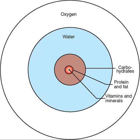
FIGURE 17-1 A nutrient target. Oxygen and six classes of nutrient are essential for life. The amount of water needed daily by an animal (in milliliters) is equal to the amount of its daily energy requirement (in Calories). Notice that vitamins and minerals are consumed in minute amounts relative to the other nutrients.
For example, many animals are able to make vitamin C from the nutrients they ingest, but guinea pigs cannot; to get vitamin C, guinea pigs must eat foods rich in vitamin C, such as fruits and vegetables or vitamin C-fortified pelleted feed. Cats cannot make the essential amino acid taurine, but dogs can, therefore cat food is fortified with taurine, but dog food iosrnot.
F this reason, you should not feed dog food to cats or, with Wιe, they will develop disorders such as blindness adniadc car disease as a result of the taurine deficiency.When animals are fed diets that contain both the essential and nonessential nutrients required for their species, they are able to synthesize all of the other additional molecules required for a healthy life. Keep in mind that the terms essential and nonessential are misleading, because both are required for life. Perhaps more accurate descriptions would be manufacturable and not manufacturable.
OXYGEN AND WATER
Oxygen is the most vital requirement for survival. Without oxygen, animal life would cease in an extraordinarily short period of' Wιe. I would last about 3 minutes without taking rauetbath, b a whale might last over an hour. Without the aobility t take oxygen into our bodies, we would quickly die. rn the priority of life’s essential requirements, water would be second only to oxygen. However, in terms of the diet, water is the most important nutrient. Most animals cuarnvinvoet s longer than a few days without it, but there are some adapted to desert conditions that can survive for several weeks. Water is obtained not only by ingesting food and drink but also by oxidizing proteins, fats, and
BOX 17-1
Summary of Nutrient Groups and Their Dietary Sources
Carbohydrates
Sugars
• Simple carbohydrates (monosaccharides and disaccharides) found in fruit, honey, sugar cane, sugar beets, and immature vegetables
Starches
• Complex carbohydrates (polysaccharides) found in grains, nuts, rice, and root vegetables such as potatoes and legumes
Cellulose
• Complex carbohydrate (polysaccharides) found in most vegetables
Proteins
• Meat, dairy products, soybeans, green leafy plants, eggs
Lipids
Neutral Fats
• Saturated-Meat, milk, cheese, cream, butter, coconuts
• Unsaturated-Vegetable oils, olive, safflower
• Phospholipids-Plasma membranes in plant and animal cells
• Steroids—eggs, butter and cream, animal fat, some chemical insecticides in the environment
• Cholesterol
Other Lipoid Substances—found in dark green leafy vegetables, root vegetables, some animal sources
• Fat-soluble vitamins
• Eicoiomoids (regulatory molecules derived from arachidonic acid)-prostaglandins, leukotrienes, and thromboxanes
• Lipoproteins
carbohydrates.
This metabolic source of water accounts for about 10% of the daily water requirement, though this is ehsiegrhter in d animals and birds.Mammals c^^wsist of about 70% water: newborns have somewhat more (75% to 80%), and adults have somewhat less (50% to 60%). Remarkably, even though an animal can survive the loss of almost all of its body fat and half of its protein, a loss of as little as 10% of its water can cause serious illness in most animals, and a 15% loss would be fatal without immediate treatment. The amount of water that is needed daily by an animal (in milliliters) is equal to the amount of its daily energy requirement (in Calories). Refer to the clinical apolication box to learn more about treating sick and dehydrated animals.
vWoalvterdis in in almost all of the metabolic processes
of the body. It is the major component of blood and is found inside all cells (intracellular) as well as outside the cell (extra- aellular). Water serves many functions in the body. It is a lourbricant f body tissues, a circulatory and transport medium, and a chemical reactant in digestion (hydrolysis). Idndiation, water is excreted as sweat and evaporated during poanting t assist in temperature regulation. Finally, it is the medium in which the biochemical reactions of metabolism
∕ j clinical application
Taurine Deficiency in Cats
Taurine is an essential amino acid in cats and is found in high quantities in meat and fish, but it is virtually nonexistent in plant-based foods, including dog food. Therefore cats that are fed dog food or home-cooked vegetarian diets develop taurine deficiency after several months. The result is a progressive, irreversible retinal degeneration that ultimately leads to blindness. In addition, taurine deficiency has been associated with dilated cardiomyopathy, a condition in which the heart enlarges because of dilation of the cardiac chambers. In some cases, the walls of the ventricles become very thin and the ability of the heart to pump blood efficiently is altered.
Cats may exhibit signs of heart failure, such as depression, shortness of breath, decreased exercise tolerance, and coughing. Fortunately, cardiomyopathy caused by taurine deficiency is reversible, and affected cats recover with nutritional supplementation.Queens that are taurine deficient may appear clinically normal but may have diminished reproductive success, including problems with abortion, early embryonic death, and malformations of neonates. An examination of the eyes of these cats usually reveals some degree of retinal degeneration.
Kittens that are born to taurine-deficient queens and fed deficient diets often die. Those that do survive exhibit neurologic signs, including cerebellar dysfunction and paresis in the hind legs, which frequently splay outward.
Occasionally, even cats that are fed diets adequate in taurine develop clinical signs of deficiency. In these cases, a biochemical disturbance in the retinal cells is thought to prevent normal taurine uptake and use.
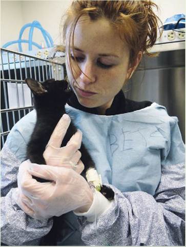
Kittens fed diets deficient in taurine are particularly vulnerable to serious illness or death. (Courtesy Joanna Bassert.)
occur, such as those involved in the growth, repair, and maintenance of cells.
CARBOHYDRATES
With the exception of lactose, which is the sugar found in milk, and a small amount of glycogen from meat, all dietary carbohydrates come from plants. Carbohydrates are divided into three categories:
• Sugars—monosaccharides and disaccharides that come from fruits, sugar cane, honey, milk, and sugar beets
• Starches—polysaccharides that come from grains, root vegetables, and legumes
• Cellulose—polysaccharides that are found in most vegetables
Glucose is the fundamental building-block molecule that results from breaking down large molecules of carbohydrates. It is used by the cell to make other molecules. Glucose is a monosaccharide, which is the simplest and smallest form of dietary carbohydrate (Figure 17-2). Fructose and galactose are also derived from the digestion of carbohydrates, but these monosaccharides are converted by the liver into glucose before they enter the general circulation.
Remember from the previous chapter that many nutrient molecules are absorbed in the small intestine and transported to the liver via the hepatic portal system. Once in the liver, they are often converted to other molecules, before they are either used by the liver itself or released into the general circulatory system for the benefit of other organs and tissues.
Glucose is readily used to make ATP through a process called glycolysis. ATP is the major fuel for the body. Although there are several types of cell that use fat as a primary source of energy, red blood cells and neurons rely almost exclusively on glucose for their energy needs. Perhaps this explains why brain-drained students crave candy bars and other high- sugar foods while studying for long periods of time. (I like espresso chocolate chip ice cream when I'm doing a lot of brain work. It has sugar and caffeine!)
The brain is exquisitely sensitive to decreases in blood glucose levels, and even temporary decreases in glucose can severely depress brain function and lead to the death of neurons. For this reason, the endocrine system keeps a vigilant watch over blood glucose levels by releasing glucose- controlling hormones from the pancreas (refer to our discussion of the endocrine system in Chapter 11). These hormones help to provide the brain and other tissues with a relatively steady level of glucose. However, as mentioned, even slight decreases in glucose can affect brain function, so if you find it difficult to make a decision at the end of the day, when you are tired and hungry, it is not your imagination. Glucose that is not immediately used by cells
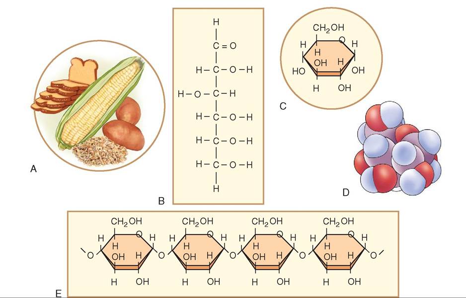
FIGURE 17-2 Carbohydrates. Carbohydrates are primarily derived from plants. A, The fundamental building block of carbohydrates is the monosaccharide. Glucose is the most important monosaccharide in the body. It may be composed of a straight chain of carbon atoms (B), or it may bend into a more stable ring (C), which in three dimensions (3-D) appears as a tight cluster of atoms (D). E, Monosaccharides link together to form disaccharides (di means 'two') or polysaccharides (poly means 'many').
is converted to glycogen and stored in the liver, or it is converted to fat and stored in adipose tissue throughout the body.
FATS AND LIPIDS
Lipids are organic molecules that are soluble in other lipids and in organic solvents, such as alcohol and ether, but are not soluble in water. Lipids are divided into four major categories:
• Neutral fats
• Phospholipids
• Steroids
• Other lipoid substances
NEUTRAL FATS
Neutral fats are commonly known as fats when they are solid and oils when they are liquid. It is important to keep in mind that fats are a kind of lipid, but not all lipids are fats. The building blocks of neutral fats are fatty acids and glycerol. Fatty acids are linear molecules classified as long-chain, medium-chain, or short-chain fatty acids depending upon the number of carbon atoms in the backbone of the molecule. Glycerol is a modified simple sugar. When fat is made in the body, three chains of fatty acid molecules are attached to a single molecule of glycerol, resulting in a molecule that looks like the letter E. Because there are three fatty acids in each molecule of fat, neutral fats are also called triglycerides or triacylglycerols. Although the glycerol component is the same in all neutral fats, the fatty acid chains vary, resulting in different types of neutral fat.
Whether a neutral fat is solid or liquid at room temperature depends upon two factors: the length of the fatty acid chains and the degree of saturation with hydrogen atoms within the chains. Fatty acids with single bonds between the carbon atoms can accommodate the greatest number of hydrogen atoms and are said to be saturated fatty acids, because the maximum number of hydrogen atoms is attached to the chain of carbon atoms. Saturated fatty acids tend to have long chains and are primarily found in meat and dairy foods, such as milk, cream, cheese, lard, and butter (Figure 17-3). Coconuts are one of the few plant sources of saturated fats.
On the other hand, fatty acids with one or more double bonds between the carbon atoms can accommodate fewer hydrogen atoms and are said to be unsaturated fatty acids, which includes monounsaturated and polyunsaturated fats. Fatty acids with short chains and those that are unsaturated are found in seeds, nuts, and most vegetables. Olive and peanut oils are rich in monounsaturated fats, and corn, soybean, and safflower oils contain a high percentage of polyunsaturated fats (Figure 17-4). Neutral fats with shortchain fatty acids and unsaturated fats are liquid at room
∕ j clinical application
Ketosis and Eclampsia: The Importance of Carbohydrates
We know how important carbohydrates are in providing animals with adequate energy levels. We also know how profoundly sensitive the brain is to even small decreases in blood glucose levels. For herbivores, the consumption of sufficient famounts o starch and cellulose from plants plays a vital role in their management, particularly if they are pregnant. The nutritional requirements in the first two trimesters of preg- lenoasnecy ar c to those needed for maintenance during the nonpregnant state. However, in the last trimester and during lactation, nutritional demands increase dramatically. Energy requirements are proportional to the number of fetuses and ahreesthig during lactation. Thus, cows carrying twins and reywiensg car twins or triplets have the highest nutritional needs. Serious metabolic disease can occur if the nutrition to trhegenseanpt or lactating animals is interrupted.
rWeghneanntp ewes and cows receive inadequate nutri
tion, ther bodies will compensate for the caloric deficiency by metabolizing their own tissues, primarily their fat reserves. Ketones are formed from the breakdown of the tissue and are released into the bloodstream, called ketonemia, and into the urine, called ketonuria. Even the breath of the animal contains ketones, and it therefore smells like acetone or nail polish remover. This condition is called ketosis. Ketosir is considered primary if the animal is fed an insufficient or unpalatable diet. It is called secondary ketosis if the animal is provided adequate untutrition b stops eating because of an illness, such as mastitis or metritis. Left abomasum displacement is the primary couse of secondary ketosis in cows. In both primary and sec- eotnodsiasr,y k the cow or ewe is not able to supply enough golmucose fr the digestion of food or from the catabolism of tissues to meet the needs of the developing fetuses or lactation rawnnd he o needs.
eWtohseisn k occurs in preparturient, or pregnant, cows and ewes, it may precipitate a dangerous and often fatal metabolic disorder known as eclampsia. Tha primary sign is hypo- gnlcyecpehmailco peathy, because the brain is adversely
yffected by the extremely low glucose level. The illness lasts 2 to 5 days,.fected animals are usually fat, as a result of overfeeding in the early part of gestation, but subsequently consume inadequate nutrition during the last trimester. Ini- tyially the are often restless and uncoordinated, as though drunk; later, they become listless, anorexic, and they walk aim- lmesestliym, seos bumping into things because of a condition
known as cortical blindness. They m»’ grind their teeth, develop unusual postures, and may exhibit muscle twitching coen the fa and ears. Finally, the animals become sternally recumbent, develop rapid, weak pulses; and do not get up. Coma and death often follow, and mortality is about 80%. Not surprisingly, blood tests show high ketone and low glucose levels. Guinea pigs, ferrets, and rabbits are also predisposed to developing eclampsia.
Postparturient ketosis usually appears a few days to weeks iavfitnerg g birth, when milk production is high. Lactation ruegqeuires h amounts of glucose to make lactose for milk. dWehqeunateina carbohydrates are consumed, glycogen
stores in the liver are used first. The metabolism of tissues, rwohmicoht eps ketogenesis, soon follows once the glycogen
svteores ha been depleted. Clinical signs of postparturient lkuedtoesis inc depression, lethargy, a staring expression, decreased milk production, weight loss, and a humped-back appearance consistent with abdominal pain. Occasionally, postparturient ketosis will cause frenzied behavior, such as circling, staggering, bellowing, head pressing, and compulsive hwiaclhking, w occurs for about an hour..s with all ketotic states, ketones are present in the blood, urine, and breath, and there is a marked decrease in blood glucose levels. Unlike eclampsia, however, postparturient ketosis is self-limiting, because the reduction of food intake eventually causes milk production and the corresponding glucose drain to stop.
By far the best and least expensive treatment for ketosis is prevention. Providing fresh, palatable feed in appropriate quantities at the appropriate times is the key to a successful rboregeradmin.g p
(From McCurnin DM, Bassert JM: Clinical textbook for veterinary technicians, ed 6, St Louis, 2006, Saunders.)
temperature (oils). In ruminants, short-chain fatty acids are found in the gas and liquid contents of the rumen and reticulum. In species such as cattle, sheep, and goats, these volatile short-chain fatty acids, derived from the microbial breakdown of' ingested plant matter, are a vital source of energy.
The liver is adept at converting one fatty acid to another, bmuet so fatty acids, such as linoleic acid, cannot be synthesized. Linoleic acid is a fatty acid component of lecithin. lThus, in al domestic species and in humans, linoleic acid is an essential fatty acid that must be available in the diet.
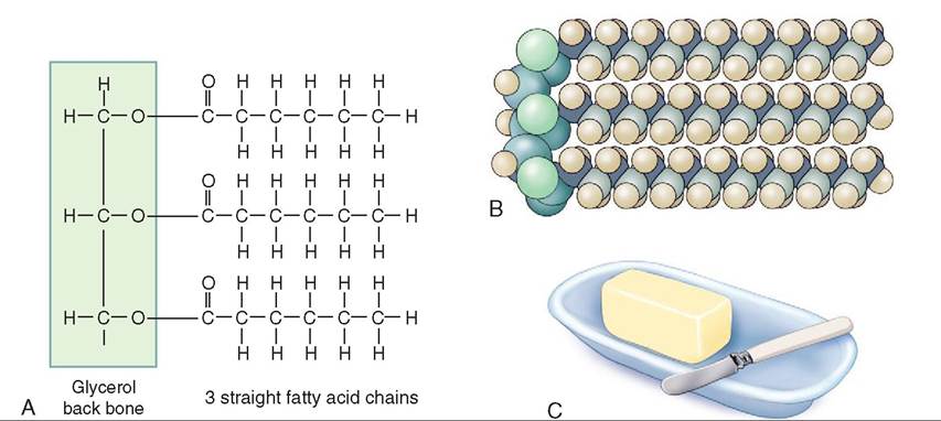
FIGURE 1 7-3 Neutral fats. A, Neutral fats are composed of triglycerides. Each triglyceride molecule includes a glycerol backbone and three fatty acid chains. These molecules together create an E shape. B, The saturated triglyceride does not have any double bonds in the molecule and can therefore hold the maximum number of hydrogen atoms, making the chains very straight. C, Saturated fats tend to come from animal fat and are found in butter, lard, and meat.
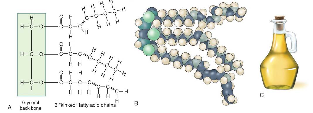
FIGURE 1 7-4 Unsaturated fats. A, Unsaturated fats contain one or more double bonds in the fatty acid chains. B, These double bonds cause the chains to bend and kink. C, Unsaturated fats tend to be liquid at room temperature and are derived from plants and vegetable oils.
Fortunately, it is found in most vegetable oils. Linolenic and arachidonic acids are also considered to be essential fatty acids. Though it is possible for the body to convert linoleic acid into arachidonic acid, the process is extremely inefficient and fes not yield adequate results for many species. Thus arachidonic acid is also considered to be an essential fcaidtt.y a
Neutral fats are amazing. They contain over twice as much potential energy by weight as proteins or carbohydrates. Fat is easily digestible, makes food taste good, staves uofnfgehr for long periods of time, and helps the body absorb the fat-soluble vitamins A, D, E, and K. Stored, subcutaneous fat is an important insulator, and fat also surrounds and cushions vital organs, such as the heart, kidneys, and eyes, hi the digestive tract, neutral fats are broken down into their fatty acid and glycerol components before being aybsorbed b the intestine and sent into the hepatic portal shyestem. T liver rebuilds neutral fats, forming many different kinds of' E-shaped triglycerides before they are sent off to other parts of the body. Not surprisingly, triglycerides are the major energy source for hepatocytes (liver cells) and skeletal mnscle cells, which are some of the most active cells iondyth. e b
PHOSPHOLIPIDS
Phospholipids are modified triglycerides derived primarily from the cell membranes of plant and animal cells. Like the triglycerides, phospholipids have a glycerol core but contain two, rather than three, fatty acid chains; therefore you could call them diglycerides. In addition, phospholipids have a phosphorus group attached to the glycerol molecule—the infamous polar head, for which the phospholipids are so well known. The polar head and the two fatty acid chains make the phospholipid molecule look like the head, shoulders, and dropped arms of a stick person (see Chapter 3).
STEROIDS
Steroids are lipids that are dramatically different from neutral fats. Unlike the E-shaped molecule typical of fats or the “head, shoulders, and arms” of phospholipids, steroids are made of four flat interlocking rings of hydrocarbons. Steroids include cholesterol, bile salts, sex hormones, and hormones released from the cortex of the adrenal gland. Of these, cholesterol is the most vital nutritionally, because all of the other steroid molecules can be made from cholesterol: it is the essential precursor. Like phospholipids, cholesterol is found in the plasma membrane and is nutritionally derived from animal products such as egg yolks, meat, and cheese. In addition, the liver is able to manufacture cholesterol; without it, animals would not be able to carry out many vital biochemical processes.
OTHER LIPOID SUBSTANCES
A diverse range of other lipoid substances, such as fat-soluble vitamins, eicosanoids, and lipoproteins, are vital for an animal's survival. Eicosanoids are regulatory molecules derived from arachidonic acid, which is a 20-carbon fatty acid found in the plasma membrane. These substances include prostaglandins, leukotrienes, and thromboxanes, which are hormones that play an important role in the inflammatory process, blood clotting, and labor contractions. Prostaglandins are also important in smooth muscle contraction and in controlling blood pressure.
TEST YOURSELF 17-1
1. What is a Calorie?
2. What are the six fundamental nutrients? Which ones generate energy when consumed?
3. Why is water so vital to the survival of an animal?
4. What are the three categories of carbohydrate?
5. What are the four major categories of lipid?
6. What is the difference between a saturated and an unsaturated fat? Why is this difference important nutritionally?
7. What is an essential fatty acid?
8. Give a specific example of a steroid.
PROTEINS
Proteins make up 10% to 30% of a cell's mass and are therefore the dominant structural material of the animal body. Proteins are used for building critical structural materials, such as keratin, collagen, and elastin for the skin; connective tissues; and muscle proteins. But not all proteins are construction materials. Many play vital and diverse roles in cell function: enzymes and hormones regulate an incredible variety of body functions; hemoglobin carries oxygen in red blood cells; and contractile proteins in muscle cells enable the body to move.
Though the roles of proteins vary, all proteins are composed of amino acids. There are 22 different types of amino acid, but they are all composed of three important functional groups: a basic amine group (-NH2), an organic acid group (-COOH), and an R group. Differences in the R group make each amino acid unique, but the acidic and basic groups are identical among all amino acids. Proteins are created when the amino acids link together, like pearls on a string (Figure 17-5). The acid group from one amino acid links to the basic group on the next, forming a peptide bond. When two amino acids link together, the resulting molecule is called a dipeptide. Three amino acids together form a tripeptide, and more than 10 amino acids link to form a polypeptide.
Proteins are huge molecules, called macromolecules. They are commonly composed of 100 to 10,000 amino acids, though any polypeptide with 50 or more amino acids is considered a protein. The type and order of the amino acids determine the structure and function of the protein. Each of the 22 amino acids is analogous to a letter in an alphabet. The letters used and the order in which letters are arranged determine a particular action or meaning. For example, if the chain reads “Have a nice day,” and only one amino acid is altered in the chain to make it read “Have a mice day,” the meaning becomes very different and possibly nonsensical.
There are 10 essential and 12 nonessential amino acids in most species. Essential amino acids must be present in the diet because the animal either cannot make them at all or cannot make them fast enough to meet the body's needs for tissue maintenance and growth. For the body to make new proteins, all of the needed amino acids—essential and nonessential—must be present in the cell in sufficient quantity and all at the same time. This is called the all or none rule. If one amino acid is missing, the protein cannot be manufactured.
Animal products, such as meat, eggs, and dairy products, contain proteins that include the largest number of essential amino acids. In fact, meat products contain all of the essential amino acids for many species and are therefore called complete proteins. However, animal-based protein is more expensive than plant-based protein, so many pet foods contain primarily plant-based proteins, such as soy. Cereals, rice, nuts, and legumes, such as beans, are protein rich, but their proteins are nutritionally incomplete, because they are low in one or more of the essential amino acids. Leafy green vegetables are also rich in essential amino acids, but have little methionine and tryptophan. When ingested together, legumes, grains, and cereals provide all of the essential amino acids for many species, including humans; these foods are therefore said to be complements of one another (Figure 17-6).
Amino acids cannot be stored. Those not immediately used to make protein are oxidized by the cell to make energy or are
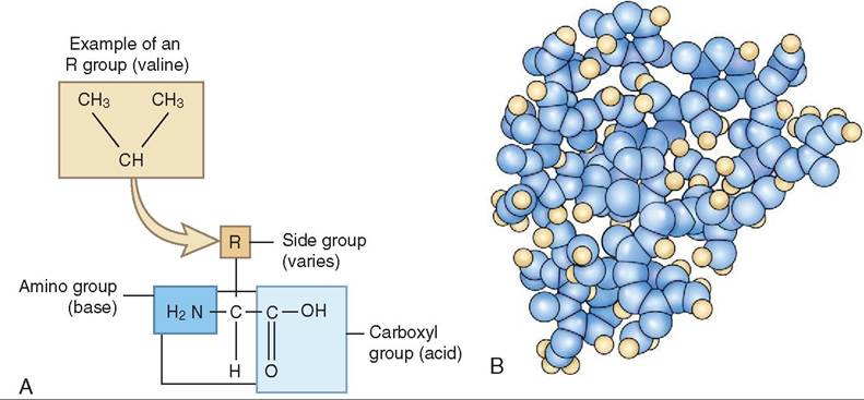
FIGURE 1 7-5 Proteins. A, Proteins are composed of amino acids, which are composed of an amino group, a carboxyl group, and a variety of side chains (P groups). Amino acids link together to form long chains. B, When the protein becomes particularly large, it coils to increase its molecular stability. Meat is an excellent source of protein, because it contains the complete assortment of amino acids needed for the body to make all of the proteins required for maintenance and growth.
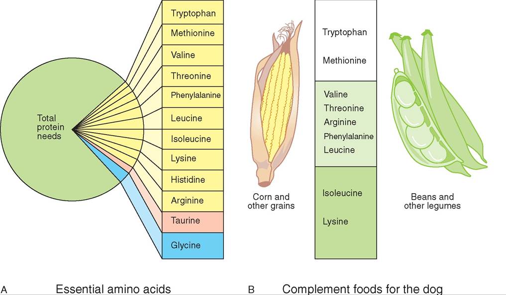
FIGURE 17-6 Protein synthesis. A, Protein synthesis depends upon all of the essential amino acids being present in sufficient amounts simultaneously. The essential amino acids make up a relatively small portion of the total protein needs of animals. Notice that the amino acids considered essential vary from species to species. The amino acids in yellow are essential for the dog. Taurine (pink) is essential for cats and is found in meat. Glycine (blue) is essential for poultry. B, Foods such as corn and grains are rich in certain essential amino acids, such as tryptophan and methionine, but lack other amino acids, such as isoleucine and lysine, which are found in legumes. When consumed together, a diet of corn, grain, and legumes contains all of the essential amino acids needed by many species, including dogs. Thus, dogs can do well on home-cooked vegetarian diets, as well as on meat-based diets, if adequate complement foods are present, but cats are strict carnivores and must consume animal proteins (because taurine is found naturally only in animal tissue). Therefore, cats should not be fed a home-cooked vegetarian diet.
converted to carbohydrates or fats. If the body does not have enough carbohydrate or fat calories in the diet to make ATP, the cell will use proteins from the diet or will catalyze tissue to make ATP. In the healthy animal, the rate of protein synthesis equals the rate of protein breakdown and loss. This is called the nitrogen balance. When the amount of nitrogen ingested in the form of protein equals the amount of nitrogen that is excreted, the body is said to be in nitrogen balance. Nitrogen from protein is packaged by the liver into a molecule called urea, which can be measured by a blood urea nitrogen (BUN) test, before it is excreted by the kidney.
A positive balance occurs when the body is incorporating more protein into tissues than it is breaking down to make energy (ATP). This happens normally during healing, and during pregnancy because of the growing fetus. It also occurs in growing animals. Certain hormones, called anabolic hormones, accelerate protein synthesis and growth. For example, pituitary growth hormone stimulates tissue growth in young animals and conserves protein in mature animals. Sex hormones trigger growth spurts. Growth and lactation increase the protein requirements of animals above what is needed for body maintenance and exertion (work or exercise). The metabolism of excess amino acids increases the workload of the liver and kidney because of the processing and excretory requirements for the urea and organic acid waste by-products. In other words, eating too much protein causes the liver and kidneys to work harder. This is why animals with kidney disease are fed a low-protein diet.
A negative balance occurs when protein breakdown exceeds the amount of protein being incorporated into tissues. This occurs during physical and emotional stress— infection, injury, debilitation—and during starvation, or when the quality of the dietary protein is poor. Glucocorticoids released during stress enhance protein breakdown and the conversion of amino acids to glucose. Cats are the consummate carnivore and are specifically adapted to a high- protein diet. They commonly manufacture proteins from the amino acids released from gluconeogenesis.
Pet foods often identify the crude protein content on the label of the can or bag of food. Most people assume that the best food for their pet contains the highest percentage of crude protein. This, however, is not necessarily true. Crude protein content gives no indication of the quality or utilization potential of the protein. A dog food, for example, may have a high protein content, but the biologic value of the protein may be low. The biologic value is the percentage of absorbable protein that is available for productive body functions. It defines the amount of amino acids available for metabolic processes.
The ideal protein content in food includes all of the essential amino acids needed to meet the specific metabolic requirements of a particular species. For example, if a protein has a missing essential amino acid, its quality may be low. However, by adding the missing amino acid, the full potential of the biologic value of the protein can be restored. This is one reason why mixed animal and plant protein sources are often complementary to each other in food formulations. In addition, the quality of proteins is improved if feeds are not overprocessed or overheated in storage, because heating can denature proteins.
In ruminants, the digestion of protein is facilitated by microbes, which break down protein derived from the grasses and grains consumed in the diet. The amino acids generated from this process are further degraded into ammonia, organic acids, and carbon dioxide. The ammonia is either absorbed through the wall of the rumen or is used by the microorganisms to make new proteins. Interestingly, the microbial-made protein has a consistent quality regardless of the quality of the nutrient source. Thus, protein in lower-quality feeds is improved by microbial metabolism. On the other hand, the usefulness of high-quality protein from feed may be lowered by microbial metabolism, so feeding high-quality protein to a ruminant may give the same results nutritionally as feeding low-quality protein. Amazingly, the rumen also has the ability to convert nonprotein sources of nitrogen, such as urea and ammonium salts, into protein. These nitrogen sources, however, must be used judiciously in feeds, because an excess or an imbalance can be toxic to the herbivore.
VITAMINS
Although consumed in minute amounts, vitamins are essential for life. Not surprisingly, the Latin word vita means life. Unlike nutrient molecules, such as carbohydrates, proteins, and fats, vitamins do not produce energy when metabolized, nor are they broken down into building-block units. Rather vitamins are, for the most part, coenzymes or parts of coenzymes. Their molecular structure is the “key” that activates an enzyme and enables it to carry out its diverse metabolic reactions. For example, during the biochemical breakdown of glucose, the B vitamins riboflavin and niacin are required. Without them, the reaction cannot be completed and glucose cannot be used to generate energy. Thus, the full use of nutrient proteins, carbohydrates, and fats is dependent upon the presence of coenzymes. However, not all vitamins are coenzymes. Vitamins A, D, and E have important and varied roles in the body. Vitamin D, for example, is converted to cal- citriol, which regulates calcium levels in the body, and a form of vitamin A, retinal, helps sensory cells in the retina of the eye to detect light.
Plants can manufacture the vitamins they require, but animals cannot. Therefore, most vitamins are not made in the body and must be consumed in the diet, but there are exceptions. Vitamin D, for example, is made in the skin, and vitamin K and biotin are made in the intestine by bacteria. In addition, the body can convert beta carotene, which is an orange pigment found in carrots and deep green leafy vegetables, into vitamin A. For this reason, beta carotene is called a provitamin and is essential for many species.
Vitamins were assigned their letters in the order that they were discovered. In addition, they also have been given a descriptive title that indicates their function in the body and have been classified as either fat soluble or water soluble (Table 17-1).
∕ j CLINICAL APPLICATION
Starvation: A Life and Death Issue
We have all marveled at the fortitude of wild animals that survive s^re winters, drought, and food deprivation. Neglected domestic animals may also endure inadequate shelter, food supplies, and access to water. For them, food deprivation and subsequent starvation are a threat to their lives. Fortunately, evolution has dictated physiologic adaptations that extend the time animals can live without food. Except for the terminal phase, starvation is reversible, and the reproductive capacity of the animal is preserved for as long as possible. Hw does the body do this?
Thrie process of starvation can be organized into three stages.
Stage I
Initially, foe body tries to balance the animal’s energy expenditure with energy intake by lowering the basal metabolic rate. In tahhye,is w t animal needs less food for maintenance but may feel weak, dizzy, and tired. Although many tissues can use nutrients other ⅛ιt glucose, certain tissues—such as blood cells, the kidney, and nervous tissue (brain and spinal cord)—are nor- lmucaollsye g dependent. The body, therefore, uses glycogen shnteores i t liver to provide glucose to these important tissues. However, foe glycogen stores are depleted after several hours, any foe body must turn to other sources of energy. Glycerol and ftcaoidtrteys a s from fat and amino acids from body proteins are catzbolized next to produce glucose. Ketone bodies are sreleased a a product of fatty acid metabolism, causing the ebvloelosd l of ketone bodies and glucose to rise.
Stage II
After 1 to 2 weeks, depending on the species, a change occurs iondythe b that allows the brain and other tissues to use ketones and glucose for energy. Stored body fat becomes the primary source of energy as the body breaks down fatty acids ientotonek bodies. This process continues until fat reserves earpeledted. The length of this stage varies among individual animals, depending on the amount of body fat that is available foolirsmca.tab Generally, this stage lasts for several weeks.
Because body fat serves primarily as an energy storage tissue, aunlaitnosr, and protector of internal organs, progressive loss iodsfispuaoese t does not threaten normal body functions or survivability until the stores are nearly depleted.
Stage III
Once fat reserves are used up, protein becomes the principal metabolic fuel. Even during the first stages of starvation, the abtoadbyolcizes protein to produce glucose. However, the level raootfatebpionlism c remains high after the fat reserves are
dneitpialelltye,d. I liver and plasma proteins are used, followed by protein torn the gastrointestinal tract, heart, and skeletal hmeusescle. T structures decrease dramatically in size, and criti- oal body functions are lost. Decreases in plasma proteins, for example, lead to changes in oncotic pressure, and fluid subse- eqaukesntly l into the abdominal cavity, causing abnormal distention called ascites. Lose of muscle between ribs and in the diaphragm may lead to respiratory failure. The heart, which can lose 50% of its mass, may simply stop beating. Gastric emptying and intestinal transit times are prolonged, and the absorption raonotfdtfeina p is impaired as the inner layers of the intestine degenerate. Diarrhea and nausea may result. In addition, malnourished animals are immunocompromised and often develop pneumonia and other infections. Only the skeletal system seems remarkably unaffected by starvation.
Treatment
Warm, intravenous fluids in conjunction with antibiotics and putarietniotenral n are important in the treatment of end-
svtaatgioens.tar In addition, malnourished animals are less aoble t maintain adequate body temperature and should be gmiven a war environment in which to recover. Good nursing coarrteanits imp in these cases, because animals will need thick, soft bedding and care for any decubital ulcers or sores tyvheat ma ha developed from prolonged recumbency. After the animal has been stabilized through the use of parenteral nutrition, high-protein liquid diets should be given by mouth initially until semisolid foods can be tolerated.
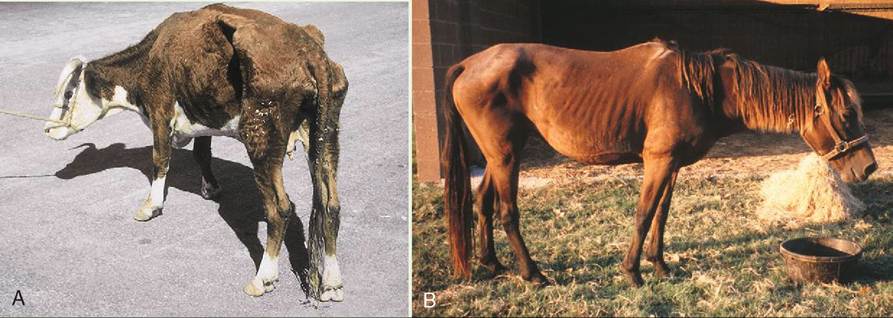
The emaciated cow (A) and horse (B) seen here stand with lowered heads, characteristic of depression and lethargy. Malnourished animals, such as these, are experiencing a negative nitrogen balance and have already metabolized all of their fat stores and much of their muscle tissue to generate the energy needed for vital metabolic functions. These animals are vulnerable to infection, particularly parasitism and bacterial pneumonias, owing to immunosuppression. (A from Hendrix CM, Robinson E: Diagnostic parasitology for veterinary technicians, ed 3, St Louis, 2006, Mosby. B from McCurnin DM, Bassert JM: Clinical textbook for veterinary technicians, ed 6, St Louis, 2006, Saunders.)
| TABLE 17-1 Summary of Vitamins, Dietary Sources and Importance | ||
| VITAMIN | DIETARY SOURCE | IMPORTANCE IN THE BODY |
| WATER-SOLUBLE VITAMINS | EXCESS IS EXCRETED IN URINE SO TOXICITIES ARE RARE | |
| B Complex Vitamins: | ||
| Vitamin B1 (thiamine) | Meat, fish, eggs, leafy green vegetables, legumes, liver | Limited amount stored in the body |
Required for the transformation of pyruvic acid to acetyl coenzyme A (CoA) in carbohydrate metabolism. Needed in the synthesis of some neurotransmitters (acetylcholine). Rapidly destroyed by heat
| Vitamin B2 (riboflavin) | Milk, meat (fish, poultry, and red meats), liver, eggs, grains, legumes | Found in the body as flavin mononucleotides (FAD and FMN), which serve as hydrogen acceptors. Also needed for oxidation of amino acids |
| Vitamin B3 (niacin or nicotinamide) | Milk, meat (fish, poultry, and red meats), liver, eggs, grains, potatoes, peanuts, leafy green vegetables, yeast, liver | The amino acid tryptophan, which is found in many food groups, is easily converted to niacin. Niacin is part of nicotinamide adenine dinucleotide (NAD+), which is important in glycolysis, and in the catabolism of fats. |
| Vitamin B5 (pantothenic acid) | Found in large quantities in internal organs such as ibdrnaeiny,, k liver, adrenal, and heart. Also found irenagiungsm,es, l eggs, and yeast. Name refers to the Greek word panthos, meaning everywhere, end referring to the diverse dietary sources | Stable when heated. Is a major component of coenzyme A. Important in the oxidation of fatty acids and in the synthesis of steroids and hemoglobin |
| Vitamin B9 (folacin or folic acid) | Lean beef, eggs, veal, whole grains, liver, orange juice, deep green vegetables, yeast. A crystalline vitamin that is bright yellow | Essential for the formation of red blood cells. Is one of many coenzymes that are part of methionine, choline, and DNA synthesis |
| Vitamin B12 (cyanocobalamin) | Found in liver, meat (poultry, fish, and red meat), dairy (but not eggs), and butter. Not found in plants. Intrinsic factor is required for absorption in the intestine. | Stored in the liver in quantities that meet bodily needs for 3 to 5 years. Essential for the synthesis of methionine and choline. Is a coenzyme in all cells in the body, particularly in the nervous system, bone marrow, and gastrointestinal tract |
| Others: | ||
| Biotin (vitamin H) | Liver, eggs, nuts, and legumes. In some species, it is generated by enteric bacteria in the gut. | Essential for reactions in the Krebs cycle. Functions as a coenzyme in a number of reactions, including carboxylation, decarboxylation, deamination, and anabolism of purines and nonessential amino acids |
| Vitamin C (ascorbic acid) | Found in fruits and vegetables, especially large quantities in citrus fruits, leafy green vegetables, tomatoes, cantaloupe, and strawberries | Important antioxidant. Needed for the formation of collagen fibers in connective tissue and the conversion of tryptophan to serotonin. Aids absorption of iron. Activates folicin (a B vitamin) |
| Fat-Soluble Vitamins: | Fat-soluble vitamins are stored for long periods of time in tissues. Excess is not excreted, making toxicity a possibility if high levels are consumed | |
| Vitamin A (retinol) | Can be formed in the body from ingested beta-carotene, which is found in dark green leafy and dark yellow vegetables. Vitamin A is found in fish oils, milk, eggs, and liver. | 90% is stored in the liver, so feeding liver routinely to dogs or cats can cause toxicity. Enough is stored to meet the body's needs for one year |
| Vitamin D (antirachitic factor or calciferol) | Formed in the skin when ultraviolet (UV) light converts 7-dehydrocholesterol. Modified to the active form in the liver and kidneys. Also found in liver oil, eggs, and fortified milk | Essential for blood clotting and bone and tooth formation. Acts as a hormone that regulates calcium levels in the blood by increasing absorption from the gut and mobilizing calcium from bone |
| Vitamin E (antisterility factor or tocopherols) | Fnhoeuant d i w germ, nuts, whole grains, vegetable oils, and green leafy vegetables. Stored in muscle and fat tissue; chemically similar to sex hormones | An important antioxidant. Prevents oxidation of unsaturated fatty acids and cholesterol. Helps prevent oxidative damage to cell membranes |
| Vitamin K (coagulation factor or quinones) | Found in leafy green vegetables, cauliflower, broccoli, pork liver, and cabbage. Also synthesized by bacteria in the large intestines of some species | Essential for the generation of clotting factors and many proteins made by the liver. Contributes to the process of oxidative phosphorylation in cells |
Water-soluble vitamins are absorbed through the gut wall when water is absorbed in the gastrointestinal tract. The water-soluble vitamins include vitamin C and eight of the B-complex vitamins. An exception is vitamin B12, however, which must bind to gastric intrinsic factor before it can be absorbed. Very small amounts of water-soluble vitamins are stored in the body, and excesses not used within an hour are excreted in the urine. Hypervitaminosis conditions involving water-soluble vitamins are therefore extremely rare.
Fat-soluble vitamins include A, D, E, and K. These bind to ingested lipids before they are absorbed with the ingesta. Thus, if fat absorption is impaired, so too is the absorption of fatsoluble vitamins. Except for vitamin K, fat-soluble vitamins are stored in the body; therefore, if an excessive amount of a fatsoluble vitamin is consumed, vitamin toxicity due to hypervitaminosis may result. For example, liver is high in vitamin A, and owners who frequently feed liver to their dogs or cats may unknowingly induce a toxic condition in their pets.
Proteins, carbohydrates, and lipids are oxidized as part of the normal metabolic processing of food. This process uses oxygen and generates some potentially harmful free radicals. Vitamins A, C, and E are potent antioxidants that disarm dangerous free radicals. Certain foods such as broccoli, cauliflower, cabbage, and Brussels sprouts are all good sources of vitamins A and C.
Antioxidants are thought to act like a bucket brigade, passing dangerous free radicals from one molecule to another until they can be excreted. For example, vitamin E returns a free radical to a less harmful state but, in doing so, vitamin E itself becomes a free radical. It is subsequently inactivated by carotenoids, and the carotenoid free radicals that are produced are then inactivated by vitamin C. Water-soluble free radicals are subsequently formed, which can be flushed from the body in urine.
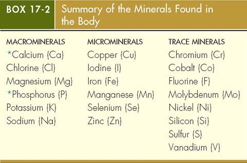
MINERALS
Minerals are inorganic substances that are essential for life, though they make up less than 4% of an animal's body by weight. Like vitamins, minerals do not generate energy, but they work with other nutrients to ensure that the body functions normally. Minerals are classified as macrominerals, microminerals, and trace elements, depending upon how much is required by the body; however, the amount of a particular mineral in the body does not tell you how important that mineral is. Iodine, for example, is needed in extremely small quantities, yet it is vital for normal thyroid function, which controls metabolic rate.
Macrominerals include calcium, chlorine, magnesium, phosphorus, potassium, and sodium. Calcium and phosphorus are the most abundant minerals in the body and together compose three quarters of the minerals by weight. These, together with magnesium salts, harden the teeth and form the rigid, hard material that gives bone its strength. Sodium and chlorine are the primary electrolytes found in blood. They are vital for maintaining normal oncotic pressures in the body and for assisting in water absorption in the kidney. Macrominerals are expressed in parts per hundred (1 pph = 10 g per kg of food).
*By far the most abundant minerals in the body.
Microminerals include copper, iodine, iron, manganese, selenium, and zinc. These are expressed in parts per million (1 ppm = 1 mg per kg of food). Iron is one of the most vital microminerals, because it is the core of the hemoglobin molecule that carries oxygen in red blood cells. Without iron, cells could not receive the oxygen needed to make ATP, and the animal would die.
Trace elements include chromium, cobalt, fluorine, molybdenum, nickel, silicon, sulfur, and vanadium. Of these, fluorine is perhaps the most well known, because of its importance in healthy teeth. Refer to Box 17-2 for a complete listing of all of the minerals.
TEST YOURSELF 17-2
1. What is the principal building-block unit of proteins? How are these units arranged?
2. What are the four basic components of an amino acid molecule? Which parts of the molecule change to create the different kinds of amino acids?
3. Some amino acids cannot be synthesized in the body and must be provided in the diet. What are these amino acids called? Can you give an example of one in cats? Can you give an example of one in birds?
METABOLISM
The cell is a dynamic, living powerhouse. It undergoes hundreds of metabolic reactions in its lifetime, building molecules and breaking down nutrients, manufacturing, packaging, and excreting. All of these biochemical events are part of the cell's metabolism. Cell metabolism is divided into two categories: catabolism and anabolism.
Catabolism is a process that involves breaking down nutrients into smaller molecules to produce energy. Energy is stored in the bonds of the ATP molecule, which can be transported to other parts of the cell where it is needed. Anabolism is a process in which the stored energy is used to assemble new molecules from the small components that are produced from catabolism. Catabolic and anabolic reactions occur simultaneously, and each must be in exquisite balance with the other so that adequate levels of energy are maintained.
CATABOLIC METABOLISM
The breakdown of large molecules into small ones is the basis of catabolism. Catabolism occurs in three stages. The first stage occurs in the lumen of the gastrointestinal tract, and stages two and three occur inside the cells of tissues; stage two occurs in the cytoplasm and stage three occurs in the mitochondria.
STAGE ONE: THE GASTROINTESTINAL TRACT
As you know, the chewed food that animals swallow is too big to pass through the wall of the intestine, and therefore it must be digested and broken down into tiny units so that it can enter the absorptive cells that line the intestinal tract. Digestion of food occurs in the stomach and in the first part of the small intestine, the duodenum, where acids and emulsifiers work together to break down the nutrient molecules into fundamental nutritional units. Water, vitamins, and minerals are also derived from food. Potatoes, for example, may be broken down into molecules of carbohydrate, and a steak may be broken down into protein and fat molecules.
Carbohydrates, proteins, and fats are further broken down, or catabolized, before being absorbed. This part of the catabolic process is called hydrolysis—hydro means water and lysis means to break down—because at least one molecule of water is used up each time a nutrient molecule is broken down. Hydrolysis is the first stage of catabolism. A large sugar molecule, such as a polysaccharide, can be broken down into disaccharides, for example, and disaccharides, in turn, can be hydrolyzed into two smaller sugar molecules, called monosaccharides:
1 Disaccharide + Water → 1 Monosaccharide +1 Monosaccharide
Through the process of hydrolysis, protein is broken down into amino acids; carbohydrates into monosaccharides; nucleic acids into nucleotides; and fats into fatty acids and glycerol (Figure 17-7). Once hydrolysis is complete, the smaller nutrient molecules are taken up by absorptive cells that line the small intestine and are transported through the cell away from the lumen of the intestine. On the basal surface of the cell, the nutrient molecules are expelled and transferred to capillaries and extracellular spaces deeper in the wall of the intestine. Here they are picked up by blood and lymph and carried to the hungry cells that make up the organs and tissues found throughout the body. Many nutrients are carried to the liver, where the nutrient molecules are either assembled into larger molecules or are further metabolized before being used by the body (Figure 17-8).
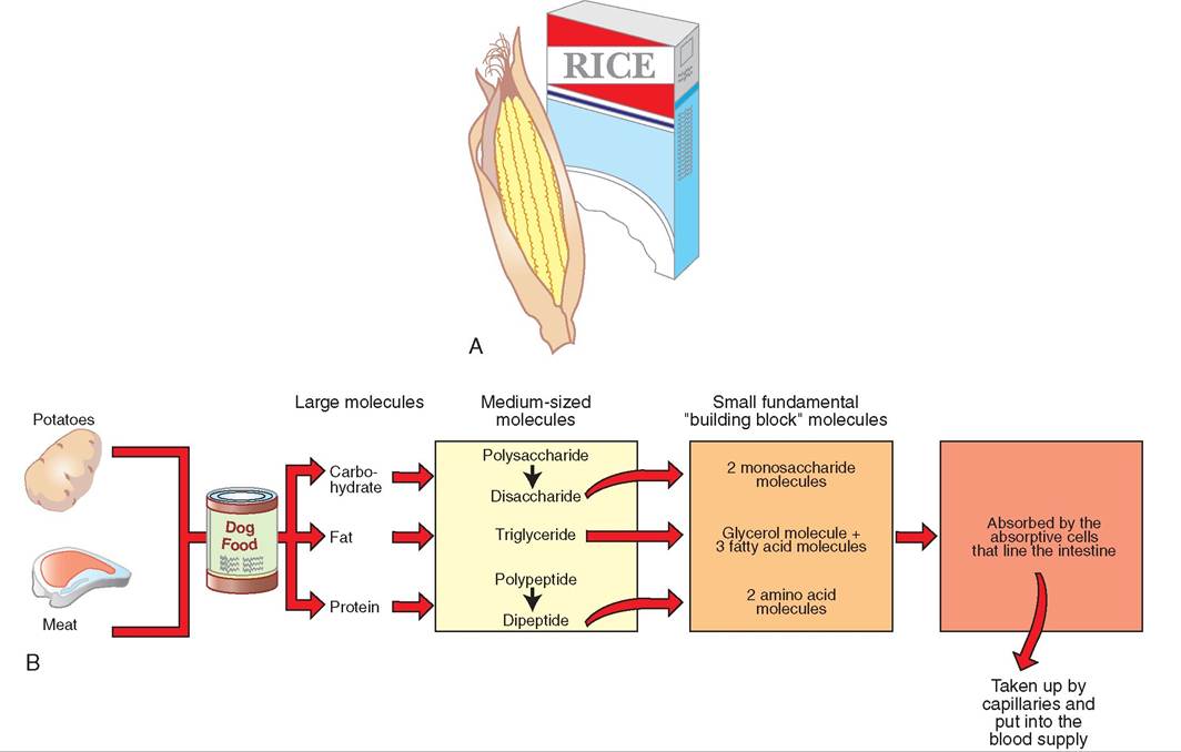
FIGURE 17-7 Building blocks. Large molecules that make up carbohydrates, fats, and proteins are broken down into fundamental building blocks to make new and different molecules. This process is the foundation of nutrition and cell metabolism.
STAGE TWO: THE CYTOSOL
The second stage of catabolism occurs inside the cell's cytosol. Amino acids, monosaccharides, fatty acids, and glycerol enter cells and are further catabolized in the cytoplasm through a process called anaerobic respiration. Because an means not, and aerobic means using oxygen, anaerobic respiration therefore is simply a metabolic process that does not use oxygen. An important molecular product of anaerobic respiration is acetyl coenzyme A (CoA), which carries a lot of the energy derived from food; acetyl CoA is transported through the cytosol to the mitochondria, where it is used in aerobic respiration, which is the third and final stage of catabolism.
STAGE THREE: THE MITOCHONDRIA
As its name implies, aerobic respiration requires oxygen; it involves the attachment of an inorganic phosphate group (PO4) to a molecule of adenosine diphosphate (ADP). The result is the formation of adenosine triphosphate (ATP), which is a critical molecule used in the cell to carry much of the energy that is derived from food; this energy is essential for making new molecules.
The mitochondrion can be viewed as an energy-producing factory; much of the energy that is derived from food is stored in the bonds of the ATP molecule. The catabolic pathways of proteins, carbohydrates, and fats are vital to the survival of the cell and therefore to the animal as a whole, because the pathways transfer the energy stored in the nutrient molecules to a small, transportable unit—ATP—which can then be used by the cell to grow, divide, heal, and maintain itself.
ANABOLIC METABOLISM
The cell uses energy in the form of ATP to manufacture a wide range of substances and to perform many vital functions. These constructive duties define anabolism, and the ATP that powers them is supplied by catabolism. Anabolic events are also called biosynthetic processes, because a biochemical substance is manufactured. Examples of anabolism are evident in many aspects of cellular life. When cells grow, for example, additional proteins are needed for the expanded cell membrane. The cytoskeleton and additional organelles are manufactured. Cell locomotion, the production and secretion of hormones, the movement of materials from one place to another inside the cell, active membrane processes, and preparation for cell division are all examples of cellular activities that require the production of biochemical substances via anabolism. With the exception of deoxyribonucleic acid (DNA), molecular substances are routinely broken down, and replacement molecules are manufactured continuously. This process is called metabolic turnover, and it represents the largest demand for protein and enzymes in the cell.
An important part of anabolism is dehydration synthesis, the effect of which is the opposite of hydrolysis. Monosaccharides, for example, are assembled—not broken down—to form chains of polysaccharides. A disaccharide, which is a chain of two monosaccharides, can be constructed as follows:
1 Monosaccharide +1 Monosaccharide → 1 Disaccharide + Water
Glycerol and fatty acid molecules are connected to form fat molecules, and proteins are created from chains of amino acids. All of these anabolic processes begin with dehydration synthesis.
Many of the anabolic cellular processes occur in the cytosol during stage two of metabolism. Here, the building blocks glucose, glycerol, fatty acids, and amino acids are incorporated into larger molecules, which may be used by the cell itself or exported and used elsewhere in the body. The liver in particular is an important organ because of its active anabolic efforts; it manufactures proteins such as albumin and clotting factors, and it manufactures and stores glycogen and fat.
Refer to Figure 17-9 for a summary of both catabolic and anabolic metabolism. Here, glycolysis is represented in blue, the Krebs cycle is in orange, and the electron transport chain appears in yellow.
TEST YOURSELF 17-3
1. What is cellular metabolism? Can you think of routine cellular processes that represent specific examples of cell metabolism?
2. Cellular metabolism is divided into two categories. What are they?
3. What is the first stage of cellular catabolism called? Is energy produced or consumed?
4. Is energy produced or consumed during an anabolic process? What is an example of anabolism?
5. What cellular process represents the largest demand for protein and enzymes?
CONTROL OF METABOLIC REACTIONS
Living cells are composed of and contain thousands of molecules. How is it possible for all of these molecules to interact in a structured and orderly fashion that maintains life for the cell? As discussed in the previous chapter, compartments are created within or on the surface of organelles, such as the mitochondria, endoplasmic reticulum, or ribosome. These “work areas” help to isolate molecules and allow chemical reactions to take place without interference. The organelles not only create separate environments for the different metabolic pathways, but also assist in storing the enzymes and cofactors required for various biochemical processes. However, grouping molecules together does not guarantee that they will react with one another. Molecules must collide with sufficient force to initiate a
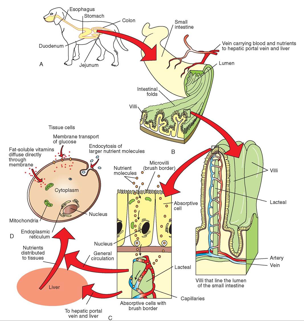
FIGURE 17-8 Summary of stage one of metabolism. A, Food is broken down in the stomach, and the digested stomach contents (chyme) pass into the small intestine. Here the food is broken down further by enzymes in the lumen of the intestine. B, In addition, the cell membranes in the microvilli brush border produce more enzymes, which further catabolize the nutrient molecules into their building-block units. Monosaccharides, fatty acids, glycerol, and amino acids are taken up by the absorptive cells. C, The nutrient molecules are then transported through the absorptive cell and are taken up by capillaries (amino acids and sugars) or lacteals (fatty acids) in the villi. Once in the blood or lymph, nutrient molecules are carried quickly away from the gastrointestinal tract. Most of the nutrient-rich blood flows directly to the liver via the hepatic portal vein. In the liver, the nutrient molecules are either incorporated into the production of larger molecules or are further broken down. From the liver, nutrients continue to travel through the general circulation to other body tissues. D, Cells throughout the body absorb the nutrient molecules from blood and lymph. Once in the cytoplasm, the molecules can undergo the second stage of metabolism.
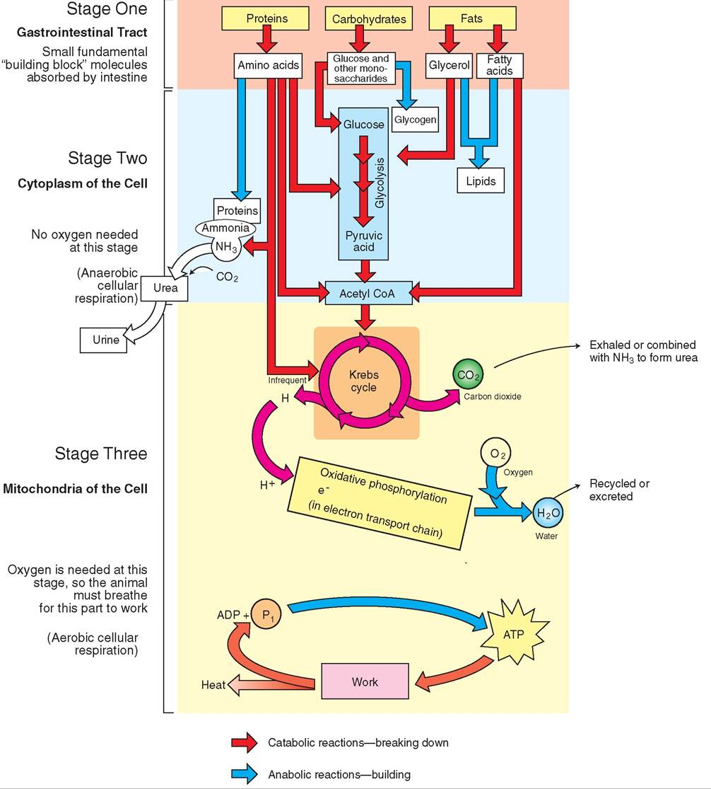
FIGURE 17-9 Summary of metabolism. Cell metabolism occurs in three stages. In the first stage, large nutrient molecules from food are degraded by enzymes and emulsifiers in the lumen of the stomach and intestine. Much smaller molecules are formed, which are absorbed through the intestine and transferred to blood and lymph. They are transported to various parts of the body. In the second stage, tissue cells absorb the small building-block molecules from the bloodstream. In the cytoplasm of the cell, the small molecules may be either used to make larger molecules (anabolism) or broken down into even smaller molecules, such as pyruvic acid and acetyl CoA (catabolism). Stage three is entirely catabolic: acetyl CoA is transported to the mitochondria of the cell, where it is broken down in the Krebs cycle. The hydrogen atoms that result are channeled into the electron transport chain, where ATP is produced via oxidative phosphorylation.
reaction. The moderate temperatures of the intracellular environment and the relative stability of intracellular organic molecules preclude forceful collisions. How, then, are molecular reactions initiated and controlled? The answer is simple: through the formation and use of specialized proteins called enzymes.
ENZYMES
Each enzyme reacts with a particular molecule called a substrate to produce a new molecule called a product. Because one enzyme reacts only with one substrate or combination of substrates, enzymatic reactions are considered highly specific. Hundreds of different biochemical reactions take place within the cell, so there must be hundreds of different enzymes available, each with the ability to locate and bond to its own special substrate. The DNA contained within the nucleus of the cell carries instructions for manufacturing all of the enzymes needed to drive these vital metabolic pathways. Metabolism therefore is a multi-enzyme sequence of events in which the product of one step is the substrate of the next. Some metabolic pathways include as many as 20 enzyme-driven steps. Although many of these pathways are linear, some are circular and all have branches leading into or out of them.
Most chemical reactions require an input of energy to get started. The energy needed to initiate a biochemical reaction is called the energy of activation. In the laboratory, the energy of activation is often supplied by heat. Heat causes molecules to become more active and to bang into one another. When molecules collide, existing bonds can be broken and new bonds can be formed. In this way, new substances are created. However, increased temperatures would be destructive to organelles and other structures within the cell. In addition, temperature changes would affect all of the chemical reactions at once and would not be selective for a particular type of reaction. The cell therefore relies on enzymes to initiate and control metabolic reactions.
An enzyme's ability to locate and bond to a particular substrate depends on the molecular shape of the enzyme. Enzymes are globular proteins that consist of one or more flexible polypeptide chains. These chains twist and coil to form a unique, three-dimensional shape that fits the special shape of the substrate molecules. When the enzyme and substrate bind together, they form a temporary enzymesubstrate complex.
The region of the enzyme molecule that binds to the substrate is called the active site. Like other portions of the enzyme, the active site is flexible and can exert pressure on the substrate that, in turn, weakens the bonds that keep the substrate molecule intact. In this way, one or more product molecules are formed, which subsequently separate from the enzyme. In biosynthetic reactions, enzymes unite smaller molecules to form one large molecule. These enzymes therefore require more than one active site, as shown in Figure 17-10. The enzyme is not altered by the reactions that it initiates and is able to move on to other substrate molecules to complete more of the same kind of reaction.
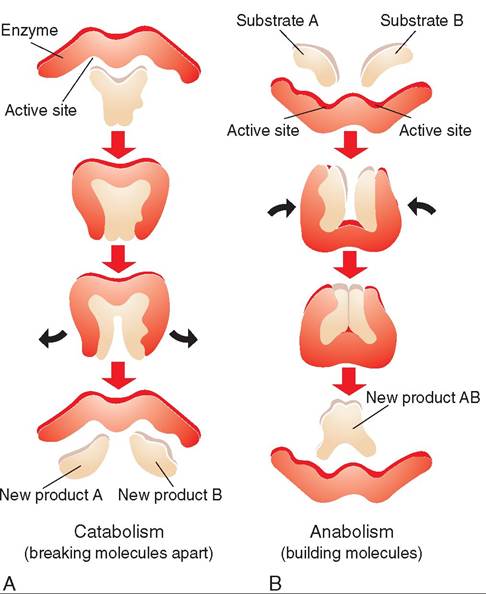
FIGURE 17-10 Enzyme activity. A, Enzymes are flexible molecules with dynamic active binding sites; they are able to bond to one substrate to produce two product molecules via catabolism. B, They can also bind to two substrates to form a single product molecule via anabolism.
Because enzymes bring reactant substrate molecules into close proximity and form temporary associations with them, they are able to speed up the rate of molecular reactions. For this reason, enzymes are also called catalysts, which are substances that speed up reactions by lowering the activation energy. Heat and various elements, ions, and chemicals can also act as catalysts. In the cell, enzymes speed up molecular reactions by a factor of a million or more. The rate at which a catalyzed reaction occurs is related to the amount of substrate and enzyme present. In general, an increase in the amount of an enzyme or a substrate causes an increase in the rate of the reaction. In addition, different enzymes have varying innate rates; that is, one type of enzyme might perform fewer reactions per second than another type of enzyme.
As mentioned earlier, each enzyme is specific for a particular reaction; for example, hexokinase is an enzyme that converts glucose to glucose-6-phosphate (G6P), and this is the only reaction that hexokinase initiates and controls. It binds with glucose and ATP to create G6P and ADP, and it repeats this reaction over and over again. The amount of enzymes needed by the cell to carry out thousands of reactions is therefore relatively small, because the enzymes are not used up during the reactions but are used multiple times to complete more reactions.
When studying cell metabolism, you can easily pick out the enzyme, because its name ends in the suffix -ase. In addition, the enzyme is usually named for the substrate on which it acts. For example, proteinases are enzymes that break down protein, lipase breaks down lipid, lactase breaks down lactose, and so on. The name of the enzyme may also indicate the kind of reaction that the enzyme initiates. For example, synthetases are enzymes that synthesize or make new substances, and transferases are enzymes that move one part of a molecule to another molecule. Phosphotransferase, for example, is an enzyme that transfers a phosphate group from one molecule to another molecule. For these reasons, enzymes may have long names, such as glucose-1-phosphate uridylyltransferase and phosphoglucomutase. Although they may be a linguistic challenge at times, the names of enzymes are very useful and may indicate the biochemical reactions that are taking place.
COENZYMES AND COFACTORS
Some enzymes are not able to complete a reaction without the assistance of another substance. Elements such as iron, zinc, or copper, for instance, are needed to complete the shape of a binding site. These nonprotein substances are called cofactors. Certain ions, such as magnesium, are cofactors in reactions that involve the transfer of a phosphate group and are therefore found in virtually all cells. The negative charges on the phosphate group are attracted to the positive charges on the magnesium cation. The attraction of these opposite charges helps to stabilize the enzyme-substrate complex. K+ and Ca2+ play a similar role in other molecular reactions.
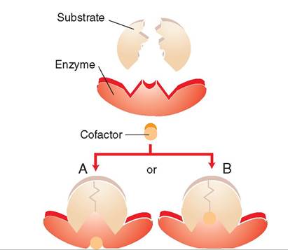
FIGURE 17-11 Cofactors and coenzymes assist in the activity of enzymes in one of two ways. A, A cofactor is added to the structure of a molecule in such a way that it indirectly changes the shape of the active site to enable a better fit between substrate and enzyme. B, A cofactor is part of the active site and plays a direct role in creating the correct shape of the active site.
Nonprotein organic substances may also act as cofactors. These substances are called coenzymes and often are, or are derived from, vitamins. They may be bound temporarily or permanently to the enzyme and are usually located near the active site (Figure 17-11). Nicotinamide adenine dinucleotide (NAD) and flavin adenine dinucleotide (FAD), for example, are commonly encountered cellular coenzymes and are critical in powering important cellular functions.
CLINICAL APPLICATION
Thermolabile Enzymes
Because enzymes are protein molecules with complicated three-dimensional structures, they are able to bend and move to accommodate bonding activities. Their shape is critical in enabling the enzyme to bond with the correct substrate. However, the shape of the binding site in some enzymes is affected by changes in the surrounding temperature. These enzymes are called thermolabile enzymes, because changes in temperature bring about changes in the structure and shape of the enzyme molecule.
For example, in the Siamese cat, a thermolabile enzyme that affects coat color functions well at cooler temperatures but is rendered nonfunctional at higher temperatures; therefore, a dark brown or black pigment is produced in the cooler regions of the body, such as the tips of the ears and the tail, face, and paws, but not in warmer areas, such as the torso, neck, and thighs. Himalayan rabbits also carry thermolabile enzymes that affect coat color. For example, Himalayan rabbits raised at temperatures of around 5° C are entirely black; those raised at moderate temperatures are white with black ears, tail, and paws; and those raised at temperatures above 35° C are completely white.
An Easy Diagnosis
It is not uncommon for cats or dogs to develop Horner’s syndrome, a condition caused by damage to a chain of nerves that extends from the chest, up the neck, and into the head and face. Possible causes include ear infections, particularly those caused secondarily by ear mites; trauma to the neck, often from misuse of choke chains; tumors in the chest; and trauma to the nerves in the armpit region. Usually only one side of the face is affected, and the most pronounced clinical signs include abnormal changes to the affected eye.
Horner’s syndrome also causes profound dilation of blood vessels in the muscles and skin of the face, an abnormality that is obvious in horses because they sweat profusely on the affected side. However, in domestic dogs and cats that do not sweat this change is usually not clinically apparent. In Siamese cats, the increased blood flow and the increased temperature in their faces change the shape of thermolabile enzymes, making them unable to function normally. The production of pigment in the hair is halted, and in cases of long-term Horner’s syndrome, the characteristic dark brown or black color of the Siamese face fades to a light tan or buff. Keep in mind that the appearance can look comical, because this condition usually affects only one side of the face. Fortunately, for most cats, Horner’s syndrome is a short-term disorder.
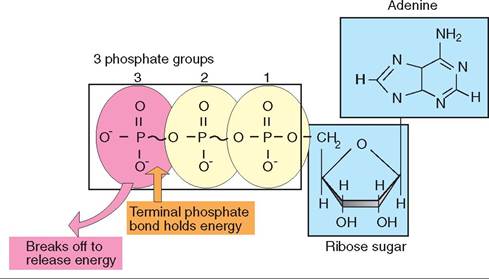
FIGURE 17-12 Conversion of ATP to ADP. When the terminal phosphate bond in an ATP molecule is broken, it releases stored energy that can be used by the cell to make other molecules. The remaining molecule, ADP, has two phosphate groups.
ENERGY FOR METABOLIC REACTIONS
Energy is required for the survival of all living cells. In mammals, energy is supplied to cells by the breakdown of nutrients, and energy is transferred to various energyholding molecules such as ATP, NADH, and FADH2. In these convenient molecular packages, energy can be stored for extended periods and easily transported to regions of the cell where energy is in demand. Energy is captured and stored in the formation of atomic bonds but is released when these bonds are broken. ATP, for example, stores energy in the terminal phosphate bond. When the phosphate group is broken off, energy is released and the molecule is transformed into ADP (Figure 17-12). The released energy is used during subsequent biochemical reactions.
The molecules that make up animal cells are adept at transforming themselves, from nutrient molecules to molecular energy stores and from substrate to product, cleaving off portions here and adding extensions there. Thus it is the molecule that stores, transforms, and uses energy in the cell through the formation and breakage of its molecular bonds.
TEST YOURSELF 17-4
1. Why are enzymatic reactions considered highly specific?
2. What is a substrate? What is a product?
3. Why is the total number of enzymes present in the body relatively low, when compared with the number of metabolic reactions?
4. What is the energy of activation in a biochemical reaction?
5. What is a catalyst? Why are enzymes considered catalysts?
6. How can you tell that a molecule is an enzyme? List three characteristics of enzymes.
7. What are some specific examples of cofactors?
8. How might vitamins play a role in enzyme-driven reactions?
9. How is energy stored in molecules? When is it released?
10. Give three examples of energy-holding molecules.
METABOLIC PATHWAYS
The goal of metabolizing nutrients derived from food is to generate the energy needed to keep the body going. The breakdown of carbohydrates, proteins, and fats each follows a different metabolic pathway, and each pathway generates different amounts of energy. As mentioned, some phases of the pathways occur in the cytoplasm and do not require oxygen (anaerobic), whereas other parts occur in the mitochondria and do require oxygen (aerobic). These pathways represent a complex series of biochemical steps that must occur in a particular sequence, and each step involves an enzyme specific for that particular step.
CARBOHYDRATE METABOLISM
Carbohydrate metabolism occurs in virtually all living cells and includes all of the catabolic and anabolic processes involving carbohydrates. In mammals, carbohydrates may be supplied either through the diet or from the breakdown of glycogen, glycerol, or, in the case of ruminants, propionate stored in the liver. In most mammals, carbohydrates provide well over half the energy required to fuel metabolic functions, such as absorption, secretion, excretion, mechanical work, growth, and repair. Refer to Figure 17-13 for an overview of carbohydrate metabolism.
Glucose is the primary carbohydrate found in blood. It is absorbed by all cells and is used to produce energy in the form of ATP. Certain types of cell, such as red blood cells and brain cells, are exquisitely sensitive to fluctuations in blood glucose levels, because they cannot derive energy from other sources. Other cells, however, such as skeletal muscle cells, can derive energy from ketones and fatty acids and therefore are not as affected by falls in blood glucose supplies. Nevertheless, when glucose is urgently needed and in short supply, the body is able to provide it quickly by mobilizing stores of glycogenic molecules in the liver.
Glucose may enter a cell either by facilitated diffusion or via active transport. Once inside the cell, glucose is further broken down to form pyruvate, or pyruvic acid. This process is called glycolysis (lysis means “break apart”) and occurs in the cytoplasm. Because glycolysis does not require oxygen, it is referred to in some texts as anaerobic respiration.
After pyruvate is formed from glycolysis, one of several pathways may follow. Most of the time, cells are fed with good quantities of oxygen via circulating red blood cells. In an oxygen-rich environment, pyruvate is transported from the cytosol to the mitochondria, where it undergoes further degradation as part of aerobic respiration. The Krebs cycle and the electron transport chain are two important components of aerobic respiration. Anaerobic glycolysis and aerobic respiration (the Krebs cycle and the electron transport chain) are collectively known as cellular respiration.
Some cells, such as skeletal muscle cells, may take a different pathway than aerobic respiration if their oxygen supply has become depleted. During vigorous exercise, for example, skeletal muscle cells rapidly consume oxygen. Once the oxygen is depleted, the cells take a different metabolic
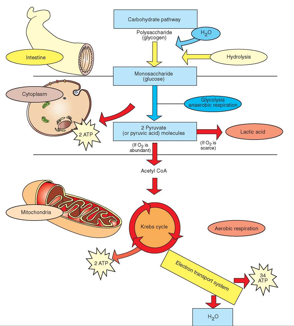
FIGURE 17-13 Overview of carbohydrate metabolism.
pathway and convert pyruvate to lactic acid. We have all experienced the effects of lactic acid production in our own muscles at one time or another, particularly after performing strenuous work that we do not normally do. Lactic acid builds up in muscles during and after exercise and causes a stiff feeling the next day. In horses, this stiffness can be severe and can cause a condition called myositis or “tying up.” ft is not uncommon for horses ridden for pleasure to “tie up” on Mondays, because many owners only ride them on weekends. For this reason, myositis in horses is sometimes called “Monday morning syndrome.”
The biochemical pathways of cellular respiration have buedeiendst extensively by biochemists and therefore are discussed in detail. However, keep in mind that the principles involved in their function and regulation are common to all pathways of cell metabolism.
ANAEROBIC RESPIRATION: GLYCOLYSIS. Glycolysis, or “sugar splitting,” occurs in all animals and includes 10 biochemical steps, each of which relies on a specific enzyme found in the cytoplasm. As mentioned, it does not require oxygen. For every molecule of glucose metabolized, the cell must use two molecules of ATP and two molecules of NAD to initiate the process. Two molecules of pyruvic acid, four molecules of ATP, and two molecules of NADH are produced. Thus, the net energy yield from glycolysis is two molecules of NADH and two molecules of ATP.
The chemical reactions of glycolysis can be summarized in three phases. Refer to Figure 17-14. In the initial phase, energy— one ATP molecule—is invested in the process of adding a phosphate molecule to glucose, which results in the formation of glucose-6-phosphate (G6P). This process is called phosphorylation, and although it costs the cell energy, it is important in preventing the glucose molecule from leaving the cell; this is because phosphorylated glucose cannot cross the cell membrane. Phosphorylation also prepares the glucose molecule for further manipulations, either in a catabolic or anabolic pathway. Subsequently, G6P is rearranged to form fructose-6- phosphate. From here, phosphorylation occurs again, using one more ATP molecule to form fructose-1,6-diphosphate.
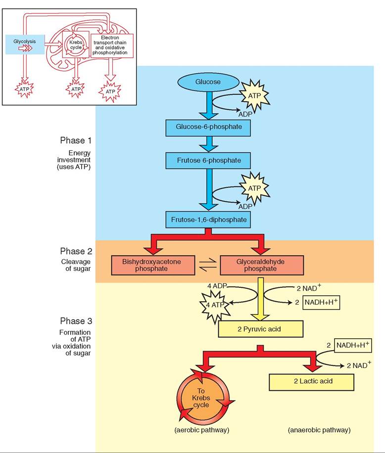
FIGURE 1 7-1 4 Glycolysis. In glycolysis, a six-carbon glucose molecule is split into a pair of three-carbon molecules of pyruvic acid (pyruvate). This process includes 10 steps and produces six energy-holding molecules: two molecules of NADH and four molecules of ATP. Given that two molecules of ATP were used in phase one, the net production of energy is two molecules of NADH and two of ATP.
During phase two, fructose-1,6-diphosphate is cleaved. A pair of three-carbon molecules is formed, but they can be one of two reversible isomers: bishydroxyacetone phosphate and glyceraldehyde phosphate. The molecules change back and forth between these two forms.
Phase three includes six steps, which result in the formation of four ATP molecules and two molecules of pyruvic acid. The net energy production by glycolysis, however, is two ATP, because two ATP molecules were used in phase one to “prime the pathway.”
Although its contribution of energy to animal cells is important, glycolysis is not the cell's primary source of ATP. A great deal of potential energy from the original glucose molecule still remains in pyruvic acid. This molecule can be further broken down in the mitochondria to form larger quantities of energy for the cell through an aerobic sequence of biochemical events.
AEROBIC RESPIRATION. When animals breathe in air, they are performing a vital function. Everyone knows that when animals stop breathing, they die. Like humans, animals can only survive for a short time without oxygen before the cells in the body—particularly the brain—start to die, because oxygen is required for the production of energy (ATP) by the cell. Many cell types cannot live without ATP for very long. Blood carries oxygen to cells throughout our bodies. After blood releases oxygen molecules to the cells, it picks up carbon dioxide molecules that have been discharged by the cell. These molecules are then circulated back to our lungs, where they are exhaled. We exhale carbon dioxide because our cells give off carbon dioxide in the process of making ATP.
Aerobic respiration occurs in the mitochondria in two stages: the Krebs cycle, also known as the citric acid cycle, and the electron transport chain. The mitochondrion is uniquely suited for the production of ATP; it is the powerhouse of the cell.
The smooth outer membrane of the mitochondrion is permeable to most small molecules, but the undulating inner membrane is more selective and permits the passage of only certain molecules, such as pyruvic acid and ATP. The folds of the inner membrane are called cristae, and they contain within their walls some of the enzymes and cofactors needed in the Krebs cycle. Other enzymes are found in the mitochondrial matrix, which is the thick solution that bathes the cristae within the mitochondria. Enzymes in the mitochondria responsible for aerobic respiration have positioned themselves on the cristae in the order in which they are needed for oxidation. This assembly-line approach is an efficient way to carry out molecular alterations and minimizes the potential for errors.
The inner membrane of the mitochondrion houses a series of cytochrome molecules, which make up the electron transport system. As electrons are passed from one cytochrome molecule to another, energy is released that is used to transport protons from the mitochondrial matrix across the inner membrane to the intermembrane space. Because protons are positive, this establishes a positive charge on the outside of the inner membrane relative to the matrix side. The electrical gradient that is established is a form of stored energy. Energy is released when protons rush back into mitochondrial matrix; this release of energy is the ultimate power source that converts ADP into ATP. Refer to Figure 17-15 for a summary of cellular respiration.
KREBS CYCLE. Pyruvic acid enters the mitochondria from the cytoplasm by passing through both the outer and inner membranes. Before it enters the Krebs cycle, it is transformed from a three-carbon molecule of pyruvic acid into a two-carbon acetyl group; this, in turn, binds to a compound known as coenzyme A to form acetyl CoA, the molecule that represents the link between glycolysis and the Krebs cycle. During this process, one molecule of carbon dioxide and one of NADH are also generated for every molecule of pyruvic acid.
Acetyl CoA enters the Krebs cycle and reacts with oxaloacetic acid to form citric acid; therefore, the Krebs cycle is also known as the citric acid cycle (Figure 17-16). As citric acid is produced, coenzyme A is released and is used repeatedly to make acetyl CoA. After seven additional steps, citric acid is converted back to oxaloacetic acid, and the entire process is repeated. Each turn of the Krebs cycle generates energy in the form of one ATP, one FADH2, and three NADH molecules. For every molecule of glucose, the Krebs cycle can run twice and produce two molecules of ATP, two of FADH2, and six of NADH. Carbon dioxide, which is formed as a by-product of respiration, diffuses out of the cell and into the bloodstream as waste and is carried to the lungs, where it is exhaled.
ELECTRON TRANSPORT SYSTEM. The final stage of cellular respiration, which produces the majority of ATP for the cell, occurs in the inner wall of the mitochondrion and is known as the electron transport system. NAD and FAD molecules released from the conversion of pyruvic acid to acetyl CoA, glycolysis, and the Krebs cycle bind to hydrogen atoms to form NADH and FADH2. The electrons in NADH and FADH2 are at a very high energy level, because they hold most of the energy once held by the original glucose molecule. In a sense, they are at the top of an energy “hill” and are carried down a chain of electron carrier molecules, collectively known as the cytochromes.
Each cytochrome molecule contains a central core of iron that accepts electrons and then releases them at a lower energy level. At each step, large amounts of free energy are released and used to pump protons from the mitochondrial matrix through the inner membrane to the intermembra- nous space, which is the space between the inner and outer membranes of the mitochondrion. This establishes a difference in electrical charge, because more positive hydrogen ions are pumped to the outside than remain on the inside of the inner mitochondrial membrane. Thus, the outside has a positive charge relative to the matrix side of the membrane. The result is the formation of potential energy that is
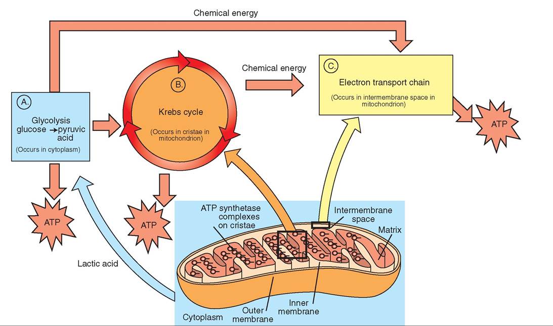
FIGURE 17-15 Summary of cellular respiration. A, Glycolysis occurs in the cytoplasm, where glucose is converted to two molecules of pyruvic acid. A net production of two ATP molecules occurs. Other chemical energy in the form of two NADH molecules is produced, which is converted to ATP in the electron transport system. B, Krebs cycle. The inner membrane of the mitochondrion has infoldings and outpouchings, called cristae, which form separate “work spaces” for the Krebs cycle. Many of the enzymes involved in ATP production, such as ATP synthetase, are built into the wall of the inner membrane. These molecules form an assembly line, which improves efficiency and helps to prevent errors. The matrix that fills the internal spaces of the cristae is rich with electron carriers, phosphates, coenzymes, and other solutes necessary for ATP production. C, The electron transport chain. The inner membrane of the mitochondrion houses a series of cytochrome molecules, which make up the electron transport system. As electrons are passed from one cytochrome molecule to another, energy is released that is used to transport protons from the mitochondrial matrix across the inner membrane to the intermembrane space. Because protons are positive, this establishes a positive charge on the outside of the inner membrane relative to the matrix side. The electrical gradient that is established is a form of stored, potential energy. Energy is released when protons rush back into the mitochondrial matrix. This release of energy is the ultimate power source that converts ADP into ATP.
available when the protons flow back to the matrix side of the inner mitochondrial membrane (Figure 17-17). The energy released during this downhill pathway is captured in the formation of ATP from ADP. At the end of the electron transport chain, oxygen accepts the low-energy electrons, joins with hydrogen ions, and forms water (H2O). Thus oxygen is the final acceptor of the electrons.
SUMMARY OF ATP SYNTHESIS. The biochemistry of glucose catabolism is complicated, so let us take a step back and look at the overall picture of what the cell is doing.
Through the breakdown of glucose, the cell produces units of usable energy called ATP. For every molecule of glucose metabolized by an animal, a maximum of 38 ATP molecules are formed, although in some cells only 36 ATP molecules are formed. Two ATP molecules are made in the cytoplasm, and the rest are made in the mitochondria.
In the cytoplasm, glycolysis gives rise to two molecules of ATP directly and two molecules of NADH. The NADH molecules are shuttled to the electron transport system in the mitochondria, where they produce six ATP molecules (three for every molecule of NADH). Thus, the total gain from glycolysis is eight ATP molecules. However, in certain cells, such as brain and skeletal muscle cells, there is an energy cost for transporting NADH into the mitochondria. In these cells, each NADH molecule produces only two ATP, rather than three; therefore, in some cells, glycolysis produces only six ATP, not eight.
Inside the mitochondria, pyruvic acid is converted to acetyl CoA before it enters the Krebs cycle. During this process, two molecules of NADH are formed, one from each of the two molecules of pyruvic acid. Each of these NADH molecules forms three molecules of ATP, so a total of six ATP molecules are derived from the conversion of pyruvic acid to acetyl CoA.
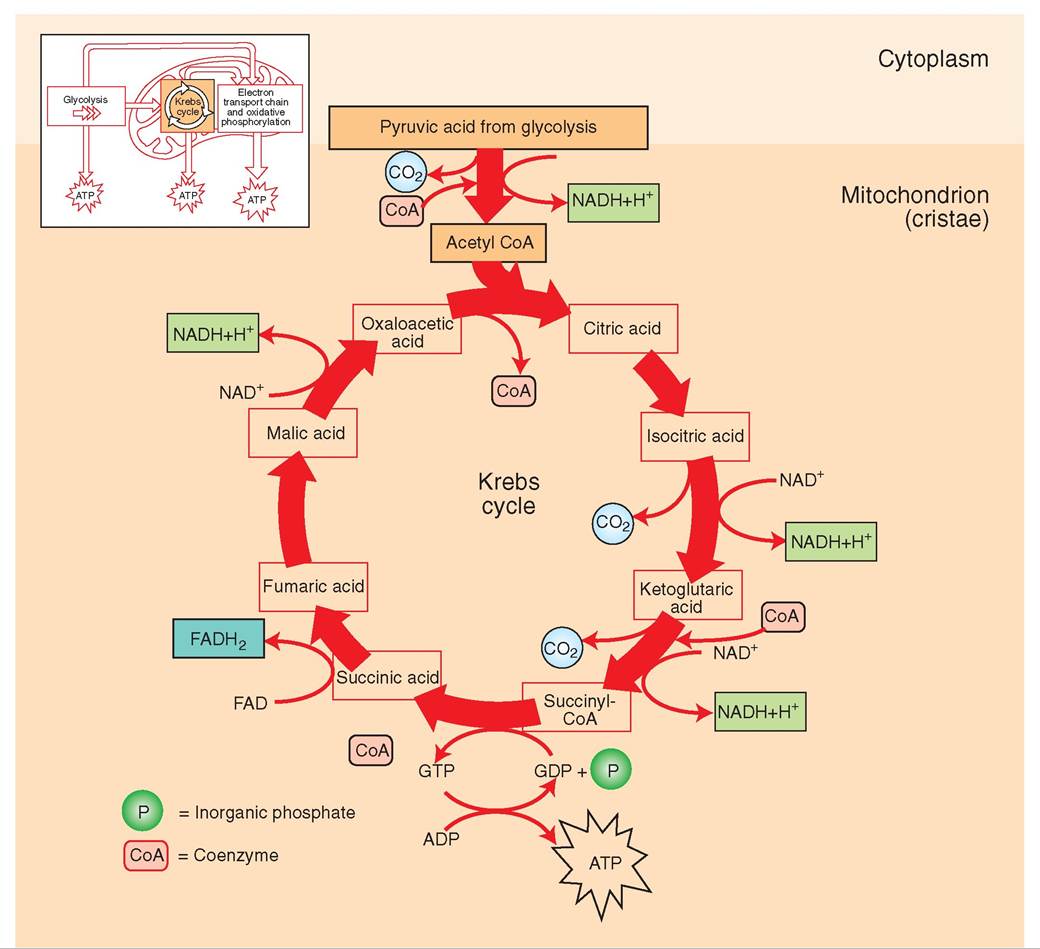
FIGURE 17-16 Simplified Krebs cycle. Pyruvic acid is converted to acetyl CoA, which in turn enters the Krebs cycle in the matrix of the mitochondrion. Carbon and oxygen are released from the cycle as waste and are ultimately exhaled by the animal in the form of carbon dioxide. For every molecule of glucose, the Krebs cycle can run twice and in doing so produces two molecules of ATP, two of FADH2, and six of NADH. From these energy-carrying molecules, hydrogen atoms are released and used in the electron transport system to produce ATP.
Also in the mitochondria, the Krebs cycle yields two molecules of ATP, two of FADH2, and six of NADH. The electron transport system converts the NADH and FADH2 molecules into 22 ATP; so the Krebs cycle produces a total of 24 ATP.
Table 17-2 provides a summary of ATP production in the cell. Notice that all but two of the 38 ATP molecules are produced in the mitochondria and that all but four ATP result from the passage down the electron transport chain of electrons carried by NADH and FADH2. Once formed, the energy-carrying ATP molecules are exported across the mitochondrial membrane, and a molecule of ADP is brought into the mitochondrion for each ATP exported.
LIPID METABOLISM
Lipids are molecules composed of carbon, hydrogen, and oxygen, which are insoluble in water but dissolve easily in other lipids or organic solvents. One common type of lipid is the triglyceride or neutral fat.
Triglycerides contain a higher number of carbon-hydrogen bonds than other nutrient molecules and therefore contain more chemical energy than either carbohydrates or proteins,
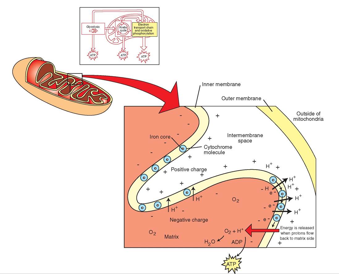
FIGURE 17-17 The electron transport system. Cytochrome molecules are located in the inner membrane of the mitochondrion. Each cytochrome molecule contains a central iron core that accepts electrons and then releases them to another cytochrome molecule at a lower energy level. At each step, a large amount of free energy is used to pump protons from the matrix through the inner membrane to the intermembrane space. This establishes an electrical charge difference (potential energy) between the matrix and the intermembrane space, because more protons are pumped to the outside than remain on the inside of the inner membrane. Energy is released when the protons flow back to the matrix side. This energy is used to convert ADP to ATP. The low-energy electrons left at the end of the cytochrome chain and the hydrogen atoms in the matrix bind to oxygen and form water (H2O).
because energy is stored in the bonds between atoms. Gram for gram, fat contains over twice as much chemicaI energy as carbohydrate and stores six times as much energy as glycogen. This fact is important, particularly in birds, because it illustrates Aat fat concentrates energy in a form that is relatively lightweight—an essential requirement for flight. Herbivores do nm ∞M∏ιe the large amount of fat that is prevalent in the diet of meat eaters. For them, the formation of fat is rdiemriavreidly p from the conversion of carbohydrates in
excess of what can be stored as glycogen.
vTehre li is the primary controller of lipid metabolism. Triglycerides can be removed from circulating blood by tvheer li and structurally altered. For example, lipids may be broken into smaller fragments that enable them to enter ltyhceoglytic pathway to form pyruvic acid, or they may be fed directly into the Krebs cycle. This process is called lipolysis.
Triglycerides, specifically, are hydrolyzed into one molecule of glycerol and three fatty acid chains. The glycerol head is further catabolized in the cytoplasm to acetyl CoA and subsequently enters the Krebs cycle; or it may be used to lsuycnothse.size g The fatty acid tails, on the other hand,
are long ⅛ns of 18 carbon atoms or more and are handled bnyzyemes found in the mitochondria.
| TABLE 17-2 Summary of Energy Production from One Glucose Molecule | |||
| CYTOSOL | |||
| Anaerobic Cellular Respiration | (Glycolysis—no oxygen needed) | ||
| PRODUCTS | ATP PRODUCTION FROM ELECTRON TRANSPORT SYSTEM (OXIDATIVE PHOSPHORYLATION) | TOTAL PRODUCTION | |
| 2 molecules of pyruvic acid (goes to mitochondria) 2 ATP 2 NADH | 6 ATP | 2 ATP 6 ATP | |
| MITOCHONDRIA | |||
| Aerobic Respiration (Oxygen Required from Inhalation) | |||
| Intermediate stage: conversion of pyruvic acid (from cytosol) to acetyl CoA | 2 NADH CO2 (exhaled) | 6 ATP | 6 ATP |
| Krebs cycle (citric acid cycle) | Two turn of Krebs cycle produce: 2 ATP 2 FADH2 6 NADH CO2 (exhaled) | 4 ATP 18 ATP | 2 ATP 4 ATP 18 ATP |
| FINAL PRODUCTION | 38 ATP | ||
Through a process known as beta oxidation, each fatty acid chain is broken into multiple two-carbon fragments. Some of these fragments are converted to acetyl CoA, whereas others are converted into compounds called ketone bodies. Later, the ketones can be converted to acetyl CoA, which can be further catabolized in the Krebs cycle to form ATP (Figure 17-18). The energy gain from the complete oxidation of an 18-carbon fatty acid chain is substantial— about 148 ATP, which is about 4 times more energy than that generated from the catabolism of one molecule of glucose. Because fat, for the most part, is not soluble in water and is therefore difficult to mobilize, it serves primarily as a reserve energy source, rather than a supply to meet immediate energy demands.
If not immediately used to produce energy, glycerol and fatty acid chains may be reconstructed into lipids and stored in fatty tissue. This is nature's way of protecting the health of animals and humans when food supplies become low.
PROTEIN METABOLISM
Proteins are found in great abundance in animals. The body of a cow, for instance, may contain several thousand different proteins, each with a unique function and a unique structure to fit its function. Proteins include structural proteins, such as hair, microtubules, and collagen; regulatory proteins, such as insulin and other hormones; contractile proteins, such as actin and myosin in muscle tissue; transport proteins, such as hemoglobin and myoglobin; storage proteins, such as those found in egg whites; protective proteins, such as antibodies; membrane proteins, such as cell receptors and membrane transport molecules; and osmoregulators, such as albumin and enzymes. Clearly
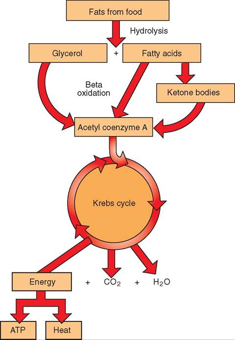
FIGURE 17-18 Triglyceride metabolism. Triglycerides from food are digested into glycerol and fatty acids. These can be further broken down to form acetyl CoA, which enters the Krebs cycle to form ATP. Excess nutritional triglycerides not immediately catabolized for energy are stored in tissue as fat.
the diversity of proteins and the functions they perform are extensive.
PROTEIN CATABOLISM. In addition to the multiple roles that proteins play in the structural and functional maintenance of the body, proteins may also be catabolized and used to make energy (ATP) (Figure 17-19). Although amino acid catabolism occurs in most tissues, it is of particular importance in the intestinal mucosa, kidney, brain, liver, and skeletal muscle, where the amino acid molecules may undergo one of two processes: deamination or transamination. These processes occur in the mitochondria (Figure 17-20).
In transamination, the amine group (-NH2) is transferred to another carbon chain to form a different amino acid. These newly constructed amino acids then diffuse across the mitochondrial membranes into the surrounding cytosol, where they become the building blocks for other proteins.
In deamination, the amine is removed from the carbon chain and becomes an ammonia molecule. Because ammonia is toxic, even in small quantities, most deamination reactions occur in the liver, where specialized enzymes are present to convert ammonia to urea, a nontoxic water-soluble molecule that is excreted in urine. After the amine has been removed by deamination, the remaining carbon chain may enter the Krebs cycle at one of several locations, resulting in the formation of ATP. Some of the pathways, for example, enter the Krebs cycle from the formation of acetyl CoA; others enter much later. As a result, deamination may give rise to a variable amount of ATP, depending on where the cycle was entered. If energy is not immediately required, the carbon chains may not enter the Krebs cycle but instead will be converted to glucose or fat.
PROTEIN ANABOLISM. The manufacture of new protein from nutrient building blocks is an important function for cells. The genetic material housed in the nucleus contains the instructions for the manufacture of thousands of different types of protein. Refer to Chapter 4 for a detailed description of the processes by which proteins are made.
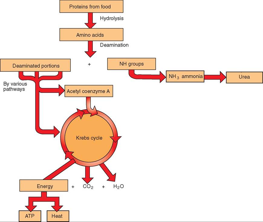
FIGURE 17-19 Overview of protein metabolism. Protein from food is digested into amino acids, but before they are completely catabolized, the amine group (NH2) is removed and converted to ammonia, which is later excreted in urine. The remainder of the deaminated portion is then converted to acetyl CoA and subsequently is used to generate ATP.
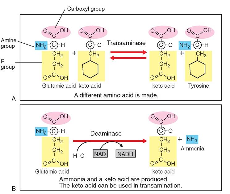
FIGURE 1 7-20 Transamination and deamination. A, During transamination, an amine group of an amino acid is transferred to auother molecule by the enzyme transaminase, and a different amino acid is subsequently formed. When stripped of its amine group, an amino acid becomes a keto acid. B, During deamination, an amine group is removed from an amino acid by the enzyme deaminase. The amino acid then becomes a keto acid, which can be reused in transamination reactions. The amine oroup is converted to ammonia, a toxin; the liver subsequently converts ammonia to a nontoxic molecule called urea.
∕j clinical application
Liver and Kidney Failure
During protein catabolism, ammonia is produced. Because ammonia is toxic, it is converted by the liver to a nontoxic molecule called urea.
Protein → (liver) → ammonia → (kidney) → urea → urine
Urea is a type of nitrogenous waste; it is a compound that contains nitrogen and is excreted. Nitrogenous waste is normally pulled out of the bloodstream by the kidneys and put into ume. When the kidney becomes diseased, however, it is sometimes unable to excrete urea effectively. The urea there- fuoilrdesb up in the bloodstream, causing a condition called uremia. Urerma can be detected by a blood test that evaluates the b Iood urea nitrogen (BUN) level of the patient. Veterinarians look for this in the blood chemistry profiles of sick animals. Sirm'larly, if the BUN is abnormally low, the veteri- hntarian mig suspect liver disease, because the liver is responsible fon converting ammonia to urea. If the liver is not functioning normally, it cannot carry out this conversion. Can you guess what might happen to the ammonia level in an animal Wh liver disease?
TESTYOURSEtf 17-5
1. W hot is themost Commoncarbohydrate found in blood?
2. Whece doescarmohoduate metabolismbegie insonsuminant animals?
3. Whot pertodcarbehyOfate metaColism oceurs tn tie cytoplasm?
4. Usdec what mo nOittonntu Iautieac iut fetmcd Cr muscle cells?
e. Howmadebiochenimolstepu are involveh in g∣o∞∣asis? Doos glycolysim res∣uire oxygen?
6. Cellglot tes3itotios is composed of whot two 30ttsU
7. How deesthommmgrage soiuctere mitocirosdri os aseint mtss proeegsoi cg∣lulorreupiration?
8. Vdhot roleOoesoxygene∣ayintSe elsstron franeeor! system?
9. Whot ^0^03^ PooosUoryIation?
10. Whot ietee mexim∣∣mrn∣ num beo cCACR moleculmdthatsac f^ehr naedfrodtSecatabaIIem etoe emo∣gculeof glucose? 1 1. Whot erctumsmommdtios cndCuamination?