The Urinary System
Angela Beal
OUTLINE
| INTRODUCTION, 446 | URETERS, 459 |
| PARTS OF THE URINARY SYSTEM, 448 | Anatomy, 459 |
| KIDNEYS, 448 | Function, 459 |
| Function, 448 | URINARY BLADDER, 460 |
| Location, 449 | Anatomy, 460 |
| Gross Anatomy, 450 | Function, 460 |
| Microscopic Anatomy, 450 | Control of Urination, 460 |
| Nerve Supply, 452 | URETHRA, 461 |
| Blood Supply, 452 | Anatomy, 461 |
| Mechanisms of Renal Action, 453 | Function, 461 |
| Urine Volume Regulation, 454 | |
| Regulation of Blood Pressure, 458 | |
| LEARNING OBJECTIVES |
When you have completed this chapter you will be able to:
1.
List and describe the functions of the kidneys.2. Describe the gross and microscopic anatomy of the kidneys.
3. List the main blood vessels associated with the kidneys.
4. Define the terms glomerular filtration, renal threshold, polyuria, polydipsia, urolithiasis, and uremia.
5. Describe the production of urine.
6. Describe the mechanisms that affect urine volume.
7. Explain the renal mechanisms that help regulate blood pressure.
8. Describe the structures and functions of the ureters and urinary bladder.
9. Describe the processes involved in urination.
10. Describe the structure and functions of the urethra.
VOCABULARY FUNDAMENTALS
Afferent glomerular arteriole a-for-ehnt glo-mear-u-lahr ahr-teer-e-ol
Aldosterone ahl-dohs-tuhr-on
Antidiuretic hormone (ADH) ahn-te-dι-u-reht-ihck hohr-mon
Anuria ahn-yar-e-ah
Azotemia a-zo-te-me-ah
Bowman’s capsule bo-mahnz kahp-sehl
Capsular space kahp-sehl-ahr spas
Collecting duct kah-lehck-tihng duhckt
Cortex kohr-tehx
Distal convoluted tubule (DCT) dihs-tahl kohn-vah-lu- tehd too-bul
Diuresis dι-u-re-sihs
Efferent glomerular arteriole e-far-ehnt glo-mear-u-lahr ahr-teer-e-ol
Erythropoietin e-rihth-ro-poy-eh-tihn
Fenestration fehn-ih-stra-shuhn
Glomerular filtrate glo-mear-u-lahr fihl-trat
Glomerular filtration rate (GFR) glo-mear-u-lahr fihl-tra-shuhn rat
Glomerulus glo-mear-u-luhs
Glycosuria glι-kos-yar-e-ah
Hilus hι-luhs
Homeostasis ho-me-o-sta-sihs
Loop of Henle loop of hehn-le
Medulla meh-duhl-uh
Micturition mihck-tuhr-ihsh-shuhn
Nephron nehf-rohn
Oliguria awl-ihg-yar-e-ah
Osmotic diuresis ohs-moh-tihck dι-u-re-sihs
Peritubular capillary peor-ih-too-bul-ahr kahp-eh-leor-e
Podocyte pod-o-sιt
Polydipsia pohl-e-dihp-se-ah
Polyuria pohl-e-yar-e-ah
Postrenal uremia post-re-nuhl yor-e-me-ah
Prerenal uremia pre-re-nuhl yar-e-me-ah
Prostaglandin prohs-tuh-glahn-duhn
Proximal convoluted tubule (PCT) prohck-sih-mahl kohn-vah-lu-tehd too-bul
Reabsorption re-ahb-sohrp-shuhn
Renal artery re-nuhl ahr-tar-e
Renal corpuscle re-nuhl kohr-puhs-ehl
Renal pelvis re-nuhl pehl-vihs
Renal uremia re-nuhl yar-e-me-ah
Renal vein re-nuhl van
Renin reh-nihn
Renin-angiotensin-aldosterone system reh-nihn ahn-je-o-tehn-sihn ahl-dohs-tuhr-on sihs-tehm
Retroperitoneal reh-tro-pear-ih-to-ne-ahl
Secretion seh-kre-shuhn
Glomerular capillary thuh glo-mear-u-lahr kahp-eh-lear-e
Trigone trι-gon
Tubular filtrate too-bul-ahr fihl-trat
Uremia yar-e-me-ah
Uresis yar-e-sihs
Ureter yar-eh-tar
Urinary calculi yar-ih-near-e kahl-kyoo-lι
Urinary stone yar-ih-near-e ston
Urination yar-ih-na-shuhn
Urolith yar-o-lihth
INTRODUCTION
An animal's body is a finely tuned machine that relies on numerous metabolic reactions to keep it alive and healthy.
These chemical reactions are of great benefit to the body but also result in the production of many by-products, some of which are useful to the body and are recycled. Other by-products, on the other hand, are of no further use to the body and can be harmful if allowed to accumulate. These potentially harmful substances are called waste products and must be eliminated from the body (Box 18-1). Some examples of metabolic waste products are:• Carbon dioxide and water from carbohydrate and fat metabolism
• Nitrogenous wastes, primarily urea, from protein metabolism
• Bile salts and pigments from red blood cell breakdown
• Various salts from tissue breakdown and excessive consumption
The body has several routes by which waste products are eliminated:
• The respiratory system removes carbon dioxide and water vapor.
• The sweat glands eliminate water, salts, and a small amount of urea.
• The digestive system removes bile salts and bile pigments.
• The urinary system removes urea, salts, water, and other soluble waste products.
The urinary system is the single most important route of waste-product removal in the body. It removes nearly all the soluble waste from blood and transports it out of the body. The urinary system is also a major route for the elimination of excess water from the body.
BOX 18-1
Summary of Waste Removal
Excretion
The urinary system's chief function is to regulate the volume and composition of body fluids and excrete unwanted material, but it is not the only system in the body that is able to excrete unnneded substances. The table below compares the excretory functions of several systems. Although all of these systems contribute to the body's effort to remove wastes, only the urinary system can finely adjust the water and electrolyte balance to the degree required for normal homeostasis of body fluids.
| SYSTEM | ORGAN | EXCRETION |
| Urinary | Kidney | Nitrogen compounds Toxins Water Electrolytes |
| Integumentary | Skin-sweat glands | Nitrogen compounds Electrolytes Water |
| Respiratory | Lung | Carbon dioxide Water |
| Digestive | Intestine | Digestive wastes Bile pigments Salts of heavy metals |
From Patton KT, Thibodeau GA: Anatomy & physiology, ed 8, St Louis, 2013, Mosby.
∕ j clinical application
Urinalysis
A urinalysis (“UA”) is the laboratory analysis of a urine sample.
It can be performed by a commercial laboratory or by veterinary pefsotιtιel within a veterinary clinic. Performing a complete urinalysis involves three major steps:1. A gross examination of the physical properties of the sample.
2. A chemical analysis of substances dissolved in the urine.
3. A microscopic examination of the solid components in the urine.
Before beginning a urinalysis, a urine sample must be obtained from the patient. There are several options for urine cdoelallelcy,tion. I the sample should be collected in a sterile manner so that any bacteria observed in the sample are derived from the urinary tract and not from contamination of the sample during collection. Sterile collection methods include urinary catheterization or cystocentesis. To perform cystocen- tesis (A), a sterile needle is inserted through the skin of the lower abdomen and into the bladder. Urine is drawn directly lfarodmde rthe b through the needle and into the sterile syringe. Nonsterile methods of sample collection include “catching” the urine in a container as it is voided (free catch method) and collecting the sample in a container as the oblmadpdresrsiesdc manually.
After collection, the physical properties of the urine are evaluated. The volume, color, odor, transparency, and specific gravity of the sample are observed and recorded (B). Nomal hutrine is lig yellow to amber in color, depending on the famount o water it contains. The odor of normal urine varies ebceitews.een sp The urine of most animals is clear or transparent; however, equine urine normally contains mucus, which gives it a cloudy appearance. Specific gravity (SG) is a reflection of the concentration of the urine and is measured using an instrument called a refractometer. Normal specific gravity measurements vary among species.
Chemical analysis of the urine sample is performed using a reagent strip (C). Each strip contains a series of pads, and edach pa is impregnated with a chemical that causes a color cohange t indicate the presence of a particular substance in itrnhinee.
ur U is placed on the reagent strip using a pipette roirngsye. The color change of the various reagent pads is observed at specific times and compared to a reference chart yprovided b the manufacturer. Chemical analysis reagent strips commonIy provide information about urine pH and the presence of protein, glucose, ketones, bile pigments (bilirubin and urobilinogen), red blood cells, and white blood cells in the sample.Lastly, a microscopic examination of the solid components (sediment) of the urine sample is performed. To prepare the isname ple, ur is placed in a test tube and centrifuged to concentrate the sediment into a pellet at the bottom of the tube. dTihme esnet is then examined microscopically for the pres- faernicoeuos v microscopic elements such as red blood cells, white blood cells, epithelial cells, tubular casts, crystals (D), aonodrgmaniicsrms such as bacteria, fungi, and parasites.
rAouthgho urinalysis is essential to identify conditions affecting the urinary system such as infections, crystalluria and urinary calculi, diabetes mellitus, and many others. Being proficient in performing a urinalysis is an important compo- fenceonmt oingb an invaluable veterinary technician.
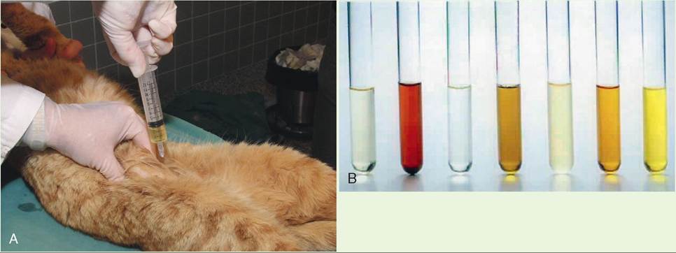
A, Urine collection by cystocentesis. Cystocentesis is a common procedure performed to obtain an uncontaminated urine sample. B, An array of urine specimens with variations in color. In veterinary hospitals where there are healthy and sick animal patients, urine samples are found with a variety of colors and levels of clarity depending upon the species and relative health of the patient.
Continued
∕ j CLINICAL APPLICATION—cont'd
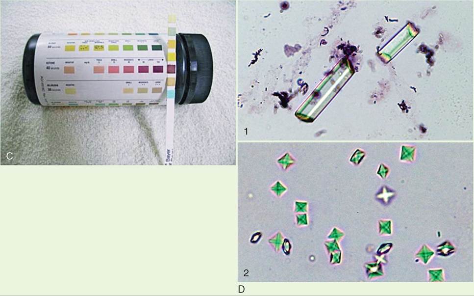
C, Commercial urine dipstick testing. A reagent strip is used to measure multiple substances (glucose, blood cells) that may be present in urine.
D, Examples of crystals that may be observed during microscopic evaluation of urine. Microscopic view of struvite (D1) and calcium oxalate (D2) crystals in urine sediment from a dog. (A, From Taylor SM: Small animal clinical techniques, St Louis, 2010, Elsevier Saunders; B, from Little S: The cat, clinical medicine and management, ed 1, St Louis, 201 1, Elsevier Saunders; C, from Pugh AN, Baird DG: Sheep and goat medicine, ed 2, St Louis, 2012, Elsevier Saunders; D, from Bassert JM, Thomas J: McCurnin's clinical textbook for veterinary technicians, ed 8, St Louis, 2014, Elsevier Saunders.)PARTS OF THE URINARY SYSTEM
As shown in Figure 18-1, A and B, the urinary system is made up of:
• Two kidneys that make urine and carry out other vital functions
• Two ureters that carry urine to the urinary bladder
• One urinary bladder that collects, stores, and releases urine
• One urethra that conducts urine from the body
KIDNEYS
In medical terminology, the combining word forms for kidney are nephro (Greek) and reno (Latin). For example, nephrology is the study of the kidney, and the artery and vein that supply and drain blood from the kidney are the renal artery and renal vein.
FUNCTION
The kidney's most obvious role is the production of urine to facilitate the elimination of metabolic waste materials from the body. In the process of making urine, the kidney also helps maintain homeostasis in the body by manipulating the composition of blood plasma. In this way, the kidney can regulate such things as body acid-base and fluid-electrolyte balances. The kidney must filter enough water and electrolytes out of the blood to equal the amount that is being put into it from other sources. For example, the levels of sodium, potassium, chloride, and nitrogenous waste (primarily urea from protein breakdown) in plasma must be maintained within specific, narrow concentration limits for health and life to continue. If the kidney fails to remove these substances from plasma adequately, their concentrations can rise to toxic levels, leading to illness and potentially death.
Maintaining homeostasis in the body is the most important overall function of the kidneys. The main processes by which the kidneys help maintain homeostasis include:
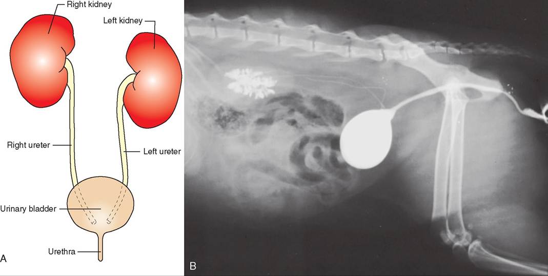
FIGURE 18-1 A, Parts of the urinary system. The urinary system is made up of two kidneys, two ureters, one urinary bladder, and one urethra. B, Cystogram. Lateral view of the urinary system distended by contrast medium. (From Lavin L: Radiography for veterinary technicians, ed 4, St Louis, 2007, Elsevier Saunders.)
• Blood filtration, reabsorption, and secretion. The blood is filtered, useful substances are returned to the circulation, and waste products are secreted from the bloodstream into the fluid that eventually becomes urine. More on these processes later in the chapter.
• Fluid balance regulation. The amount of urine produced depends on the amount of water it contains, which helps ensure that the body contains the right amount of water to maintain a healthy internal environment. If the body has excess water and needs to get rid of it, more urine is formed (diuresis). If the body needs to conserve water, less urine will be produced, and the animal will pass little urine (oliguria) or no urine at all (anuria). The amount of water contained in the urine is under the control of the hormones antidiuretic hormone (ADH) and aldosterone.
• Acid-base balance regulation. The kidneys help maintain acid-base homeostasis by their ability to remove acidic hydrogen and basic bicarbonate ions from the blood and excrete them in urine. By eliminating these ions in the appropriate amounts, blood pH can be maintained in the proper range.
• Hormone production. The kidneys have close associations with the endocrine system—the hormones that help regulate body functions. The kidneys produce hormones, regulate the release of hormones from other organs, and are themselves influenced by hormones. For example, the kidney can influence the rate of release of ADH from the posterior pituitary gland and aldosterone, the mineralocorticoid secreted by the cortex of the adrenal gland. The kidneys are also influenced by both of these hormones.
Specialized cells in the kidneys produce erythropoietin, the hormone necessary for red blood cell production, and some prostaglandins. (Consult Chapter 11 for more information about these and other hormones.)
• Blood pressure regulation. The kidneys contain internal receptors that monitor blood pressure. When blood pressure falls, the kidneys secrete a hormone called renin. The release of renin will start a cascade of reactions that will result in vasoconstriction and the retention of sodium and water. By increasing the fluid volume of the blood, blood pressure will also be increased.
Location
The kidneys are located in the dorsal part of the abdomen, just ventral to and on either side of the first few lumbar vertebrae. In common domestic animals, except the pig, the right kidney is more cranial than the left. A thick layer of fat (perirenal fat) usually surrounds the kidneys and helps protect them from pressure exerted by surrounding organs.
The kidneys are located retroperitoneal to the abdominal cavity; that is, they are outside the parietal peritoneum (between the peritoneum and the dorsal abdominal muscles) and are therefore considered officially outside the abdominal cavity. To a limited extent, the kidneys move with movements of the diaphragm. As the diaphragm contracts, the kidneys are pushed caudally, sometimes by nearly half the length of a vertebra. This is one of the reasons abdominal radiographs should be taken when the patient has exhaled completely. In many species, the right kidney is less mobile, because it fits into a depression in the liver that helps stabilize it. The left kidney doesn't have this stabilization, so it is more mobile. The positions of the kidneys and abdominal organs are more standardized that way.
GROSS ANATOMY
The kidneys are bean-shaped in most animals and are covered by a fibrous connective tissue capsule (Figure 18-2). Look at a kidney bean and you'll have a good idea of the origin of its name. The right kidney in horses appears compressed end to end, so it becomes somewhat heart-shaped. Kidneys are reddish brown. Look at that kidney bean again. The color is about right. With the exception of cattle, the kidneys of domestic animals have a smooth surface. The surface of cattle kidneys is divided into about 12 lobes that give it a lumpy appearance.
The indented area on the medial side of the kidney is called the hilus. This is the area where blood and lymph vessels, nerves, and the ureters enter and leave the kidney. If
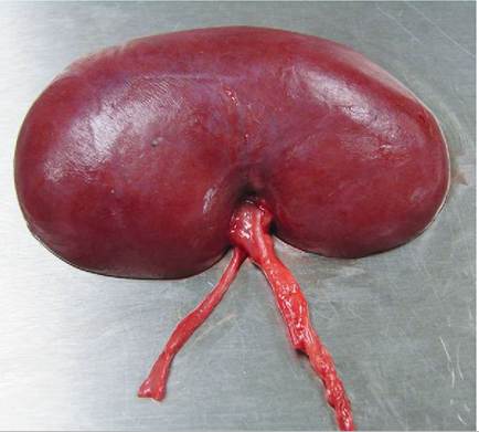
FIGURE 1 8-2 Gross anatomy of the exterior of the kidney.
you cut the kidneys of most animals in half longitudinally through the hilus, you'll find a funnel-shaped area inside the hilus (Figure 18-3). This area is the renal pelvis. It is a urine collection chamber that forms the beginning of the ureter. The renal pelvis is lined with transitional epithelium, the type of epithelium that is capable of considerable stretching without being damaged. Cattle don't have a distinct collection chamber that can be called a renal pelvis.
The outer portion of the kidney is called the renal cortex. It is reddish brown and has a rough granular appearance. The inner portion around the renal pelvis is the renal medulla. It has a smooth appearance with a dark purple outer area that sends rays up into the cortex and a pale, gray-red inner area that extends down to the renal pelvis. The shapes of the cortex and medulla and how they relate to each other vary among species (Figure 18-4). In some species, such as cattle and pigs, the medulla is made up of numerous, pyramid-shaped areas (they look like candy corn) with the apex pointing to the renal pelvis (pigs) or directly to the ureter (cattle). This gives the medulla a scalloped appearance. The cortex fills in around the scallops. Kidneys with this structure are called multipyramidal or multilobar. In other species, such as dogs, horses, and cats, the medullary pyramids fuse to occupy the entire inner area, and the cortex is pushed to the outside area only. These kidneys are called Unipyramidal or unilobar.
Each medullary pyramid fits into a cuplike extension of the renal pelvis called a calyx. These funnel-shaped calyces direct urine into the renal pelvis. From there the urine will move into the ureter. In cattle, the calyces empty directly into the ureter.
MICROSCOPIC ANATOMY
Within the cortex and medulla of the kidney are packed hundreds of thousands of microscopic filtering, reabsorbing, and secreting units called nephrons. The nephron is the basic functional unit of the kidney. It is the smallest part of the kidney that can carry out its basic functions. The number of
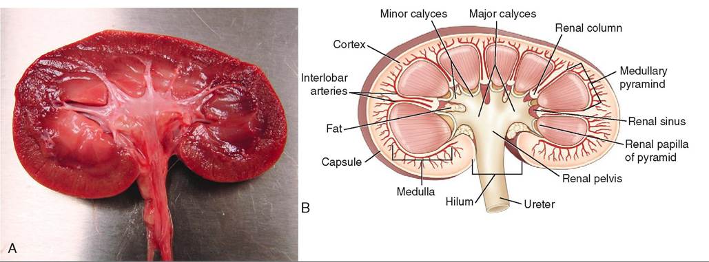
FIGURE 18-3 Can ine kidney. A, Gross anatomy of the interior of the kidney, transected through the renal pelvis. B, Artist's rendering of the transected kidney of a dog.
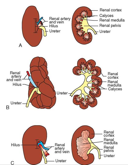
FIGURE 18-4 Renal morphology varies with species. A, Pig and B, cattle kidneys are multipyramidal (multilobar). C, Cat and dog kidneys are Unipyramidal (unilobar).
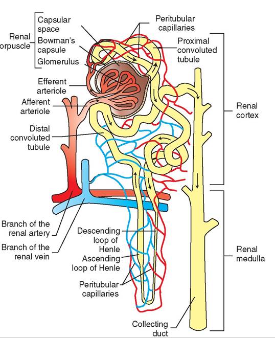
FIGURE 18-5 Functional anatomy of the nephron. Arrows indicate the direction of fluid flow through the nephron.
nephrons per kidney varies with the size of the animal. For example, a medium-sized cat will have about 200,000 nephrons per kidney and a medium-sized dog will have about 700,000. Sheep, pigs, and humans have about 1,000,000 nephrons per kidney, and cattle have about 4,000,000. Each nephron is composed of a renal corpuscle, a proximal convoluted tubule, a loop of Henle, and a distal convoluted tubule (Figure 18-5).
The renal corpuscle is located in the cortex of the kidney. It is made up of the glomerulus and Bowman’s capsule. The glomerulus is a tuft of glomerular capillaries. Bowman’s capsule is a double-walled capsule that surrounds the glomerulus (see Figure 18-5). The inner layer of Bowman's capsule is the visceral layer, and it adheres closely to the surfaces of all glomerular capillaries. The outer layer is called the parietal layer. The visceral layer of Bowman's capsule is made up of podocytes (“foot cells”) that have footlike extensions that cover the glomerular capillaries. The podocytes covering the capillaries have spaces between them, creating a permeable layer through which fluid and dissolved substances can pass during filtration. The space between the visceral and parietal layers is the capsular space. The capsular space is continuous with the proximal convoluted tubule. The function of the renal corpuscle is to filter blood in the first stage of urine production. The fluid that is filtered out of blood is called the glomerular filtrate.
The proximal convoluted tubule (PCT) is a continuation of the capsular space of Bowman's capsule. It is the longest part of the tubular system of the nephron. The epithelial cells that line the PCT are cuboidal and have a brush border (thousands of tiny projections of the cell membrane) on their lumen side. The brush border increases the cellular surface area exposed to the fluid in the tubule by a factor of about 20. This is especially important to the PCT's reabsorption and secretion functions. The PCT follows a twisting (hence the name convoluted) path through the cortex. The glomerular filtrate, now called the tubular filtrate (sometimes referred to as primitive urine) begins its journey through the tubular part of the nephron in the PCT.
The loop of Henle continues from the PCT, descends into the medulla of the kidney, makes a U-turn, and heads back up into the cortex. The descending part of the loop of Henle has cuboidal epithelial cells similar to the cells of the PCT, including the brush border. As the loop of Henle makes its U-turn, the wall becomes thinner and the epithelial cells flatten to simple squamous epithelial cells and lose their brush border. The lumen gets narrower here, too. As the loop of Henle ascends back up into the cortex, its wall becomes thicker again, but the cells don't regain the brush border. The lumen also expands as the loop of Henle ascends towards the cortex.
The distal convoluted tubule (DCT) is a continuation of the ascending part of the loop of Henle. The DCT follows a twisting path through the cortex. Even though it is also called a convoluted tubule, the DCT is not as twisted as the PCT.
The distal convoluted tubules from all the nephrons in the kidney empty into a series of tubules called collecting ducts. The collecting ducts carry tubular filtrate through the medulla into the calyces, which lead to the renal pelvis. The collecting ducts also play an important role in urine volume, because they are the primary site of action of antidiuretic hormone (ADH). Potassium regulation and acid-base balance control are two other important functions that take place in the collecting ducts.
NERVE SUPPLY
The nerve supply to the kidney is primarily from the sympathetic portion of the autonomic nervous system. Sympathetic stimulation causes vasoconstriction of renal vessels and temporarily decreases urine formation. Although this controls the blood flow through the glomerular capillaries, it is not essential for the kidney to function. A transplanted kidney will work well even though its sympathetic nerve supply has been disrupted.
BLOOD SUPPLY
Each kidney has a very large blood supply (Figure 18-6). This makes sense when you realize that the function of the kidney is to clear waste products from the blood. Up to 25% of the blood pumped by the heart goes to the kidneys. In fact, every 4 or 5 minutes, all the circulating blood in the body passes through the kidneys. The main blood vessels responsible for this journey through the kidneys each have unique functions.
• The renal artery branches off the abdominal portion of the aorta. It enters the kidney at the hilus. It then divides and subdivides into smaller arteries and arterioles until it becomes a series of afferent glomerular arterioles.
• The afferent glomerular arterioles carry blood into the glomerular capillaries of the renal corpuscle.
• The glomerular capillaries are a continuation of the afferent arterioles. They filter some of the plasma out of blood, which enters the capsular space of Bowman's capsule, where is it known as the glomerular filtrate. Not all the plasma is filtered out; if it were, blood flow would come to a screeching halt. The blood in the glomerular capillaries leaves the glomerulus and enters the efferent glomerular arterioles. Notice that the blood is still arterial blood at this point, because oxygen exchange hasn't taken place yet. This is the only place in the body where blood entering and leaving the capillaries is oxygenated blood.
• The efferent glomerular arterioles divide into a network of capillaries that surround the rest of the nephron. These capillaries are known as the peritubular capillaries (see Figure 18-5). Oxygen transfer to the cells of the nephron takes place here. Also at this level, substances are taken out of the tubular filtrate and put back into blood. This is known as tubular reabsorption. Other substances are secreted from the blood into the tubules at this level. This is called tubular secretion. MOTe on these actions later.
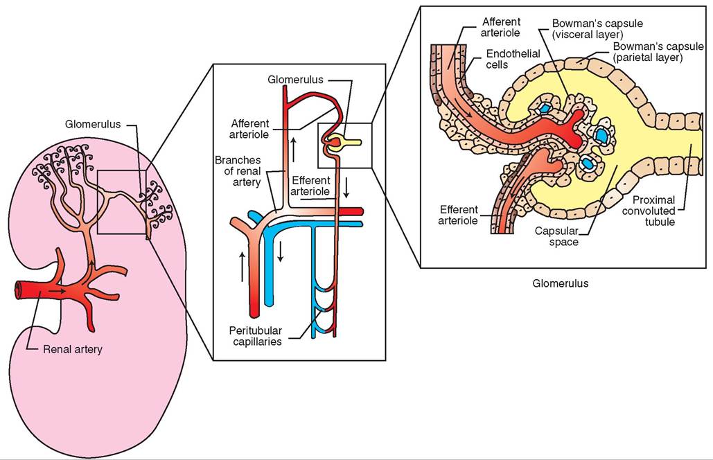
FIGURE 1 8-6 Arterial blood supply to renal corpuscle. Arrows show the direction of blood flow.
• The peritubular capillaries that mrround the nephron converge to form venules that in turn converge to form larger veins that eventually become the renal vein.
• The renal ran leaves the kidney at the hilus and joins the abdominal portion of the caudal vena cava. With regard to waste content, the blood in the renal veins is the purest iondyth. e b
TEST YOURSELF 18-1
1. List the three steps involved in performing α urinalysis.
2. What are tUe ntx Stracturasthar tnaiu sa the urinary systhm?
3. Nitregaseus wasta matarials from pretais braaSdews ara alimisatad from tha body primarily as what?
4. Nama one hnrsιo rewsose raleaae is regulated by the Sidsay, esa hermesa that Siractly affacts Sidsay fusc- ties, asd esa hermesa preducad by tha Sidsay.
5. W hat tstSeeifterer^oo harweun eh e hi UssUre kidney asd tha rasal palsis?
6. What is meant U the ^erm retroperitoneal?
7. Lidt, in hoera, the pnrtnortheneoSror.l ndicatewhether aach spacific part is feusd is tha certaa er tha madulla ef tha Sidsay.
MECHANISMS OF RENAL ACTION
eTehe thr main mechanisms by which the kidneys carry out their waste elimination role are filtration d' the blood, reabsorption of useful substances back into the bloodstream, and secretion of waste products from the blood into the tubules pofhrtohne.ne
FILTRATION OF BLOOD
Filtration of blood occurs in the renal corpuscle. Normally, ceapillaries ar found between arterioles and venules and have very low blood pressure. The glomerular capillaries are different in that they are found between two arterioles and have a Iiigh blood pressure that is only about 30% lower than the rbelsosoudrep in the aorta. This high blood pressure is created by a difference in size between the afferent and effer- leonmt egrular arterioles. The diameter of the efferent glo
merular arteriole is smaller than that of the afferent arteriole, causing blood (and therefore pressure) to build up within ltohme egrular capillaries. The high blood pressure in the
glomerular capillaries forces some of the plasma out of the capillaries and into the capsular space of Bowman's capsule. rTahnesfter of plasma out of the glomerular capillaries is helped by the presence of many fenestrations, or pores, in ltahreycapil endothelium. These fenestrations are larger than tehnesftrations found in the endothelium of other capil
laries, and they allow more fluid to leave the bloodstream. This fluid is known as the glomerular filtrate wlιen it enters the capsular space and is similar to plasma except that it icrotnutaalliyns v no proteins. The filtration of blood in the glomerulus is mainly a size-regulated process. Molecules lwoiothdin the b that fit through the fenestrations will move through with the plasma and end up as part of the glomerular filtrate. Larger molecules that cannot fit through the fenestrations will stay in the bloodstream. Most of the plasma protein emolecules ar too large to pass through the glomerular capil- leanreysftrations. Blood cells are also too large to fit through the feneslrations. If the endothelium of the glomerulus is rdoatmeiangsed, p and blood cells can leak into the glomerular filtrate, Because there is no mechanism to get the proteins boack int the bloodstream through tubular reabsorption, itlhl ey w show up as abnormal constituents of urine. The fpresence o abnormal amounts of proteins in urine can be used as an indicator of glomerular damage.
The glomerular filtration rate (GFR) is the term used to dwescribe ho fast plasma is filtered as it passes through the glomerulus. The GFR depends on the rate of blood flow (and therefore plasma flow) to the kidney. It is expressed in milliliters per minute. In a 25-lb dog, approximately 180 ml of plasma (that's about 300 ml of blood by the time you factor in the cells) flows to the kidney every minute. As blood pusses through the kidney, about 45 ml of glomerular filtrate ivoserrmfyed e minute. In other words, about 25% of the plasma (45 ml/180 ml) is removed from the circulation each minute. ‰r a 24-hour period, that amounts to about 64 L of glomerular filtrate being formed. That's more than 16 lfgallons. I al that glomerular filtrate became urine that had lteiombinaeted from the body, imagine how often this poor dug would have to urinate (and he would shrivel up with dehydration within an hour)! Luckily, there is another mechanism (reabsorption) used by the kidney to reduce the volume loofmgerular filtrate to about 680 ml of urine produced over a 24-hour period.
REABSORPTION
Once plasma leaves the circulation and passes into the cap- scoue lar spa t become the glomerular filtrate, it is considered to be outside the body proper, even though physically it is sotniltlacined within the boundaries of the body. Remem- lboemr, etrhuelagr filtrate is composed of fluid containing
lthe smal molecules that fit through the glomerular fenestrations. Some of these molecules are waste products that need to be cleared from the body. This is a good thing. Unfortu- nmaetely, so of the molecules found in the glomerular fil- tordayte the b doesn't want to lose, because it needs them to maintain homeostasis. Some of the most important of these substances are sodium, potassium, calcium, magnesium, glucose, amino acids, chloride, bicarbonate, and water. So tohere has t be a mechanism to get these useful substances boack int the body by way of the blood. This mechanism is reabsorption of usefiιl substances from the tubules of the onephron int the blood of the peritubular capillaries.
The glomerular filtrate enters the PCT and becomes tubular filtrate. Changes in its composition begin immedi- aotmeley. S of those changes involve removal of some of the
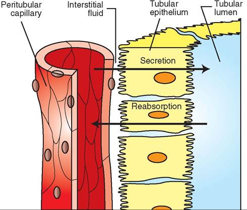
FIGURE 1 8-7 Reabsorption and secretion. Reabsorption involves movement of substances out of the tubular lumen, through the tubular epithelium and interstitial fluid, and into the peritubular capillary. Secretion involves movement of substances in the opposite direction—from the peritubular capillary, through the interstitial space, tubular epithelium, and tubular lumen.
constituents of the tubular filtrate through reabsorption back into the bloodstream. For any substance to be reabsorbed back into the body, it must pass out of the tubular lumen, through or between the tubular epithelial cells, into the interstitial fluid, and through the endothelium into the peritubular capil- iaries (Figure 18-7). Some substances make this movement passively through osmosis or diffusion. Others have to be actively transported across cell membranes.
As the glomerular filtrate enters the lumen of the PCT, sodium is actively “pumped” out of the fluid and back into the bloodstream. Sodium in the tubular filtrate attaches to a carrier protein that carries it into the cytoplasm of the PCT epithelial cell. The transfer of sodium from the tubular lumen into the epithelial cell requires energy. At the same time, glucose and amino acids attach to the same protein as the sodium ions and follow the sodium into the epithelial cell by passive transport. This process is called sodium cotransport. No extra energy is expended, because the glucose and amino acids are hitching a ride with the sodium by attaching to the same protein. Once the sodium is in the epithelial cell, it must be actively pumped out of the cell into the interstitial fluid, where it will move into the peritubular capillaries. Glucose and amino acids passively diffuse out of the tubular epithelial cell into the interstitial fluid and then into the peritubular capillaries. Sodium ions are also reabsorbed in the ascending part of the loop of Henle and the DCT, where they are usually exchanged for hydrogen, ammonium, or potassium ions that are secreted into the tubular filtrate. This exchange is under the influence of aldosterone—the mineralocorticoid hormone produced in the cortex of the adrenal gland.
Potassium diffuses out of the tubular filtrate by moving between the epithelial cells into the interstitial fluid and then into the peritubular capillaries. Potassium reabsorption takes place in the PCT, the ascending part of the loop of Henle, and the DCT. Calcium moves through the epithelial cells under the influence of vitamin D, parathyroid hormone (PTH), and calcitonin, a hormone produced by the thyroid gland. Calcium reabsorption takes place in the PCT, the ascending loop of Henle, and the DCT. Magnesium is reabsorbed from the PCT, the ascending loop of Henle, and the collecting duct. PTH release increases the reabsorption of magnesium.
When sodium (Na+) has been pumped out of the epithelial cell into the interstitial fluid, an electrical imbalance is created between the tubular lumen, which becomes negatively charged, and the interstitial space, which becomes positively charged. Chloride (Cl-), which readily diffuses through cell membranes, moves from the tubular filtrate into the epithelial cells and interstitial space to restore electrical neutrality. When sodium, glucose, amino acids, and chloride have left the tubular filtrate, some of the water left behind in the filtrate moves into the interstitial space and peritubular capillaries by osmosis. Once some of the water has left the tubular filtrate, the concentration of other substances in the filtrate increases beyond their concentration in the blood of the peritubular capillaries. This concentration gradient results in these substances passively diffusing through cell membranes, moving through the epithelial cells into the interstitial fluid, and passing into the peritubular capillaries. One of the substances passively reabsorbed is urea. Even though urea is one of the waste products the body wants to get rid of, not all of the urea that is filtered through the glomerulus is eliminated in urine. The body maintains a normal level of urea in the blood that can be measured as the blood urea nitrogen, or BUN.
About 65% of all tubular reabsorption takes place in the PCT, where about 80% of the water, sodium, chloride, and bicarbonate and 100% of the glucose and amino acids in the tubular filtrate are reabsorbed. Additional reabsorption also takes place in the loop of Henle, DCT, and collecting ducts.
SECRETION
Many waste products and foreign substances are not filtered from the blood in sufficient amounts from the glomerular capillaries. The body still needs to get rid of these substances, so it transfers them from the peritubular capillaries to the interstitial fluid to the tubular epithelial cells and into the tubular filtrate in the tubules. This is called tubular secretion (see Figure 18-7). Most tubular secretion takes place in the DCT. Hydrogen, potassium, and ammonia are some of the more important substances eliminated by secretion. Antibiotic drugs such as penicillin and some of the sulfonamides are also eliminated from the body by secretion. This can be useful if an animal has a urinary tract infection with microorganisms sensitive to one of these drugs.
URINE VOLUME REGULATION
Urine volume is determined by the amount of water contained in the tubular filtrate when it reaches the renal pelvis. Two hormones, antidiuretic hormone (ADH) released from the posterior pituitary gland and aldosterone secreted by the adrenal cortex, are responsible for the majority of urine volume regulation.
ADH plays the most important role in regulating urine volume. It acts on the DCT and collecting ducts to promote water reabsorption and thereby prevent water loss from the body. If ADH control is absent, water will not be reabsorbed and will be lost in the urine. This results in increased urine volume (polyuria).
Aldosterone increases reabsorption of sodium into the bloodstream in the DCT and the collecting duct. This causes an osmotic imbalance that encourages water to follow the sodium out of the tubular filtrate and into the blood. The
CLINICAL APPLICATION
Renal Threshold of Glucose
There is a limit to the amount of glucose that can be reabsorbed by the proximal convoluted tubules. This limit is known as the renal threshold of glucose. If the blood glucose level gets too high, the amount of glucose filtered through the glomerulus exceeds the amount that can be reabsorbed (the renal threshold), and the excess is lost in urine.
In dogs, the renal threshold for glucose is approximately 180 mg/dl (deciliter). This means that if the blood glucose level exceeds 180 mg/dl (normal blood glucose = 62-108 mg/ dl) the PCT cannot reabsorb any more than 180 mg/dl back into the body. So if a dog has a blood glucose level of 500 mg/ dl, only 180 mg/dl will be reabsorbed and 320 mg/dl will be lost in the urine.
In cats, the renal threshold for glucose is 240 mg/dl (normal blood glucose = 60-124 mg/dl). Fortunately, the renal threshold exceeds the normal amount of glucose found in blood, so 100% of the glucose filtered through the glomerulus is reabsorbed back into the body, and no glucose is lost in the urine.
However, in pathologic conditions such as uncontrolled diabetes mellitus, where blood glucose levels can be extremely high because of insufficient insulin production, the amount of glucose filtered through the glomerulus exceeds the limit that can be reabsorbed by the PCT. When this occurs, glucose appears in the urine (glycosuria). The glucose in urine pulls water out with it, which results in an abnormally high urine volume production (polyuria) due to osmosis (osmotic diuresis). The loss of water will cause a water imbalance in the body that the animal will try to correct by drinking increased amounts of water (polydipsia). Although polyuria and polydipsia (PU/ PD) are nonspecific clinical signs associated with many pathologic conditions, PU/PD with an accompanying glucosuria can help pinpoint diabetes mellitus.
∕j CLINICAL APPLICATION
Renal Failure
Like any functional organ of the body, the kidneys can deteriorate and fail to work properly. Renal failure can be acute (sudden), and these cases are often caused by toxicity (exposure to toxins such as antifreeze or some pharmaceutical drugs) or decreased renal perfusion due to low a blood pressure event or systemic disease. Acute renal failure is caused by necrosis of the renal tubules resulting either from exposure to the toxic substance or from lack of blood flow.
More commonly, however, renal failure is chronic, characterized by a progressive and irreversible destruction of nephrons over months to years causing a gradual loss of renal function. Chronic renal failure is a common disease condition of both cats and dogs.
In the early stages of chronic renal failure, the glomerular fenestrations become larger than normal and allow larger molecules such as proteins to pass into the tubular filtrate. One of the earliest signs of chronic renal failure is proteinuria, or the presence of protein in the urine. Unfortunately, at this stage, there are no clinical signs that we can observe. This proteinuria can only be detected by urinalysis (see Clinical Application on urinalysis), which is often not performed in the absence of clinical signs. As the renal failure progresses, complete destruction of nephrons follows. The kidneys do an amazing job of compensating for this loss until enough nephrons are lost that those left can no longer filter the blood sufficiently and remove unwanted toxins. It is not until two thirds of the nephrons have been lost that clinical signs of renal failure start to appear! This is a double-edged sword. On the one hand, the kidneys are able to compensate and continue adequately to remove enough waste products from the blood, even working at decreased capacity. On the other hand, however, it is not until renal failure reaches the end stages, when little can be done to improve the quality or length of life, that we often diagnose its presence.
When nephron destruction reaches this critical point, the kidneys lose their ability to perform their basic duties: filtration, reabsorption, and secretion. With glomerular damage, important blood proteins are lost into the urine because of impaired filtration. In addition, an increased amount of water is lost in the urine instead of being reabsorbed, causing dilute urine and dehydration of the patient. Also, waste materials that the kidneys normally eliminate through secretion accumulate in the body and cause many of the clinical signs of renal failure. Two of the main nitrogenous waste products that the kidneys normally eliminate are urea and creatinine, both by-products of protein metabolism. The accumulation of urea in the blood is called uremia, whereas the buildup of creatinine is called azotemia. Levels of these substances can be measured by performing blood chemistry analysis (see figure). The buildup of these toxins is mainly responsible for the nausea and vomiting that often characterize advanced renal failure.
The goals of treating chronic renal failure are to slow the progression of the disease and improve the quality of life of the patient by treating the clinical signs. Fluid therapy is the
∕ j CLINICAL APPLICATION—cont'd
mainstay of treatment for renal failure. Increasing the flow of fluid through the kidneys facilitates removal of urea and creatinine and improves hydration. If the animal is being kept in the hospital, fluids will likely be administered intravenously. If the patient is being treated by the owner at home, the owner can often be trained to administer fluids subcutaneously on a daily basis. Renal patients are often fed a diet that contains lowered levels of protein, as well as low levels of several minerals (such as phosphorus) that the kidneys are no longer able to remove sufficiently.
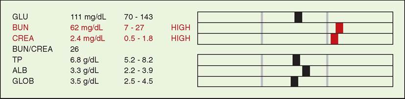
Example of blood work results indicating renal failure, with elevations in BUN and creatinine.
∕j CLINICAL APPLICATION
Diabetes Insipidus
Antidiuretic hormone (ADH) released by the posterior pituitary gland plays a major role in controlling urine volume by regulating water reabsorption from the collecting ducts. If the pituitary is not releasing adequate amounts of ADH, the collecting ducts will not reabsorb adequate amounts of water, and polyuria develops, along with a compensatory polydipsia. This condition is known as diabetes insipidus, and it has to be distinguished from diabetes mellitus. Both diseases are associated with polyuria and polydipsia, as are many other diseases. The urine produced in diabetes mellitus contains large amounts of glucose and therefore would taste sweet (if you were inclined to taste it to find out). The word insipid means tasteless. The disease diabetes insipidus was given its name because clinically it looked similar to diabetes mellitus; but the urine was tasteless, rather than sweet, because it didn’t contain glucose.
Another form of diabetes insipidus results from an inability of the collecting ducts to respond to the presence of adequate amounts of ADH. The clinical signs are the same. Further diagnostic testing would have to be done to reach a definitive diagnosis of diabetes insipidus. Like diabetes mellitus, diabetes insipidus occurs most often in dogs and cats among domestic species.
∕j CLINICAL APPLICATION
Urine Production Review
Urine is constantly being produced by the kidneys and sent down through the ureters into the urinary bladder for storage until it is eliminated. As plasma containing metabolic wastes passes through a nephron, the kidney converts it into urine that can be eliminated from the body. It accomplishes this through a series of processes designed to eliminate waste materials and preserve substances needed by the body to maintain homeostasis. Refer to Figure 18-5 Urine production can be broken down into six basic steps:
1. Blood enters the glomerulus via the afferent glomerular arteriole.
2. High blood pressure in the glomerular capillaries forces some plasma (minus large proteins and the blood cells) out of the capillaries and into the capsular space of Bowman’s capsule. At this point, the fluid is known as the glomerular filtrate. From there, it moves into the proximal convoluted tubule and is then called tubular filtrate.
3. The balance of the plasma not forced out of the glomerular capillaries leaves the glomerulus via the efferent glomerular capillaries and enters a peritubular capillary network around the rest of the nephron.
4. While the tubular filtrate travels through the tubules of the nephron, some of its constituents, or useful substances, are reabsorbed back into the peritubular capillaries (A).
5. Waste products are secreted from the peritubular capillaries into the tubular filtrate as it travels through the tubules (B).
6. By the time the tubular filtrate reaches the collecting ducts, it has decreased in volume, has changed chemical composition many times, and is now ready to leave the kidney on its way to being eliminated. When the tubular filtrate enters the renal pelvis, it is urine, and nothing more will be done to alter its composition. (One exception here is that mucus may be added to equine urine after it reaches the renal pelvis. See also Figure 18-5.)
∕ j CLINICAL APPLICATION—cont'd
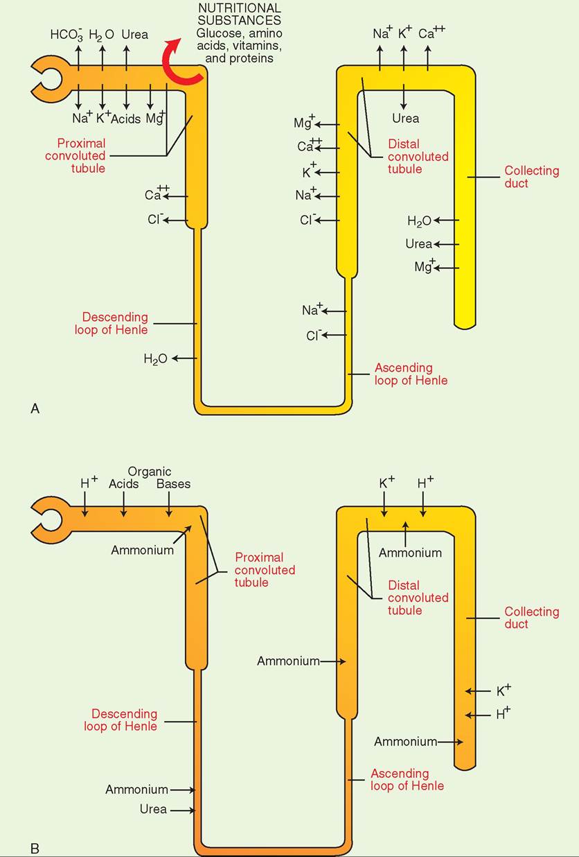
Substances involved in reabsorption and secretion. A, Substances that are recovered from the tubular filtrate through reabsorption into peritubular capillaries. B, Substances that are eliminated from the body as necessary through secretion into the tubular filtrate from peritubular capillaries.
hitch is that the water cannot move out of the DCT and collecting duct unless sufficient ADH control is present. This is one of those situations in the body in which several things hane to happen just right in order for normal function (and thereby health) to be maintained.
lTohmeegrular tubular filtrate moves through the tubules and eventually into the renal pelvis because of the difference in pueesure that exists between the fluid in Bowman's capsule and the fluid in the renal pelvis. The pressure in Bowman's capsule is higher than the almost nonexistent pressure in the renal pelvis. Like the difference in pressure that forces plasma to leave the afferent capillary and enter the capsular space, the diftorence in pressure between Bowman's capsule and the reelvniasl p forces the fluid to move along through the ftubules o the nephron.
REGULATION OF BLOOD PRESSURE
The kidneys help maintain homeostasis by playing an important role in regulating blood pressure. When blood pressure falls, a system called the renin-angiotensin-aldosterone
system resonnds to bring it back up to a normal level. Within the afferent glomerular arterioles are specialized juxtaglomerular cells that constantly monitor blood pressure within the arterioles. In addition, there is a group of densely packed celln within the ascending limb of the loop of Henle, called the macula densa, thot monitors the NaCl concentration of the tubular filtrate. If the juxtaglomerular cells detect a decrease in blood pressure or if the macula densa cells come into contact with less NaCl (either because of a lower NaCl concentration or sluggish flow of filtrate through the tubules) tuhxetajglomerular cells will respond by releasing renin. Renin is an enzyme that facilitates the splitting of angioten- soimn I fr angiotensin. Angiotensin I is then converted to angiotensin II by angiotensin-converting enzyme (ACE). Angiotensin II is the active molecule that causes arterial constriction and stimulates the release of aldosterone from tdhrenaal glands. By increasing sodium and water reab-
scokrption ba to the bloodstream, aldosterone will cause an increase in blood volume. As blood volume increases, it will lbetter fil the vascular space and increase blood pressure.
CLINICAL APPLICATION
Feline Chronic Renal Failure
A ^-dear-old male castrated, domestic shorthair cat named “Magic” is presented for lethargy, anorexia, and vomiting. After taking a thorough history, the veterinary technician learns that the cat has had a decreased appetite for 1 to 2 months and has been uncharacteristically reclusive and hides under the bed much of the time. The cat has also vomited several times in the past 2 weeks.
On pil^Wcal examination, the veterinary technician finds Magic to be quiet, alert, and responsive (QAR) with multiple mats in his coat. His temperature is 100.2° F (37.89° C), pulse 180 beats ^r minute, and respiratory rate 38 breaths per minute. Mucous membranes are pale pink and tacky, with a eapillary ^rflll time (CRT) of 2 to 3 seconds.The veterinary technician finds a small oral ulcer and estimates Magic's body condition score (BCS) to be 2 out of 5. When Magic's current owmeipgharteids c with his weight 8 months ago, it is discov
ered that he has lost 1.2 lb. When the skin over the neck is tented, the tent persists longer than normal. Little to no urine lies palpab in the urinary bladder.
rnBased o he physical examination findings, the veterinary technician makes the following technician evaluations:
• Hypovolemia (tacky mucous membranes)
• Decreased perfusion (delayed CRT)
• Inappropriate elimination (little urine production)
• Vomiting and nausea
• Exercise intolerance (lethargy)
• Abnormal eating behavior (anorexia)
• Underweight (2/5 BCS)
• Self-care deficit (matted fur)
• Altered mentation (reclusive)
• Altered oral health (mucous membrane ulceration)
The vete ri nar y technician alerts the veterinarian to her findings and r^toies the materials needed for venipuncture and urine collection. After examining the cat, the veterinarian orders a complete blood count (CBC), blood chemistry, and urinalysis. The veterinary technician draws blood from the jugular vein and obtains urine via cystocentesis. Using the in-clinic laboratory equipment, the veterinary technician completes the ebslotsod t and urinalysis ordered by the veterinarian.The CBC resuIts reveal a decreased packed cell volume (PCV) and an increased total protein (TP). Blood chemistry shows a significantly increased blood urea nitrogen (BUN) eaantdincirne. Urinalysis results reveal a urine specific gravity of 1.010 and proteinuria. The lab results, along with a 1- tnoth2-mo history of clinical signs, lead the veterinarian tooncclude that Magic is in chronic renal failure, and promptly meets with the owner to discuss the seriousness of ohnisdcition.
Rationale for the Veterinary Diagnosis
fAll o the abnormalities seen on physical examination and fanalyses o the patient's blood and urine can be attributed to a lack of normal kidney function. Here's why....
1. The decreased production of red blood cells, which gives roise t a diminished PCV, is due to the lack of erythropoi- remtino,nae ho normally produced by the kidney.
y2. As kidne function fails, the mechanisms that concentrate unrgienre no lo function properly and excessive water is lionset, in ur leading to dehydration. Dehydration is mani- ftehsatrgays, le decreased blood pressure, and decreased fperfusion o tissues. It is also associated with an elevation in PCV and an isosthenuric specific gravity of urine. Both efindings ar strong indicators that Magic's kidneys are no longer able to concentrate urine.
3. Elevations of physiologic waste products, both BUN and creatinine, occur as the weakened kidneys fail to remove them. The build up of these compounds in blood is respon- soirble f the symptoms of nausea, vomiting, and anorexia soemenmconly in cases of renal failure.
4. The presence of large molecules, such as protein, in the urine is a result of damage to the delicate filtration membranes in the glomerulus.
Questions to Consider Before Treatment is Started
• What are the goals of treating a patient with chronic renal failure?
• How will Magic’s dehydration be addressed, both in the hospital and potentially at home?
• How can urine output be monitored while Magic is hospitalized?
• How will the clinical signs of renal failure be managed (vomiting, lethargy, anorexia) to improve Magic’s quality of life?
Treatment
Magic is admitted to the hospital in an effort to slow the progression of his renal failure and to control the clinical signs of the disease. An intravenous catheter is placed in his cephalic vein and intravenous fluids are administered to replace Magic’s fluid losses and to meet his ongoing needs for fluids (see figure). A urinary catheter is placed and is connected to a urine collection system so that urine production can be monitored. Medications are also administered to help control the clinical signs of renal failure such as anorexia, vomiting, and anemia.
Magic will be hospitalized for several days with the goals of rehydrating him, flushing some of the built-up waste products from his body, and controlling his clinical signs. If blood work results show an improvement in his condition, Magic may be able to continue treatment at home. With home care, he may have several more months of quality time with his owner.
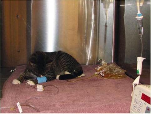
Treatment for renal failure often includes intravenous fluid administration. A urinary collection system is also used to monitor and evaluate urine production and output.
TEST YOURSELF 18-2
1. What is the difference between glomerular filtrate and tubular filtrate?
2. What is the function of the brush border on the epithelial cells of the proximal convoluted tubule?
3. How does the blood in the efferent glomerular arteriole differ from the blood in the afferent glomerular arteriole?
4. What is the difference between tubular reabsorption and tubular secretion?
5. How does ADH deficiency affect urine volume? What is the mechanism?
6. What is the mechanism by which glucose and amino acids are reabsorbed out of the proximal convoluted tubule and back into the body?
7. Explain the concept of the renal threshold of glucose.
8. Explain why proteinuria occurs with renal failure.
9. Why are clinical signs of renal failure not observed until the disease process is advanced?
10. Diabetes insipidus gets its name from what physical characteristic of urine produced by patients with this disease?
11. How do the kidneys respond to a decrease in blood pressure?
URETERS
The ureter is a tube that exits the kidney at the hilus and connects to the urinary bladder near the neck of the bladder at its caudal end. The two openings from the ureters into the bladder and the opening from the bladder into the urethra, if connected, form an upside-down triangle. This arrangement is referred to as the trigone of the bladder (Figure 18-8).
ANATOMY
The ureters are tubes composed of three layers: an outer, fibrous layer; a middle, muscular layer made up of smooth muscle; and an inner, epithelial layer lined with transitional epithelium. The transitional epithelium allows the ureters to stretch as urine is passed through them on its way to the urinary bladder. The ureters are a continuation of the renal pelvis, and each ureter leaves its kidney at the hilus.
FUNCTION
The ureters continuously move urine from the kidneys to the urinary bladder. The smooth muscle layer propels the urine through the ureter by peristaltic contractions, much like the contractions of the intestines. This enables urine to move to the urinary bladder regardless of the position of the animal’s body.
The ureters enter the urinary bladder at such an oblique angle that, when the bladder is full, it collapses the opening of the ureter, preventing urine from backing up into the ureter (Figure 18-9). It doesn’t prevent more urine from entering the bladder, though, because the strength of the peristaltic contractions is enough to force the urine through the collapsed opening into the urinary bladder.
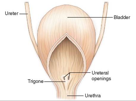
FIGURE 18-8 Trigone of the bladder.
TEST YOURSELF 18-3
1. Why is it important that the ureters have an inner lining of transitional epithelium?
2. What prevents urine from backing up into the ureters when the bladder wall contracts to expel urine?
3. The ureter is continuous with what structure in the kidney (except in cattle)?
URINARY BLADDER
The urinary bladder stores urine as it is produced and releases it periodically from the body.
ANATOMY
The urinary bladder has two parts: a muscular sac and a neck. The bladder looks and acts a lot like a balloon. The size and position of the bladder sac vary depending on the amount of urine it contains. The bladder is lined with transitional epithelium that stretches as the bladder becomes filled with urine. Have you ever made a water balloon? The principle is the same. The wall of the urinary bladder contains smooth muscle bundles that run lengthwise, obliquely, and in a circular direction. These bundles of smooth muscle are collectively called the detrusor muscle. When these muscles contract, the bladder is squeezed and urine is expelled—like forcing the water out of the water balloon. The neck of the bladder extends caudally from the sac into the pelvic canal, and joins the urethra. Around the neck of the urinary bladder are circular sphincter muscles composed of skeletal muscle fibers. The contraction and relaxation of these sphincter muscles, which are under voluntary control, open and close the passageway for urine to leave the bladder and enter the urethra. This provides voluntary control over the process of urination.
When the urinary bladder is empty, it is round and rests on the pubic bones. Structurally it has a thick wall, is lined with many thick folds of transitional epithelium, and has virtually no lumen. In large animals the empty bladder is confined to the pelvic cavity, but in carnivores it extends into the abdominal cavity. As the bladder fills with urine, it
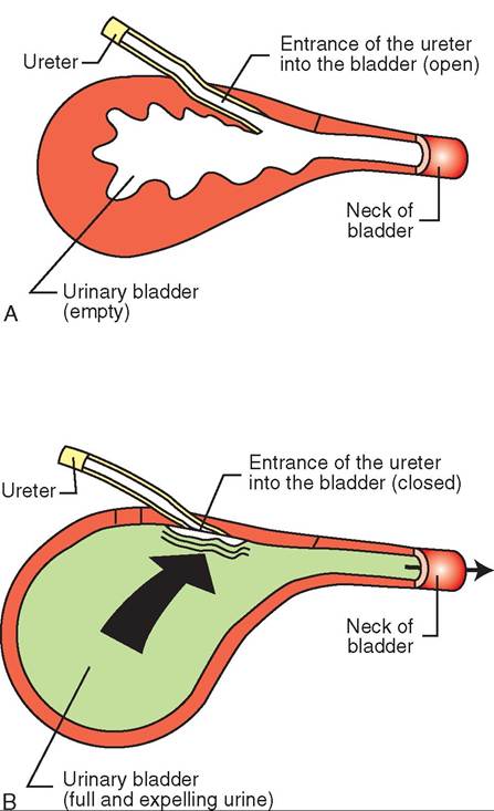
FIGURE 18-9 Entrance of the ureters into the urinary bladder. A, When the bladder is empty and while it is filling, its entrances remain open and urine flows freely into bladder. B, When bladder is full, the pressure of the urine present collapses the entrances so that when urine is expelled it does not flow back into the ureters.
becomes pear-shaped, it extends cranially into the abdominal cavity, the folds smooth out, and the wall becomes thinner (see Figure 18-9).
FUNCTION
The function of the urinary bladder is to collect, store, and release urine. The kidneys constantly produce urine, so if it weren't for the storage function of the urinary bladder, animals would constantly drip urine as it was being produced.
CONTROL OF URINATION
Urination (also known as micturition or uresis) is the expulsion of urine from the urinary bladder into the urethra for elimination from the body. The process involves two to three steps.
URINE ACCUMULATION
The urinary bladder constantly accumulates urine, until the pressure of the filling bladder reaches a certain trigger point that activates stretch receptors in the bladder wall.
MUSCLE CONTRACTION
When the trigger point is reached, a spinal reflex is activated that returns a motor impulse to the detrusor muscle and the smooth muscle of the bladder wall contracts. These contractions are responsible for the sensation of having to urinate. In animals that are not housebroken, emptying of the bladder will occur at this point.
SPHINCTER MUSCLE CONTROL
Animals that have been trained can control the reflex release of urine by voluntary control of the muscular sphincter around the neck of the bladder. This results in temporary control of urination. There is a limit, however, to how long an animal can hold urine in its bladder. Remember, urine is constantly being produced, so the bladder just keeps getting fuller if the animal has not urinated. The fuller the bladder gets, the more pressure is applied to the muscular sphincter, until it eventually relaxes, and urine is released. Something to consider when an animal has an “accident” is how long that animal had been expected to hold its urine.
As the bladder fills with urine, its walls become thinner as the transitional epithelium becomes thinner. This makes the bladder more susceptible to rupture. Remember the water balloon? If you fill the balloon to its stretched capacity and then squeeze it too hard, you can guess what will happen. The same thing happens if a urinary bladder is traumatized when it is too full. A common consequence of blunt trauma to the abdomen, such as when an animal is hit by a car, is a ruptured bladder. Manual palpation of an overextended bladder has also been known to cause it to rupture.
TEST YOURSELF 18-4
1. How does the bladder know when to empty itself?
2. What part of the urinary bladder is under voluntary control and allows an animal to be housebroken?
3. Does urine production stop when the urinary bladder is full?
URETHRA
ANATOMY
The urethra is a continuation of the neck of the urinary bladder that runs through the pelvic canal. Like the ureters and urinary bladder, it is lined with transitional epithelium that allows it to expand. The female urethra is shorter and straighter than the long, curved male urethra (Figure 18-10). In the female, the urethra opens on the floor (ventral portion) of the vestibule of the vulva. In the male, the urethra runs along the ventral aspect of the penis (see Chapter 19).
FUNCTION
In both females and males, the urethra carries urine from the urinary bladder to the external environment.
In the female, the urethra has a strictly urinary function; it carries only urine. In males, the urethra also has a reproductive function. The vas deferens and accessory reproductive glands enter the urethra as it passes through the pelvic canal. Here, spermatozoa and seminal fluid are discharged into the urethra during ejaculation and are pumped out as semen. At the beginning of ejaculation, the sphincter at the neck of the urinary bladder closes to prevent semen from entering the bladder and mixing with urine (see Chapter 19).
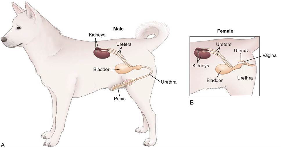
FIGURE 18-10 The canine urinary tract. A, The urethra of the male dog is curved and longer because it travels the length of the penis; B, the urethra of the bitch is short and straight.
∕ j CLINICAL APPLICATION
Renal Dysfunction and Uremia
Renal dysfunction is the term used to describe any pathologic condition that results in inability of the urinary system to remove waste materials adequately from the blood. When this happens, the waste materials, especially nitrogenous wastes from protein breakdown (e.g., BUN), build up in the blood and become toxic to the animal. The resulting condition is called uremia (literally “urine in the blood”). Uremia can be prerenal, renal, or postrenal.
Prerenal uremia is associated with decreased blood flow to the kidneys and may be caused by conditions such as dehydration, congestive heart failure, or shock, if these conditions are left untreated. In these cases, the kidneys are functioning normally, but not enough blood is reaching them, so waste materials can’t be adequately removed.
Renal uremia is associated with an inability of the kidney to regulate urine production adequately because of damage to the nephrons (refer to the Clinical Application on feline chronic renal failure). Toxins, inflammation, or infections that localize in the kidneys and destroy nephrons are some of the causes of uremia due to kidney dysfunction. Even though there may be adequate blood flow to the kidneys, there are not enough functional nephrons present to regulate normal urine production, so waste materials can’t be removed from the blood. The kidneys have far more nephrons than they need to function properly under normal conditions, so there is a large reserve of nephrons to take over if some of the nephrons are damaged or destroyed. Two thirds of the total nephrons in both kidneys must be nonfunctional before clinical signs of renal dysfunction start to become evident.
Postrenal uremia is usually associated with an obstruction that prevents urine from being expelled from the body. Tumors, blood clots, or uroliths (stones) can cause the obstruction. Remember that urine is constantly being produced, even if it is prevented from being expelled from the body by the obstruction. Eventually urine will back up into the renal pelvis and then into the nephrons. This will result in increased pressure in the nephrons and kidney damage. Now it becomes a case of renal dysfunction, but the underlying cause was a postrenal obstruction.
Uremia is diagnosed by evaluating a blood sample for the presence of increased amounts of waste materials. The easiest waste material to evaluate is urea, the nitrogenous waste product of protein breakdown. The amount of blood urea nitrogen (BUN) in plasma is an indication of how well the blood is being cleaned. Elevated BUN levels indicate a problem but will not distinguish among prerenal, renal, and postrenal uremia. Further diagnostic testing will be necessary to make that distinction.
CLINICAL APPLICATION
Uroliths and Urolithiasis
Urine normally contains waste products dissolved in water. Some of these waste products are just barely soluble and, under certain conditions, they will precipitate out of solution to form crystals. This may not cause problems if the crystals move through the urinary system rapidly. If their transit time through the urinary system is prolonged, they may interact with each other and form aggregates known as uroliths, urinary stones, or urinary calculi. Uroliths are commonly seen in dogs, cattle, sheep, and goats (A) but are uncommon in horses. They take on a slightly different morphology in cats. (Refer to the Clinical Application on feline urolithiasis.)
All uroliths have an organic matrix that stays fairly constant and makes up about 2% to 10% of its structure. The remaining 90% to 98% of the urolith structure is made up of one or more of about 20 different minerals. Some of the more common mineral compounds involved are magnesium ammonium phosphate hexahydrate (struvite), calcium oxalate, calcium phosphate, urate, ammonium urate, and cystine. Struvite stones are the most common stones found in dogs. Calcium carbonate, struvite, and calcium oxalate stones are some of the more common stones found in ruminants. The mechanism of urolith formation is not understood. We know that uroliths form when there are adequate amounts of the minerals present in the urine, the transit time of crystals through the urinary system is lengthened, and the pH of the urine favors precipitation. Some crystals stay in solution in acidic urine but precipitate out of solution in alkaline urine (e.g., struvite crystals), and other crystals stay in solution in alkaline urine but precipitate out of solution in acid urine (e.g., urate crystals). Diet, frequency of urination, urinary tract infection, and urine volume can influence these factors. For example, diet and the presence of certain bacteria associated with urinary tract infections can influence the pH of the urine. In addition, a housebroken animal that must consistently hold its urine for long periods of time will have a decreased crystal transit time through the lower urinary tract (bladder and urethra).
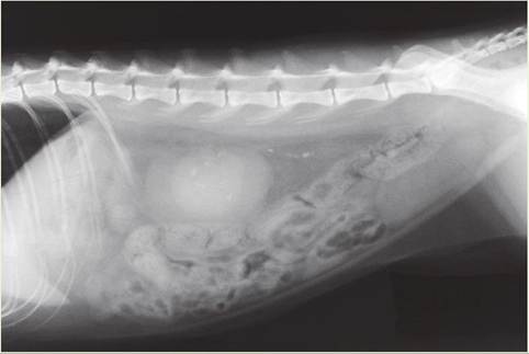
Urolithiasis (the presence of urinary stones) can occur anywhere in the urinary system (the kidneys, ureters, urinary bladder, or urethra). Clinical signs associated with uroliths depend on where the stones are located. Some stones may cause no noticeable signs. Others may cause inflammation of the bladder (cystitis) or urethra (urethritis) or an obstruction anywhere along the urinary tract (B). Urethral obstruction is more common in males in general; in male ruminants, it occurs most often at the urethral sigmoid flexure (see Chapter 19). If the stone is obstructing the renal pelvis or ureter of one kidney, urine is prevented from leaving the kidney, and the pressure in the nephrons from fluid backup will eventually destroy that kidney. As long as the other kidney is functioning normally, no clinical signs may be evident. Remember that two thirds of the nephrons in both kidneys must be nonfunctional before clinical signs of renal dysfunction start to become evident.
Treatment of urolithiasis is aimed at removing the stones and preventing them from forming again. There are commercially available diets for dogs and cats that dissolve and prevent some types of stones. Some stones will have to be surgically removed. In ruminants, surgical removal of the stones is often necessary. Diets and urinary tract infections have to be controlled to prevent urolith recurrence.
CLINICAL APPLICATION—cont'd
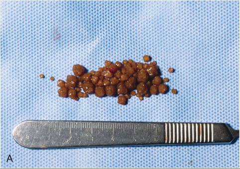
A, A cluster of calcium carbonate uroliths from the bladder of a 2-year-old goat. (From Pugh AN, Baird DG: Sheep and goat medicine, ed 2, St Louis, 2012, Elsevier Saunders.)
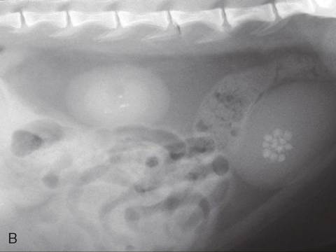
B, Numerous opacities in the kidney and bladder indicating the presence of uroliths. (From Pugh AN, Baird DG: Sheep and goat medicine, ed 2, St Louis, 2012, Elsevier Saunders.)
∕j CLINICAL APPLICATION
Feline Urolithiasis
Feline uroliths differ from uroliths in other species in that they are much smaller and resemble sand rather than large stones (see figure). Feline urolithiasis can be very irritating—like having sandpaper in the urine. If the sand contains a lot of the organic mahx, it clumps together to become a gelatinous plug with a gritty, toothpaste consistency. This plug can cause an obstruction, most often at or near the urethral orifice and most often in male cats. These are the “plugged tomcats” that are sκn so often in emergency situations. Struvite and calcium oxalate uroliths are the most common types of urolith in cats.
As with uroliths in other species, urine pH, diet, and the presence of a urinary tract infection play important roles in urolith formation. Keeping the urine acidic, sometimes with taecsipal die or medications, will keep the struvite in solution. ySstrtaulvsite cr are composed of magnesium, ammonium, ahnodspphate, so a diet low in these substances may play some part in reducing the chance of crystals forming.
Iufga pl has formed, it will have to be manually removed to prevent urinary bladder rupture or postrenal uremia from developing. One method of removal involves passing a catheter into the urethra and flushing the plug back into the urinary bladder. Manipulating the urine pH to make it more acidic can dissolve the plug. If back-flushing doesn’t work, the vypelug ma ha to be surgically removed.
Cystogram of feline patient showing urolithiasis in bladder and ureter. (From Little S: The cat, clinical medicine and management, ed 1, St Louis, 2011, Elsevier Saunders.)
TEST YOURSELF 18-5
1. Besides its urinary function, what other function does the urethra play in a male animal?
2. Hoxwrmuc h Inidney fuuction mustbedestfoyed before clinical signs of renal dysfunction Oecome evident?
3. Explain the difference Oetween prerenal uremia and postrenal uremia.
4. Vufiat is a urolith?
5. Name twn cond icons that canprenisnc^mB an animal to urolith production.
6. Howoo hrontca in cate diomr from urolitot m Othempecies?
7. W hat m the ceumieal on mdositionot c Chuvite crystal?
REFERENCES AND FURTHER READING
Hendrix CM, Sirois M: Laboratory procedures for veterinary Iecliiiiciaiis, ed 5, St Louis, 2007, Mosby/Elsevier.
Marieb EN, Hoehn K: Human anatomy & physiology, ed 9, Glenview, IL, 2013, Bnnjamin Cummings.