4 Oesophageal stricture in a cat
Initial presentation
Regurgitation
Signalment: 9-year-old male neutered Siamese cat, body weight 3.5 kg
Case history
The cat had been healthy until 3 weeks prior to admission, when he had been anaesthetized for a routine dental cleaning.
Since then he had been regurgitating food and froth, but not bile. The regurgitated food appeared undigested. He was also retching.He was de-wormed regularly and vaccinated yearly. He had arthritis and he was treated with meloxicam for this condition. He was an indoor and outdoor cat, but had no known exposure to toxins. He was fed canned cat food and chicken, but had a poor appetite during the 3 weeks prior to presentation. He was able to prehend and swallow his food and could drink water, although he may have been drinking less than usual.
His stools were normal in appearance and he was able to urinate and defecate normally. He was on no other medications or supplements.
Physical examination
The cat was bright and responsive. His body condition score was 4/9 and he was estimated to be about 5% dehydrated. Mucus membrane colour was pink and capillary refill time was less than 2 seconds.
Thoracic auscultation revealed normal heart and lung sounds, with a heart rate of 190 beats per minute (bpm) and a respiratory rate of 30 breaths per minute. There was no evidence of abnormalities or pain on abdominal palpation. Rectal temperature was 38.0° C.
Problem list and discussion of problems
• Regurgitation
Regurgitation usually indicates an oesophageal disorder.
Differential diagnosis
Oesophagitis
• Obstructive oesophageal disorders
• foreign body
• stricture
• neoplasia
• peri-oesophageal masses
Oesophageal neuromuscular disorders
• Megaoesophagus, including systemic disorders affecting swallowing, such as myasthenia gravis and dysautonomia
• Oesophageal motility abnormalities
Case work-up
The cat was admitted to the hospital for investigative procedures and rehydration with crystalloid fluids administered intravenously.
Minimum data base
Haematology and serum chemistry were performed and results were all within the reference ranges.
Imaging
Plain radiographs of the cat’s thorax were unremarkable. While a barium swallowing study could have been performed to further evaluate the oesophageal function, this test carries the risk of aspiration of the barium and in this case it was elected to perform endoscopy rather than the swallowing study.
Clinical tip
While thoracic radiographs are useful to evaluate the lungs and many other oesophageal disorders, strictures may not be visible on a plain radiograph and even liquid barium may pass through a stricture rapidly enough that it is not detected. Barium mixed with food may allow visualization of a narrowed area of the oesophagus at the point of the stricture, but may or may not show multiple areas of strictures if the food is regurgitated when it reaches the first area of stricture. Barium swallow studies, whether liquid or liquid mixed with food, do carry a risk of aspiration pneumonia, especially if the patient must be put in lateral recumbency to obtain the radiograph.
Endoscopy
A severe concentric stricture extending throughout most of the thoracic oesophagus was found (Fig 4.1). The width of the narrowed oesophagus was about 3 to 4 mm. Gentle dilation with a balloon catheter was performed, stretching the affected part of the oesophagus to about 1 cm. After dilation with the balloon, a couple of tears were noted in the mu-
cosa and a severe oesophagitis was noted to be present (Fig 4.2). A gastric ulcer was also found in the cardia of the stomach.
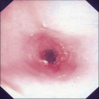
Fig 4.1
Endoscopic view of oesophageal stricture
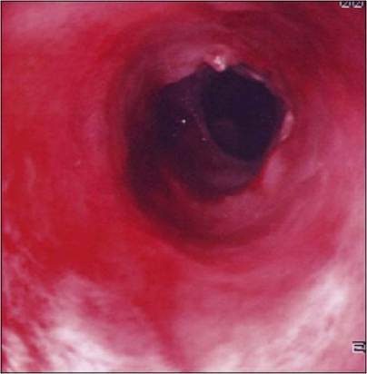
Fig 4.2
Endoscopic view of oesophageal stricture after dilation, showing mucosal tearing and oesophagitis
Nursing tips
The main areas of nursing support for this case were analgesia and nutrition.
Observation of difficulty or pain while swallowing would indicate that more attention be paid to analgesia. Generally nutritional support with liquid foods is initially indicated after balloon dilation or in other cases of severe oesophagitis. If the patient becomes able to eat food with a more solid or semi-solid consistency, they should be fed these foods. The presence of food in the oesophagus may decrease the risk of re-stricturing. If the animal is unable to take any nutrition orally a gastrostomy tube may need to be placed; however, fasting or ‘oesophageal rest’ is not indicated otherwise. There are already saliva and other secretions in the oesophagus so it does not ‘rest’.Follow-up
Probably due to the extent and severity of the oesophageal stricture in this cat, the strictures reformed and his oesophagus was repeatedly balloon dilated on about a weekly basis 11 more times (Fig 4.3). After the 12th balloon dilation, he was able to eat soft foods successfully and has done well, although continues to need to eat soft food.
Fig 4.3
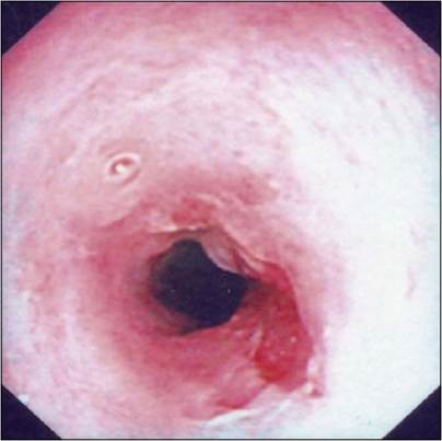
Endoscopic view of re-stricturing of oesophagus after dilation
Medical therapy
Post-dilation treatment in this cat was mostly for the oesophagitis and the gastric ulcer.
The goals of therapy for oesophageal strictures were to decrease any ongoing reflux as the damage to the oesophageal mucosa can decrease the tone of the lower oesophageal sphincter, worsening the condition, to resolve the inflammation and to decrease the risk of repeat stricture formation.
Gastric acid inhibitors such as ranitidine were used as they decrease the output of gastric acid and pepsin. Human patients with oesophagitis report a decrease in symptoms when administered histamine H2 receptor antagonists. These medications are adequate for mild to moderate oesophagitis, but may not be sufficiently effective for severe oesophagitis.
Proton pump inhibitors such as omeprazole produce greater and more long lasting suppression of gastric acid than do the H2 antagonists and are frequently used for human oesophagitis patients. Abrupt discontinuation of omeprazole is more likely to be associated with a relapse of the condition in humans; while this has not been studied in dogs or cats, a cautious approach to discontinuation of the drug may be indicated.Sucralfate has a mucosal protective effect on the oesophageal mucosa. While it has been stated that it is only effective in an acid environment (i.e. during active reflux), others have indicated that it may be effective in a neutral pH environment as well as an acidic one.
Injection of corticosteroids into the dilated area via the endoscope is also recommended; however, it was not done in this cat as the necessary instrumentation was not available. Some clinicians use systemic corticosteroid administration after balloon dilation, although there is no evidence that this is effective.
Some clinicians also use antibiotic therapy in cases of oesophagitis; however, there are also no studies showing any added benefit for them. If aspiration pneumonia is present, appropriate antibiotics should be used and possibly if systemic corticosteroids are used prophylactic antibiotics should also be used.
Colchicine (0.03 mg/kg po q 48 hour) was used in this cat to attempt to decrease fibrosis and re-stricturing. The usefulness of this drug in these cases has not been studied. Colchicine inhibits collagen synthesis. Potential side effects of colchicine include vomiting, diarrhoea and abdominal pain.
Buprenorphine (0.01 mg/kg iv q 8 hour) was used for post-procedural analgesia in this cat. Initially liquid foods were fed and when these were tolerated, feeding with very soft food was started.
Surgical therapy
Surgical techniques for the treatment of fibrous oesophageal strictures have been described, but the outcome is likely to be less successful than balloon dilation in cases without neoplasia.
While the prognosis remains guarded for animals with oesophageal strictures, many animals do well after balloon dilation; in one study 88% of patients had a successful outcome.Case discussion
Balloon dilation is the current treatment of choice for oesophageal strictures. Although many animals require repeated dilation before the oesophagus remains large enough for them to eat without regurgitating, repeating the procedure 12 times is exceptional (and also required a very dedicated owner and veterinary surgeon). Most veterinary patients require three dilations for adequate alleviations of an oesophageal stricture.
Pathogenesis
Severe oesophagitis and oesophageal strictures after anaesthesia are generally due to gastro-oesophageal reflux. Factors affecting the risk of oesophagitis include the tone of the lower oesophageal sphincter, the volume of gastric contents refluxed, the content of the refluxed material, how long it is present within the oesophagus and the healing ability of the oesophagus.
Some premedications such as atropine and xylazine used prior to general anaesthesia are associated with increased risk of gastro-oesopha- geal reflux. A pre-anaesthetic fast longer than 24 hours is also associated with an increased frequency of reflux, likely due to the more acidic nature of the contents. Alkaline reflux with a pH greater the 7.5 can also cause oesophagitis; however, it is thought to usually be less severe than that caused by acidic reflux.
When oesophageal injury due to reflux involves the deeper layers of the oesophagus, scarring and stricture are more likely. Strictures are most likely with circumferential injury and inflammation or that affecting opposing sides of the oesophagus. Inflammation stimulates lymphocytes, fibroblast and macrophage activity and formation of collagen. The initial response occurs immediately and collagen formation over several days. The signs of a stricture may be present within days to a couple of weeks after the injury.
Prognosis
Since the development of balloon dilation, the prognosis for patients with oesophageal strictures has improved. In one study a successful outcome occurred in 88% of patients treated with balloon dilation, with most animals able to eat canned, mashed or dry food without regurgitation.