Phenobarbitone responsive retching in a dog
Initial presentation
Gagging, retching and vomiting for 10 days
Signalment: 6-year-old neutered male Jack Russell terrier x poodle, body weight 6.6 kg
Case history
This dog had been referred for a suspected upper respiratory disorder or oesophageal foreign body based on his clinical signs and diet.
He had been keen to eat, although according to the owner it appeared painful for him to eat and his food intake was decreasing. He was able to prehend and swallow food, but vomited 5 to 30 minutes after eating. Active abdominal contractions were present prior to and during the vomiting; the vomitus contained bile, digested and undigested food. The frequency of the episodes was increasing to several times per day.His faeces were normal and he was defecating, drinking and urinating normally. He was fed dry food mixed with some canned food, plus table scraps including pork bones and chews. His vaccinations and de-worming programme were up to date.
He had lost weight and was becoming tired more quickly on his walks.
Physical examination
The dog was dull but responsive and his body condition score was 4/9. Mucus membranes were pink but tacky and he was estimated to be about 6% dehydrated. His capillary refill time was less than 2 seconds. Oral examination revealed hypersalivation, halitosis and mild gingival tartar.
He was trembling and palpation of the ventral neck resulted in a marked pain reaction, with spasms of the neck, bruxism and vocalization. The submandibular salivary glands were mildly enlarged and very firm.
Thoracic auscultation revealed normal heart and lung sounds, with a heart rate of 80 beats per minute (bpm) and a respiratory rate of 24 breaths per minute. Rectal temperature was 36.7° C.
Problem list and discussion of problems
• Vomiting with severe gagging and retching
Differential diagnosis
Differential diagnoses for the vomiting with gagging and retching may include some of the following categories of disorders.
• Oral/pharyngeal/oesophageal disorders are included in this case as the vomiting was accompanied by signs possibly relating to the upper alimentary tract. In some cases, patients with these disorders will also vomit.
• salivary gland necrosis
• oesophagitis
• foreign body within the pharynx, oesophagus or trachea
• gastro-oesophageal reflux
• Disorders of the stomach
• foreign body
• gastritis
• ulceration
• chronic partial dilation-volvulus
• neoplasia
• Disorders of the small intestine
• foreign body
• inflammatory bowel disease
• neoplasia
• intussusception (unlikely due to age and signs)
• Dietary causes
• dietary sensitivity
• dietary indiscretion
Case work-up
The dog was admitted to the hospital and administered intravenous crystalloid fluids to correct the dehydration.
Minimum data base
Haematology, serum chemistry and routine urinalysis were performed.
Haematology results showed a mature neutrophilia with a neutrophil count of 14.89 ? 109∕l (reference range 3.6-12.0 ? 109∕l) and a monocytosis (2.1 ? 109∕l; reference range 0-1.5 ? 109∕l), consistent with inflammation or infection.
Serum chemistry revealed a hyperalbuminaemia of 40.8 g/l (reference range 26-35 g/l), consistent with dehydration. Hypokalaemia with a potassium concentration of 3.1 mmol/l (reference range 3.6-5.6 mmol∕l) was also noted and corrected with the addition of potassium chloride to the intravenous fluids.
Urinalysis showed a high specific gravity of 1.041, again consistent with dehydration and showing good renal concentrating ability; the rest of the urinalysis was unremarkable.
Imaging and histopathology of the biopsy
Thoracic and abdominal radiography showed no abnormalities. Abdominal ultrasound showed no abnormalities.
Upper gastrointestinal endoscopy revealed erosions and erythema of the distal oesophagus near the lower oesophageal sphincter (Fig 5.1).
No foreign bodes or masses were observed. The gastric and duodenal mucosa appeared normal and histopathology of pinch biopsies taken from these areas were reported as normal tissue.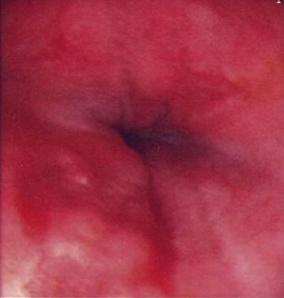
Fig 5.1
Endoscopic view of distal oesophagus showing oesophagitis
Surgical biopsies were taken of one of the enlarged salivary glands. Histopathology of this gland showed some evidence of necrosis.
Diagnosis
The results for this case rule out many causes of gagging and vomiting and are most consistent with salivary gland necrosis, also termed phenobarbitone responsive hypersialism. In some cases the histopathology of the salivary gland will be normal. This dog also had evidence of oesophagitis.
Pathophysiology and epidemiology
This disorder is poorly understood but is thought to be a form of limbic epilepsy. While it has been reported in a wide variety of dogs, young- to middle-aged Jack Russell terriers and wirehaired fox terriers appear to be predisposed.
Medical treatment
Treatment with phenobarbitone at 2 mg/kg twice a day orally was initiated. The dog responded within 48 hours with a resolution of the brux- ism, oesophageal spasms, hypersalivation and vomiting.
Treatment for the concurrent oesophagitis was also started using ranitidine (2 mg/kg po q 12 hours), sucralfate (1 ml po q 8 hours) and buprenorphine (20 μg∕kg iv q 6 hours).
Follow-up
The dog was sent back to the referring veterinary surgery for assessment of clinical improvement and serum phenobarbitone concentration assay, which was 23 μg∕ml. While this is on the low end of the therapeutic range for idiopathic epilepsy, it was adequate for controlling the signs in this dog. A commonly used reference range for serum phenobarbitone concentrations is 15-45 μg∕ml; however, some neurologists feel that values below 20 μg∕ml may not control clinical signs and with values over 40 μg∕ml (or even over 35 μg∕ml) there is a risk of hepatotoxicity.
Because of this risk, monitoring of liver parameters was also recommended, as for any dog on phenobarbitone.Clinical tip on serum phenobarbitone concentrations
The most useful single sample is the trough phenobarbitone concentration, i.e. immediately before a dose is due. If it is not practical to collect a sample at this time then try to take subsequent samples at the same time in the dosing cycle. Some authors advocate the use of peak phenobarbitone levels (2-4 hours post-pill) as well as trough levels (prior to dosing) to check the maximal serum concentration. A study has suggested that in most cases this is not necessary.
Prognosis
The prognosis in cases which respond to therapy is excellent. In most reported cases it has been possible to slowly wean the dogs off the phenobarbitone starting from 3 months post-diagnosis with cessation of medication within 6 months from diagnosis.