Oesophageal foreign body in a dog
Initial presentation
Dysphagia, regurgitation
Signalment: 8-month-old entire male Labrador retriever, body weight 20 kg
Case history
The dog had been regurgitating for about 2 days.
He was still interested in eating, but appeared to be uncomfortable when trying to swallow. After swallowing, undigested food was frequently regurgitated, usually within 5 minutes. He was able to swallow water, although occasionally appeared to also regurgitate water. His faeces had been normal, although he had not passed any faeces for 1 day before admission.Prior to this he had been a healthy puppy, had received his initial course of vaccinations and had been de-wormed twice with fenbendazole. His regular diet was a premium quality puppy food and he was given treats of human food. The owners were suspicious that the dog could have eaten a bone that he scavenged from the rubbish.
Physical examination
At presentation, the dog was quiet but alert. He had a body condition score of 5/9 and was estimated to be about 6% dehydrated. His mucous membranes were pink and slightly tacky; his capillary refill time was about 3 seconds. Halitosis was noted during the oral examination and
there was evidence of dried saliva around his mouth. A gag reflex was present, the dog could move his tongue normally and the oral cavity appeared normal.
Thoracic auscultation and abdominal palpation revealed no abnormalities and his lymph nodes were of normal size. His rectal temperature was 38° C, respiratory rate 40 breaths per minute and heart rate was 80 beats per minute.
Problem list and discussion of problems
The dog’s problems were regurgitation and halitosis. The discomfort in trying to swallow appeared to be due to oesophageal dysphagia and was thought to be related to the cause of the regurgitation. There was also evidence of drooling, with the dried saliva present around his mouth.
The dehydration was thought to be secondary to his swallowing problems.Differential diagnosis
Causes of regurgitation include:
• Pharyngeal disorders
• pharyngeal obstructive disorders, such as foreign bodies, tonsillar neoplasia or retropharyngeal lymphadenopathy
• pharyngeal neuromuscular disorders, such as myasthenia gravis, cranial nerve (IX, X) neuropathies, brain stem or cerebellar disorders, cricopharyngeal achalasia, botulism, rabies (although rabies is unlikely in the UK)
• Oesophageal disorders
• oesophagitis
• oesophageal obstructive disorders, such as foreign bodies, stricture, neoplasia, vascular ring anomalies, perioesophageal masses
• oesophageal neuromuscular disorders, such as megaoesophagus or motility disorders
• oesophageal diverticula
• hiatal hernia
Differential diagnoses for halitosis include:
• Diet related cases, such as food remaining in the mouth, pharynx or oesophagus, and coprophagia
• Cheilitis
• Oral cavity or pharyngeal disorders such as foreign bodies or inflammatory lesions
• Nasal cavity or sinus disorders with inflammation or necrosis
• Dental disease
• Oesophageal diseases with food remaining in the oesophagus
• Malassimilation
• Systemic disorders such as uraemia or liver disease
Drooling can be due to excessive saliva production or failure to adequately swallow saliva. Some dogs also drool in anticipation of feeding and some cats while purring. With the concurrent history of regurgitation, the drooling in this dog was thought to be due to a swallowing problem or oesophageal discomfort.
Case work-up
Initial treatment included administration of intravenous crystalloid fluid therapy to correct the estimated 6% dehydration.
Minimum data base
Haematology showed a packed cell volume of 0.516 l/l (reference range 0.39-0.55 l/l). While this is within the reference range, it is higher than expected for a Labrador retriever and the increase was likely due to dehydration.
Serum chemistry results, including electrolyte values, were within the reference ranges except for urea which was just above the upper end of the reference range at 8.1 mmol/l (reference range 1.7-7.4 mmol/l). The serum creatinine was within the reference range at 111 pmol/l (reference range 40-132 pmol/l). Elevations in urea can be due to haemoconcentration from dehydration, but can also occur due to renal causes or from bleeding into or from the gastrointestinal tract.The urine specific gravity of this dog was 1.047, indicating good urine concentrating ability and consistent with dehydration, and as the serum creatinine was not elevated, renal or post-renal causes of the elevated urea were unlikely.
Imaging
Thoracic and abdominal radiographs showed a foreign body of mineral density in the caudal oesophagus at the level of the diaphragmatic hiatus (Figs 6.1 and 6.2) and another foreign body of mineral bone density inside the stomach. There was no evidence of aspiration pneumonia and no evidence of mediastinitis or pneumomediastinum, so the foreign body did not appear to have penetrated the oesophagus.
Figs 6.1
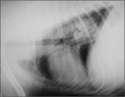
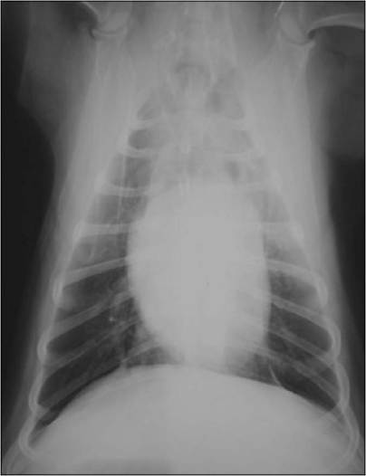
Figs 6.1 and 6.2
Lateral and ventrodorsal thoracic radiographs showing radiopaque foreign body in distal oesophagus
(courtesy of Dr Tobias Schwarz)
Diagnosis and treatment
Endoscopy
After re-hydration the dog was anaesthetized and oesophagoscopy was performed. A bone foreign body was visualized (Fig. 6.3).The foreign body was removed using retrieval forceps (Fig 6.4). The oesophageal wall showed ulcerations and mucosal tearing from the presence of the bone (Fig 6.5).
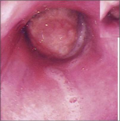
Fig 6.3
Endoscopic view of bone stuck in the oesophagus
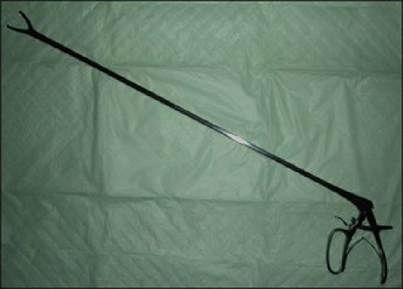
Fig 6.4
Retrieval forceps used to remove oesophageal foreign bodies
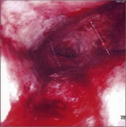
Fig 6.5
Endoscopic view of the oesophagus after removal of the bone, showing trauma and tearing of oesophageal mucosa
(courtesy of Alison Ridyard)
Medical management
After removal of the bone, the dog was treated with sucralfate suspension (4 ml po q 8 hours), ranitidine (2 mg/kg po q 12 hours) for its antacid and promotility effects and clavulanate-potentiated amoxicillin (15 mg/kg po q 12 hours).
He was continued on intravenous fluids at a maintenance rate. At 24 hours post-procedure the dog was offered water and then later a liquid food, which he was able to swallow without discomfort or regurgitation.Outcome
The dog was discharged 48 hours after removal of the bone, with instructions to continue the medications and feed soft food for a week. At a re-visit 2 weeks later, the dog was eating well and was able to swallow comfortably with no regurgitation.
Discussion and epidemiology
Oesophageal foreign bodies are a frequent clinical problem in dogs and cats. They are more common in dogs than in cats, possibly because of the more discriminating eating habits of cats. Small breeds of dogs, especially West Highland white terriers and Yorkshire terriers are predisposed, although Bernese mountain dogs were over-represented in one study.
The most common oesophageal foreign bodies found in dogs are bones or bone fragments, such as found in this dog, and fish hooks. Other foreign bodies found in dogs include coins and hard food (e.g. uncooked or partially cooked potatoes, hard fruits), whereas toys or other play objects are more commonly found in cats. Many other objects have been found, including fabric, magnets, sticks, needles and just about anything an animal can attempt to swallow. Dental chews are increasingly being recognized for their potential to cause oesophageal obstruction.
Many foreign bodies are regurgitated or pass into the stomach or intestines, but those that are too large to pass through the oesophagus cause mechanical obstruction. The severity of oesophageal damage is dependent upon foreign body size, angularity or sharp points and the duration of obstruction. The most common locations are within the thoracic inlet, the base of the heart or the diaphragmatic hiatus, as these are the areas of least distensibility within the oesophagus. The pressure of the object can lead to pressure necrosis of the oesophageal wall, causing perforation or subsequent stricture.
In many cases there is a history of foreign body ingestion. In some cases the ingestion of the object goes unnoticed, particularly those associated with scavenging or rubbish ingestion. The onset of clinical signs depends upon the severity of oesophageal obstruction. Animals with complete oesophageal obstruction are often presented with acute signs, whereas animals with incomplete obstruction may be presented within days to weeks after the initial ingestion of the foreign body. Clinical signs include regurgitation, excessive salivation, odynophagia, anorexia, dysphagia, retching and respiratory distress.
Bone foreign bodies can occasionally be palpated if they become lodged in the cervical oesophagus, but definitive diagnosis requires radiography. Radiodense foreign bodies can be detected with survey radiography, but confirmation of radiolucent foreign bodies will require administration of contrast agents. Iodine contrast agents should be used instead of barium if oesophageal perforation is suspected. A foreign body can be confirmed and often removed during endoscopy.
The most important differential diagnoses would include oesophageal stricture, neoplasia, hiatal hernia and gastro-oesophageal intussusception. Each of these conditions can be differentiated with radiography and/or endoscopy.
Oesophageal foreign bodies should be removed promptly. The longer the foreign body is lodged in the oesophagus the greater the chances of oesophageal mucosal damage, ulceration and perforation. Rigid or flexible fibreoptic endoscopic retrieval should be the initial approach to treating an oesophageal foreign body although fluoroscopic-guided retrieval has been described. A rigid endoscope is most useful in retrieving large foreign bodies, particularly bones or bone fragments. Large grasping forceps are passed through the rigid endoscope to retrieve the foreign body. Large foreign bodies that cannot be safely removed through the mouth can occasionally be pushed into the stomach and removed by gastrotomy.
Smaller foreign bodies are best managed with a flexible fibreoptic endoscope and basket, tripod or snare retrieval forceps. Flexible endoscopes are particularly useful in retrieving fish hooks. In one study, removal of oesophageal foreign bodies using an endoscope was successful in 90.2% of the dogs.Careful assessment should be made of the damage to the oesophagus via the endoscope after foreign body removal. Survey radiographs should also be taken after removal to check for pneumothorax or pneumomediastinum secondary to oesophageal perforation.
Affected animals may be fasted for 24 hours after foreign body removal. Longer periods of fasting may be required if the oesophagus is necrotic, in which case, placement of a gastrostomy tube at the time of endoscopy facilitates feeding bypassing the oesophagus.
Nursing tip on feeding post-oesophageal foreign body removal
Patients who have had an oesophageal foreign body removed are likely to initially have a painful oesophagitis. Liquid foods or canned food which has been made more liquid in a blender may be easier to swallow. If water is added to a canned food, the caloric content should be determined to make sure it is adequate. As these diets may be new to the patient and especially if the patient has not been eating adequately for the past 3 days or longer, the amount of food initially fed should not be more than one-third of the resting energy requirement per day. The amount can be gradually increased over 3 days. If the patient is reluctant to eat or swallows with difficulty, reassessment of the oesophagus and possibly the use of more analgesia may be necessary.
Specific therapy for oesophagitis should include oral sucralfate suspensions (0.5-1.0 g po q 8 hours). Suspensions of sucralfate are more therapeutic than intact tablets. Anti-inflammatory doses of glucocorticoids have been recommended by some clinicians in those animals at risk for oesophageal stricture; however, there is no evidence that oral administration of glucocorticoids is of any benefit. In animals which have formed an oesophageal stricture, local oesophageal injections of corticosteroid (e.g. triamcinolone) via an endoscope after balloon dilation have shown some benefit and possibly this would be beneficial in animals at risk for oesophageal stricture, although it could also possibly delay healing. The risk of stricture is greatest in animals with a 180 degree or greater transmucosal ulceration. Broad-spectrum antibiotics should be considered in animals with severe ulceration and/or small perforations.
Minor oesophageal tears or lacerations less than 1 cm in length can usually be managed conservatively. Surgery is indicated if endoscopy fails or if there is evidence of larger oesophageal perforation. Gastro- tomy is preferred to oesophagotomy for distal oesophageal foreign bodies because of the poorer healing properties of the oesophagus and the potential for stricture formation. However, oesophagotomy is indicated in those cases where the foreign body could not be removed through gastrotomy. Surgery is also indicated to repair oesophageal perforation.
Prognosis
The prognosis for most cases with oesophageal foreign bodies is generally good, especially if they are removed immediately, and one study reported 92% of dogs had no complications after discharge (although this study included gastric foreign bodies, which have a better prognosis). A worse prognosis is associated with foreign bodies that are large, have sharp points or are retained for a prolonged period of time. Immediate complications include complete obstruction or laceration, or aspiration pneumonia from the regurgitation. Late complications, which can occur a week or more after foreign body removal, include perforation, haemothorax, fistulation and diverticula, segmental hypomotility, or stricture formation.
More on the topic Oesophageal foreign body in a dog:
- Oesophageal foreign body in a dog
- Foreign Body Periondontitis: Hair Tooth
- Persistent right aortic arch in a dog
- Phenobarbitone responsive retching in a dog
- Table of Contents
- 4 Oesophageal stricture in a cat
- 1 Swallowing and regurgitation
- Medications used in the treatment of feline hepatic lipidosis
- 11 A foreign body in the small intestine of a dog
- Multiple choice questions