Olfaction: The sense of smell
Both smell and taste are chemical senses involving chemoreceptors. Olfaction involves the detection of volatile chemicals in solution.
Anatomy of olfactory receptors
The olfactory epithelium is located in the roof of the nasal cavity along the inferior surface of the cribriform plate of the ethmoid bone and extending along the superior nasal concha and upper part of the middle nasal concha.
It consists of olfactory receptors, supporting cells, and basal stem cells.Olfactory receptors are bipolar neurons (Fig. 11.1). At their apical end, the dendrite forms a knob from which several long cilia project. These cilia lie flat on the nasal epithelium and are covered with a thin mucus layer produced by the supporting cells and olfactory glands. Unlike other cilia, these remain stationary. At their basal end, the axon from the olfactory receptors projects through the cribriform plate and into the olfactory bulb.
The supporting cells are columnar epithelial cells surrounding the olfactory receptors. They provide physical support and cushioning, nourishment, and electrical insulation for the olfactory receptor cells.
Anatomy and Physiology of Domestic Animals, Second Edition. R. Michael Akers and D. Michael Denbow. © 2013 John Wiley & Sons, Inc. Published 2013 by John Wiley & Sons, Inc.
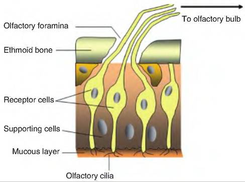
Fig. 11.1. Olfactory receptors. Olfactory receptor cells are interspersed among supporting cells. The olfactory cilia, embedded in the mucous layer of the olfactory epithelia, detect odorants causing the development of a receptor potential in the olfactory cells. Axons project from the olfactory receptors forming the olfactory nerve (cranial nerve I).
They also contain a yellow-brown pigment that gives the olfactory epithelium a yellow tint.
Basal stem cells are found between the supporting cells. They continually undergo cell division producing new olfactory receptors. The olfactory receptors live for approximately 1 month before dying and being replaced. This makes these neurons unusual, since most neurons are long-lived and are not replaced.
Olfactory (Bowman's) glands are found in the connective tissue that supports the olfactory epithelium. These glands produce mucus that is carried to the surface of the olfactory epithelium and that dissolves odorants, that is, chemicals that stimulate the olfactory hairs.
The facial nerve (cranial nerve VII) innervates supporting cells and olfactory glands. When these structures are stimulated by certain chemicals, nerve impulses in the facial nerve can result in stimulation of the lacrimal glands in the eyes and nasal mucous glands. This can cause tearing and a runny nose.
Physiology of olfaction
Odorants dissolve in the mucus membrane of the olfactory epithelium. They bind to protein receptors on the olfactory cilium membrane, stimulating G proteins and resulting in the activation of adenylate cyclase. Adenylate cyclase then catalyzes the production of cyclic adenosine monophosphate (cAMP), which causes Na+ channels to open. The influx of Na+ results in depolarization of the olfactory receptor and the production of a receptor potential.
Olfactory pathway
The unmyelinated axons of the olfactory receptors constitute the first-order neurons, which pass through the numerous olfactory foramina in the cribriform plate of the ethmoid bone. These axons collectively form the right and left olfactory nerves (cranial nerve I). They terminate on secondary neurons located within the olfactory bulbs located just below the frontal lobes of the cerebrum. Neurons from the olfactory bulb extend posteriorly in the olfactory tract, projecting to the lateral olfactory area in the temporal lobe. The olfactory area is part of the limbic system. The olfactory neurons also project to the hypothalamus and other limbic areas, thus explaining how smell can evoke various memories and emotions.