Open-Mouth View
A commercially available U-shaped acrylic head rack can be used to facilitate positioning (Figure 4-4, A). Without a positioning device, medical-grade adhesive tape can be used to separate the mandible from the maxilla (Figure 4-4, B).
With the animal in dorsal recumbency, the head is acutely flexed toward the thoracic inlet. The vertical primary beam is directed rostroventral to caudodorsal and centered immediately ventral to the hard palate. The hard palate and mandible are 30 degrees from the vertical plane. This will highlight both tympanic bullae with minimal superimposition from the surrounding structures. The endotracheal tube should be removed or secured against the mandible. To avoid increased bullae opacity due to superimposition, the tongue should be pulled rostrally and secured to the mandible on the midline. The normal bullae are noted as thin-walled structures with a lucent center ventral to the base of the skull (Figure 4-4, C). Increasing the angle of the hard palate relative to the primary beam can be used as an alternative to the open-mouth projection (Figure 4-4, D). This projection is easier to perform because it is a closed-mouth view that highlights the most caudal surface of the tympanic bullae. Caution should be taken in assessing abnormal findings on this projection because its clinical value has not been studied as extensively as the open-mouth view.2 Normal bullae are thin-walled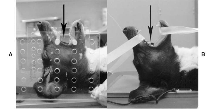
Figure 4-4
A, Patient positioning to obtain a rostral 30-degree ventral-caudodorsal open-mouth oblique projection. The arrow indicates the trajectory of the primary beam. The positional device is known as an acrylic head rack, which is radiolucent on radiographs. B, Patient positioning to obtain a rostral 30-degree ventral-caudodorsal open-mouth oblique projection without the aid of a positional device. Medical-grade adhesive tape is used to keep the mouth open.
The arrow indicates the trajectory of the primary beam.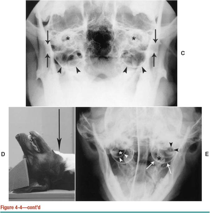
C, Rostral 30-degree ventral-caudodorsal open-mouth oblique radiograph of a normal dog. The tympanic bullae (arrowheads) are located ventral to the petrosal portions of the temporal bones (asterisks). The external acoustic meatus (arrows) is partially obscured by the overlying coronoid process of the mandible. D, Patient positioning to obtain a rostroventral-caudodorsal closed-mouth oblique radiograph. There is an increased angle of the hard palate in relationship to the primary beam represented by the arrow. This projection is technically easier to perform, but its clinical value has not been studied as extensively as the open-mouth projection. It highlights the caudal aspect of the tympanic bullae. E, Rostral 30-degree ventral-caudodorsal open-mouth oblique radiograph of a normal cat. An osseous septum (white arrowheads) separates the dorsolateral compartment (white asterisk) from the larger ventromedial compartment (black asterisk). The white arrows indicate the walls of the medioventral compartment. The external acoustic meatus (black arrowheads) is visualized overlying the dorsolateral compartment. structures with a lucent center ventral to the base of the skull (see Figure 4-4, C). Their walls are of uniform thickness. They are symmetrical in size, shape, and opacity when compared with one another.
In cats an osseous septum divides the bullae into two separate but communicating tympanic cavities—a smaller dorsolateral compartment and a larger ventromedial compartment (Figure 4-4, E). There is less variation in the thickness of the bulla walls between breeds in cats. The external acoustic meatus is sometimes superimposed on the dorsolateral compartment.