Abnormal Radiographic Findings
Otoscopic evaluation is the method of choice to evaluate the external ear canal. However, radiographs can reveal narrowing of its lumen by soft-tissue proliferation from extraluminal masses in cases of neoplasia or by inflammatory tissue, exudates, or debris in cases of otitis externa or trauma (Figure 4-5, A).
Dystrophic calcification can be seen associated with chronic otitis externa (Figure 4-5, B).Diseases affecting the middle ear, such as otitis media, neoplasia, and cranio- mandibular osteopathy, as well as polyps can be evaluated with a bullae series. Radiographic findings are nonspecific; therefore the list of differential diagnoses
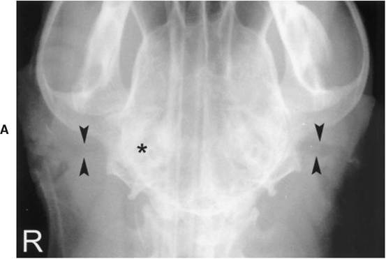
Figure 4-5
A, VD radiograph of a 4-year-old Doberman diagnosed with chronic bilateral ear infections. Both external acoustic canals (arrowheads) are narrowed and somewhat tortuous. The right canal is smaller and less defined than the left. There is an increased opacity associated with the right tympanic bulla and petrosal portion of the temporal bone (asterisk). Compare the canals with the normal external acoustic canals depicted in Figure 4-3,C.
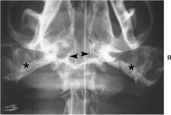
Figure 4-5—cont'd
B, VD radiograph of a 2-year-old Bulldog diagnosed with chronic bilateral otitis. Radiographs were taken prior to a total ear canal ablation. There is exuberant bilateral dystrophic calcification of the external acoustic canals (asterisks). The visible walls of the tympanic bullae (arrowheads) appear normal.
should be generated in light of the clinical history and not radiographic findings alone.
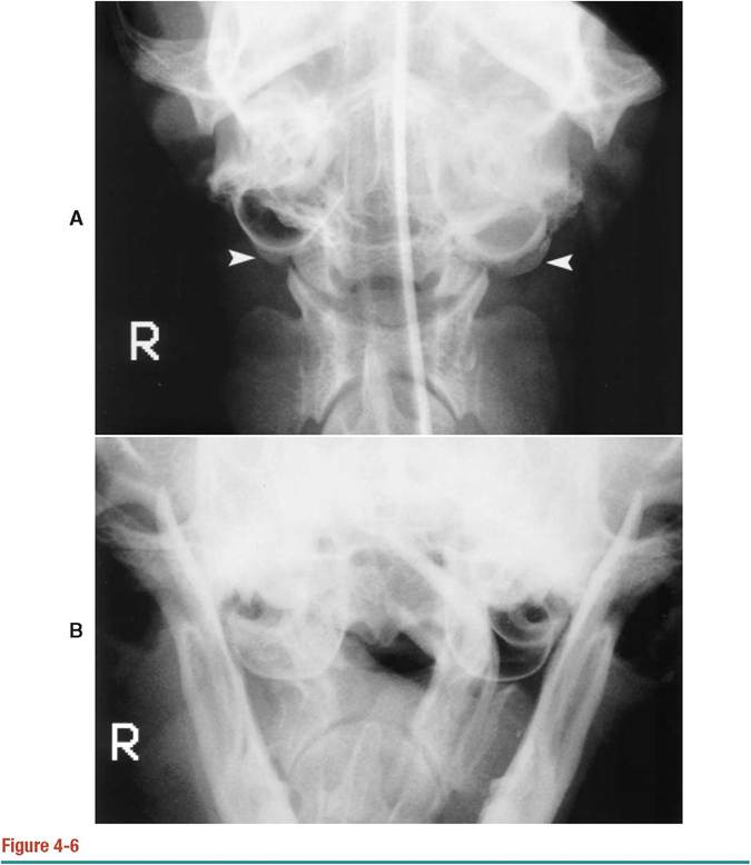
Common findings in otitis media include thickening of the wall of the bullae, increased soft tissue opacity within the bullae, and increased size of the bullae (Figure 4-6).
In the large majority of cases it is not possible to differentiate a fluid- filled bulla from one with a thickened wall. If the process is chronic, the increased opacity is likely the result of both thickening and fluid accumulation. Rare mineral concretions within the bullae, also known as middle ear otoliths, have been reported in four dogs.3,4 Middle-ear otolithiasis may be associated with nonactive or active cases of otitis media. If the otitis media is secondary to otitis externa, narrowing and mineralization of the external acoustic canal can also be seen.Common findings associated with neoplasia affecting the middle ear include soft-tissue swelling, which may or may not obliterate the external acoustic canal; lysis of the wall of the bullae; and increased opacity of the bullae without lysis (Figure 4-7). Less commonly, ill-defined periosteal reactions arising from the bullae and surrounding bones can be seen. Neoplasia of ceruminous glands, squamous cell carcinomas, and anaplastic carcinomas have been diagnosed among others.5,6
Increased opacity of the bulla as the result of thickening of the walls can also occur in cases of invading nasal polyps7 and craniomandibular osteopathy (Figure 4-8).
A, Rostroventral-Caudodorsal closed-mouth oblique radiograph of a 14-year-old cat diagnosed with a nasopharyngeal polyp. There is bilateral thickening of the caudal aspect of the walls of the bullae (arrowheads). The left bulla is increased in opacity, which can be the result of fluid or a mass within the bulla or the result of the sclerosis and thickness of the wall. B, Rostroventral- caudodorsal open-mouth oblique radiograph of an 11-year-old cat diagnosed with otitis media, which presented with right-sided head tilt and circling. The right bulla is mildly enlarged with a generalized increase in opacity, which is compatible with a diagnosis of otitis media.
However, radiographically it is not possible to determine whether the increased opacity is the result of fluid or a mass within the bulla. Otoscopic examination reveals generalized thickening of the external and middle ear.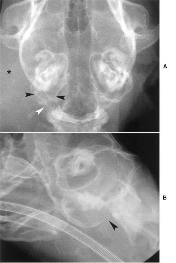
Figure 4-7
A, Ventrodorsal radiograph of a 10-year-old cat diagnosed with a ceruminous gland carcinoma. There is lysis of the caudal aspect of the occipital bone (white arrowhead), thinning and lysis of the caudal aspect of the wall of the bulla (black arrowheads), and an increased soft tissue opacity obliterating the external acoustic canal (asterisk). B, Oblique radiograph of a 10-year-old cat diagnosed with a ceruminous gland carcinoma. There is lysis of the most caudal aspect of the tympanic bullae (black arrowhead).
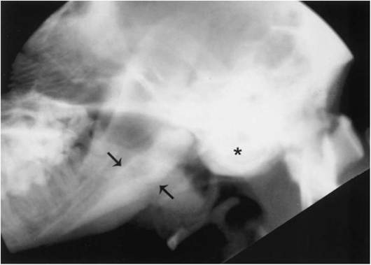
Figure 4-8
Lateral radiograph of the middle ear of a 1-year-old Scottish Terrier diagnosed with cranio- mandibular osteopathy. There is marked increased opacity associated with the tympanic bullae (asterisk), which is the result of thickening of the walls. The visible cortex of the mandibular body (arrows) is also thickened. VD radiographs (not shown) confirmed bilateral bullae thickening. Compare the bullae with the normal bullae in Figure 4-1, C.
Positive Contrast Canalography
This technique uses nonionic iodine-based contrast material to assess the integrity of the tympanic membrane as well as the anatomy of the external acoustic canal.1,8 The technique is more accurate than otoscopy for detecting iatrogenic rupture of the tympanic membrane in normal dogs and can be used to assess stenosis of the external acoustic canal. Its usefulness in cases of otitis media remains uncertain, as inflammatory secretions may block the flow of contrast material and prevent it from filling the canal completely.8
Technique and Normal Radiographic Findings
The canals should be gently cleaned before the study.
The animal is placed in sternal recumbency, and 1 cc of iohexol (300 mg iodine per cc) is placed within the lumen of the canal. After a massage of its vertical and horizontal portions, the canal is slowly filled with contrast until it reaches the level of the tragus. A final massage is then performed to ensure adequate distribution of the contrast in the canal. To avoid leakage of contrast, a cotton swab is placed to plug the vertical portion of the ear canal. A DV view is taken, followed by the rostrocaudal open-mouth projection. The resultant radiographs (Figure 4-9) are compared with the precontrast survey study.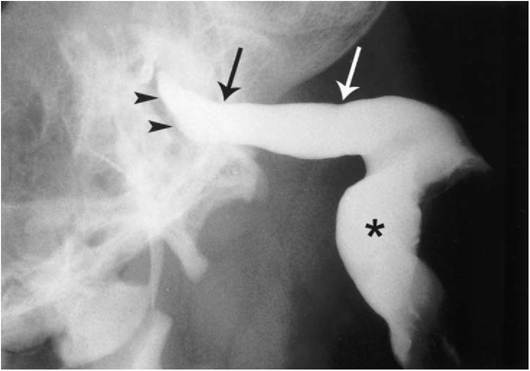
Figure 4-9
DV radiograph of a normal canalogram in a 5-year-old mixed-breed hound dog. There is complete filling of the canal by contrast material. The proximal (blackarrow) and distal borders (white arrow) of the annular ligament are noted as slight indentations in the diameter of the canal. The slightly concave border that abruptly stops the flow of contrast into the tympanic bulla represents the tympanic membrane (arrowheads). It is oriented at an oblique angle to the axis of the canal. The vertical portion of the ear canal (asterisk) is also noted.
After the study is completed, the ear is flushed with saline solution and dried. No side effects have been reported as the result of the procedure.1,8
The diameter of the proximal end of the annular cartilage tends to be smaller than the distal end of the annular cartilage. On the DV view these ends can be noted as minor indentations in the wall of the canal (see Figure 4-9). The tympanic membrane is a straight or slightly concave border overlying the tympanic bullae. It is oriented at an oblique angle in relationship to the longitudinal axis of the horizontal ear canal.
Abnormal Radiographic Findings
An intact tympanic membrane should prevent the flow of contrast material into the tympanic bullae; therefore, any leakage of contrast into the middle ear is considered diagnostic of a ruptured tympanic membrane. Stenosis of the ear canal has been documented in Shar-Peis and in Pugs.1 Although canalography can effectively determine the degree of ear canal stenosis, additional studies are needed to establish fully stenosis as a predictor of chronic inflammatory ear disease. The average diameter of the proximal end of the annular cartilage in stenotic canals is 2.6 ± 0.8 mm. In dogs in which the tympanic membrane is visible otoscopically, the range is 4.1 ± 0.7 mm.1
More on the topic Abnormal Radiographic Findings:
- Abnormal Findings
- Abnormal Bony Density or Structure
- Abnormal Cytology
- Description and Pathophysiology of the ART-Related Abnormal Fat Distribution
- Physical Findings
- Some Findings
- Instrumented gait analysis has evolved into a recognized objective evaluation that is important in surgical and rehabilitation therapy planning for the child with an abnormal walking pattern.
- Evaluation of Findings and Results Differentiated According to Demographic Variables
- DISCUSSION AND FINDINGS
- Findings and Discussions
- Results and Findings
- FINDINGS AND RECOMMENDATION
- Technique and Normal Findings
- Key Findings and Conclusions
- Report Findings and Recommendations
- Plan of the Study and Principal Findings
- INITIAL EVALUATION AND CLINICAL FINDINGS