Computed Tomography (CT)
CT provides cross-sectional imaging of the ear. By eliminating superimposition of surrounding bone, CT can clearly depict the anatomy of the inner, middle, and external ear. The CT anatomy of the middle and inner ear has been described by comparing transverse CT images in reference to a standard anatomy textbook.9 CT is slightly more sensitive than radiography in the diagnosis of otitis media.10 CT can detect subtle soft tissue changes before they are apparent on radiographs, due to its higher soft-tissue contrast resolution.
However, CT is more expensive and less available to general practices than radiography. It requires a higher degree of technical expertise to operate a CT unit than a routine x-ray room, as well as a complete knowledge of the acquisition protocols and associated image post-processing software to generate diagnostic images.Technique and Normal CT Findings
As with conventional radiography, assessment of the images relies on symmetry; therefore, careful positioning of the patient under anesthesia in the CT gantry is crucial. Most new units have laser-positioning guides to ensure that the longitudinal axis of the head enters the gantry at a 90-degree angle to the primary beam. Head-positioning devices can also facilitate positioning the hard palate parallel to the CT table (Figure 4-10). Higher kilovolt potential (kVp) and mA settings than conventional radiographs are used to reduce artifacts and noise. In dogs, a kVp of 120 and an mA of 200 are used with a full field of view and a 512 matrix. In cats, an mA of 150 with a half field of view and a 512 matrix are used. Contiguous or overlapping transverse slices 1 to 3 mm thick extending from the middle third of the nasal cavity to the foramen magnum are generated. Either the helical or axial mode of scanning can be used. A cursory look at adequately positioned images should reveal symmetry between contralateral anatomical landmarks in the head (Figure 4-11).
Both frontal sinuses, temporomandibular joints, zygomatic portions of the temporal bones, occipital brain lobes, and atlantooccipital joints should appear similar in size and shape when they are being imaged in a slice (see Figure 4-11). Two different window settings, which determine the range of tissues that will appear gray on the image, are used to depict fully the anatomy of the middle and inner ear. Wide window-width settings are used to highlight bone, whereas narrow window widths centered at a soft tissue level are used to highlight soft tissues (Figure 4-12).11 A typical window for bullae is 3200 at a level of 500, whereas a soft-tissue window width is 375 with a level of 40. Subtle changes commonly noted in cases of otitis media such as sclerosis and thickening of the bulla could be missed if a wide window width is not used. On the other hand, small amounts of fluid within the bulla can be missed if a narrow window width centered at soft-tissue settings is not used. Intravenous injection of iodinated contrast material, such as meglumine diatrizoate, should be performed to enhance inflammatory and neoplastic lesions, at a dose of 1 cc per pound of body weight (375 mg iodine per cc).Like conventional radiography, CT can identify the major anatomical landmarks of the external and middle ear. A major advantage of CT over conventional radiography
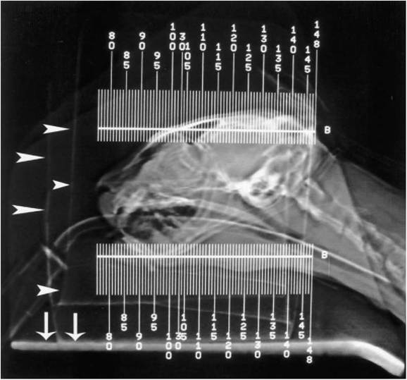
Figure 4-10
Scout image generated by the CT unit. The head of the cat is positioned in a U-shaped head holder (arrowheads) that facilitates positioning. The holder elevates the head over the CT table (arrows), maintaining the area of interest in the center of the unit's gantry. Each numbered line represents a single transverse slice. In this patient, approximately 20 slices are required to cover the ear anatomy. The tracheal tube does not need to be removed.
is the detailed visualization of the structures of the middle and inner ear, such as the tympanic membrane, the auditory ossicles, the cochlea, the vestibular aqueduct, and the semicircular canals (Figure 4-12, C).
An artifact known as beam hardening, however, often obscures the area of the pons. This artifact can be identified as streaklike black bands generated by the petrosal portions of the temporal bones. Beam hardening is the result of absorption of low-energy x-ray photons by the highly dense bone. Visualization of the middle and inner ear structures requires careful patient positioning, generation of thin slices, wide window settings, and reconstruction of images using bone algorithms.9Abnormal CT Findings
Evaluation of CT images follows criteria similar to those of conventional radiography; both techniques use x-ray absorption in tissues to generate images.
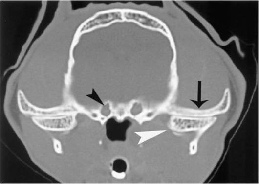
Figure 4-11
Transverse CT image at the level of the temporomandibular junction. A well-positioned patient should show symmetry between the right and left halves of the skull. Both temporomandibular joints show simultaneously in the image. The zygomatic portions of the temporal bone (arrow) and condyles of the mandible (white arrowhead) are similar in size and shape. The oval foramina (black arrowhead) are also symmetric.
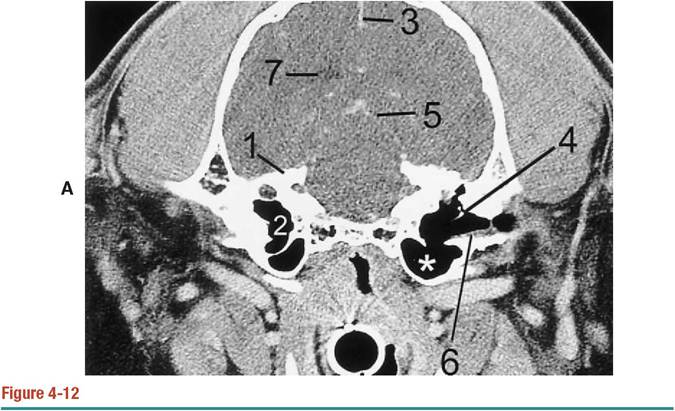
A, Post-contrast transverse CT image of a normal cat at the level of the bullae using a soft- tissue window. The septum bulla divides the bulla into a smaller dorsolateral (2) and a larger ventromedial (asterisk) compartment. The anatomical detail of the petrosal portion of the temporal bone and its contents (1) is obscured when compared with image 4-12, B. Only a faintly visible malleus (4) is seen. However, cerebral landmarks such as the falx cerebri (3), contrast enhanced meninges (5), and portions of the lateral ventricles can be identified. There is a small amount of cerumen overlying the horizontal portion of the external acoustic canal (6).
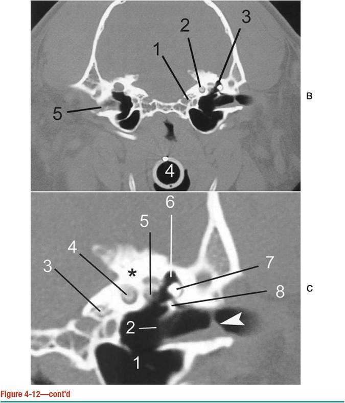
B, Transverse CT image depicted in 4-12, A, with a bone-tissue window. Compare the detail of the middle ear with image 4-12, A.
The epitympanic recess (3), cochlea (2), and carotid canal (1) are now visualized. By using the proper window, structures such as the external acoustic meatus (5), noted here with a small amount of cerumen, become apparent. The endotracheal tube is indicated with the number 4. C, Transverse CT image at the level of the inner ear in the patient depicted in images 4-12, A and B. There is a small amount of cerumen overlying the horizontal portion of the external acoustic canal (arrowhead). The septum bullae (1) and tympanic membrane (2) are faintly visible as linear areas of increased density. The carotid canal (3) is medial and ventral to the cochlea (4). The vestibular window (5) is medial to the incus (7) and malleus (8), which are located within the epitympanic recess (6). The petrosal portion of the temporal bone (asterisk) also contains the vestibular aqueduct and semicircular canals (not pictured).Diseases such as otitis,10 neoplasia,12 nasopharyngeal polyps,13 and Craniomandibu- lar osteopathy can be assessed with CT.
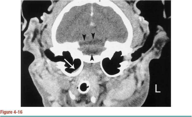
Common findings in otitis include thickening of the external acoustic canal, with or without mineralization (Figure 4-13). Also, enlargement, thickening, and sclerosis of the walls of the bulla (Figure 4-14), as well as sclerosis of the petrosal portion of the temporal bone, can be seen (Figure 4-15). Unlike radiography, CT can differentiate fluid within the bullae from thickening of the walls (Figure 4-16). An exception to the latter arises when the bullae are completely obliterated by soft-tissue density. Use of contrast can help differentiate whether the density is fluid or mass because masses tend to enhance with intravenous injection of contrast material. However, this author has seen inflammatory exudates associated with otitis externa and media that have enhanced after contrast medium administration. An inflamed external acoustic canal can be seen as a contrast-enhancing tubular structure (see Figure 4-13).
If the enhancement is limited to the canal, it is possible to be more confident in a diagnosis of otitis than when the enhancement extends out of the canal, such as in cases of neoplasia.Abnormalities noted in otitis can also be seen associated with neoplasia or osteomyelitis. Therefore, findings should be interpreted in light of the clinical history. However, neoplasia can be placed at the top of the list of differential diagnoses when
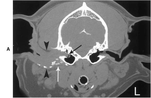
Figure 4-13
A, Transverse view of the bullae of a 5-year-old male Beagle diagnosed with chronic otitis externa imaged with a bone-tissue window. Otoscopic examination of the middle ear was not possible. The diameter of the right external acoustic canal (arrowheads) is increased. A soft- tissue density has completely replaced the air in the canal. Scattered foci of dystrophic mineralization are also noted (white arrow). The tympanic bullae are considered normal. However, there is a bulge at the level of the right tympanic membrane (black arrow), which suggests a compromise of its integrity.
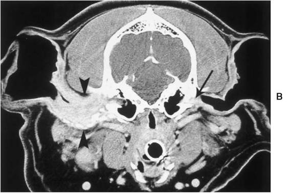
Figure 4-13—cont'd
B, Soft tissue window after injection of contrast material of the dog imaged in image 4-13, A. The obliteration of the lumen is the result of severe thickening of the walls, which exhibit well-defined contrast enhancement, likely the result of the chronic inflammatory changes (arrowheads). Cerumen or inflammatory exudates are also noted lateral to the left tympanic membrane (arrow).
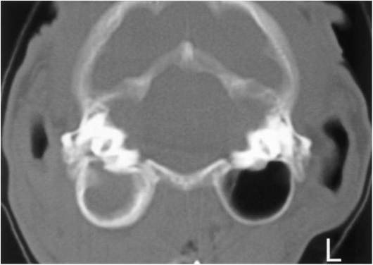
Figure 4-14
CT image of a 6-month-old cat diagnosed with otitis media. There is generalized thickening of the walls of the right bulla. The bulla is filled with dense material, which represents fluid. The affected bulla is mildly enlarged compared with the contralateral one, which is considered normal.
The petrosal portions of the temporal bones are symmetrical.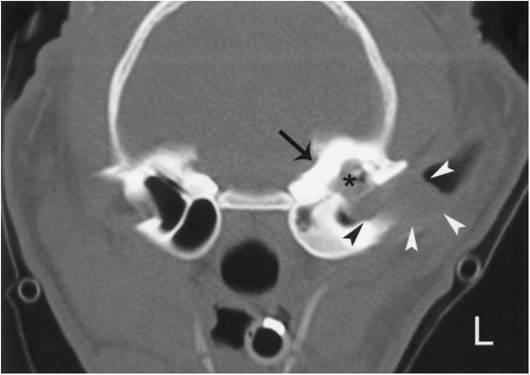
Figure 4-15
CT image at the level of the bullae of an 18-month-old-male cat diagnosed with a polypoid mass. The mass (arrowheads) is noted as a homogeneous space-occupying dense structure at the level of the external acoustic meatus and extending into the middle ear. The petrosal portions of the left temporal bone (arrow) as well as the bulla wall are thicker and sclerotic when compared with the contralateral normal right. The left septum bulla is no longer present and the epitympanic recess (asterisk) is wider, likely the result of pressure bone atrophy caused by the mass.
CT image at the level of the bullae of a castrated male cat diagnosed with otitis and a fibrosarcoma of the most rostral third of the mandible. There is a minimal volume of dense fluid (arrow) on the dependent portion of the right bulla. The corresponding radiographs of the bullae were considered within normal limits. The black streaks (arrowheads) are known as beam hardening, which is a common CT artifact generated by the petrosal portions of the temporal bones. The artifact hampers full visualization of the pons.
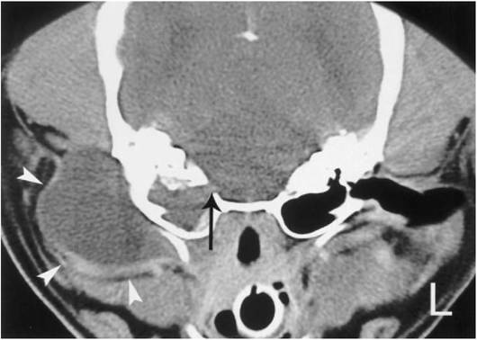
Figure 4-17
Transverse CT image of a 7-year-old Chihuahua. There is a large homogeneously dense mass lateral to the right bulla (arrowheads). Lysis of the medial aspect of the petrosal portion of the temporal bone is also noted (arrow).
there is a space-occupying lesion (Figure 4-17) of variable enhancement characteristics, extension of the lesion into the caudal fossa, and lysis of the tympanic bullae (Figure 14-18).
More on the topic Computed Tomography (CT):
- Selecting the appropriateimaging method, correctly applying the technique selected, and accurately interpreting the examination are the key steps in imaging ear disorders in dogs and cats.
- Diagnostic Tests
- Myringotomy
- Lung Cancer
- NEUROIMAGING
- SCIWORA
- Empiricalanalysis
- The existing evidence on volatility and growth
- Boon Andrew. The Ethics and Conduct of Lawyers in England and Wales. Hart Publishing,1999. — 808 p., 1999
- Griffiths-Baker Janine. Serving Two Masters: Conflicts of Interest in the Modern Law Firm. Hart Publishing,2002. — 227 p., 2002
- Grisso T.. Evaluating Competencies: Forensic Assessments and Instruments. 2nd edition. — Springer,2002. — 564 p., 2002
- Luban David. Legal Ethics and Human Dignity. Cambridge University Press,2007. — 350 p., 2007
- Ayupova Z.K.. Theory of state and law: textbook. - Almaty: Kazakh University,2015. - 192 pages., 2015
- Allen Danielle, Benkler Yochai et al. (eds.). A Political Economy of Justice. The University of Chicago Press,2022. — 416 p., 2022
- Barnes Rudolph C.. Military Legitimacy: Might and Right in the New Millennium.Frank Cass,1996. — 198 p., 1996
- Bedner Adriaan (ed.).. Real Legal Certainty and its Relevance: Essays in Honor of Jan Michiel Otto. Leiden University Press,2018. — 261 p., 2018