Magnetic Resonance Imaging (MRI)
The reader is referred elsewhere11 for an overview of the basic concepts of MRI technology. MRI does not use ionizing radiation (x-rays); therefore, it is considered a noninvasive imaging modality.
MRI manipulates the spinning behavior of hydrogen protons within a strong magnetic field to produce an image. Changes in this behavior are achieved by sending radio-frequency pulses to the hydrogen protons. The hydrogen protons in turn send radio frequencies back to a receiver antenna. A computer uses the signal from the hydrogen protons to form a gray-scale image. MRI provides cross-sectional imaging of the ear. By eliminating superimposition, the structures medial to the external acoustic canal can be seen clearly. Unlike CT, the area of interest can be imaged in an infinite number of planes without relying on slice reconstruction and without changing the position of the animal in the gantry. The image quality of the CT reconstructions is less desirable (Figure 4-19, A) than the equivalent MRI plane. MRI offers better soft-tissue contrast resolution than CT or conventional radiographs.14 Assessment of the pons is not hampered by the presence of beam-hardening artifacts commonly noted on CT (Figure 4-19, B). On the other hand, tissues with minimal hydrogen protons such as bone, air, or areas of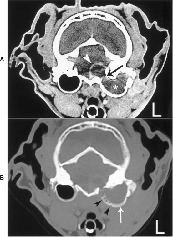
Figure 4-18
A, Transverse CT image of a 10-year-old spayed female cat presented with a unilateral persistent drainage from the ear. There is a space-occupying mass of heterogeneous enhancement (asterisk) causing enlargement of the left bulla. There is ringlike enhancement associated with the left half of the pons (arrowhead) and lysis of the medial aspect of the petrosal portion of the temporal bone (arrow). The beam-hardening artifact overlying the caudal fossa lesion precludes full assessment of the pons.
B, CT of the cat depicted in Figure 4-18, A, imaged with a bone-tissue window. Permeative lysis (arrowheads) of the medial wall of the left bulla as well as thickening of the ventral wall (arrow) become apparent. The aggressive process not only extends into the caudal fossa but also medial to the affected bulla. No further diagnostics were performed due to the poor prognosis.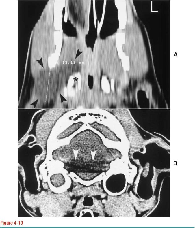
A, CT dorsal plane reconstruction of the dog depicted in Figure 4-17. There is a hypodense mass (arrowheads) located lateral and rostral to the right bulla (asterisk). The mass extends laterally to the right hemimandible. The spatial resolution of this image is poor compared with the MRI image of another dog in Figure 4-23, A. B, Transverse CT image at the level of the bullae. The beam hardening artifact (arrowheads), generated by the dense petrosal portion of the temporal bones, hampers full visualization of the intracranial structures. The right bulla is fluid filled. MRI images offer a better view of the area due to the lack of artifacts as well as to higher soft-tissue contrast resolution.
calcification will have little signal on MRI and are represented as a signal void or black on the resultant image. Therefore the normal petrosal portion of the temporal bone, the bulla wall, and the air within the bulla will appear black on MRI images and cannot be assessed with the same degree of detail as with CT (Figure 4-20). Ferromagnetic foreign material such as gun pellets, hemoclips, and identification
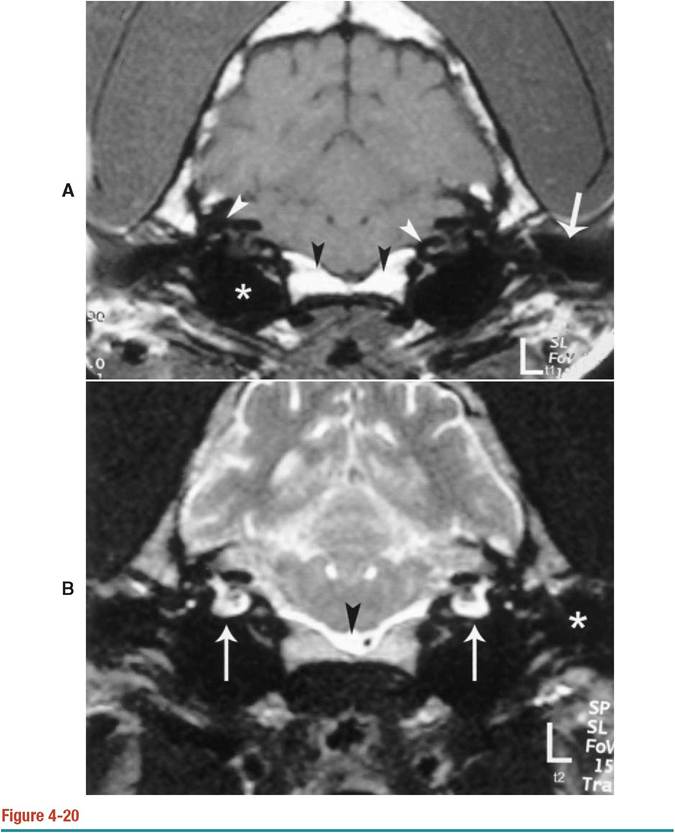
A, T1-weighted transverse image of the normal bulla of a dog. There is a large signal void, which represents the air and the walls of the bullae (asterisk). The petrosal portions of the temporal bone are also represented as irregular structures without signal (white arrowheads). Portions of the external acoustic canal (arrow) and the basisphenoid bone (black arrowheads) are also included. B, T2-weighted transverse image of the normal bullae of a dog. The hyperintense signal of the intralabyrinthine fluid associated with the cochlea and semicircular canals is noted (arrows). Low cellular fluid such as the cerebrospinal fluid is hyperintense on this sequence (arrowhead). The arrows are overlying the bullae.
chips close to the areas of interest may render the study nondiagnostic due to abrupt changes in the magnetic field, which distort and degrade the image.14 Finally, MRI is less available and significantly more expensive than CT or conventional radiography. Obviously, a higher degree of expertise and training is required to run an MRI unit than an x-ray room.
More on the topic Magnetic Resonance Imaging (MRI):
- Selecting the appropriateimaging method, correctly applying the technique selected, and accurately interpreting the examination are the key steps in imaging ear disorders in dogs and cats.
- Imaging investigations
- 19 Neurologic Diseases in Pregnancy
- Physics
- Basic principles of radiotherapy
- Placenta accreta (morbidly adherent placenta)
- Cervical cancer
- Adenomyosis
- Pelvic Mass
- Imaging of the Tympanic Bulla