Physics
Ultrasound
Ultrasound imaging has been used for medical purposes for several decades. Ultrasound refers to sound waves greater than 20,000 cycles/sec, a frequency greater than that which the human ear can appreciate.
In obstetrics and gynaecology, frequencies of 2-12 million cycles/ sec (MHz) are used. The ultrasound probe consists of two elements: a transducer and a receiver.Understanding the physical principles underlying ultrasound technology can help optimize image quality, and improve diagnostic capabilities. This information is also essential for ensuring this technology remains safe for the woman and the fetus.
Sound waves are described in terms of frequency, which is the number of repetitions (i.e. cycles) per second. The unit for measuring them is the Hertz (Hz). Another characteristic is wavelength.
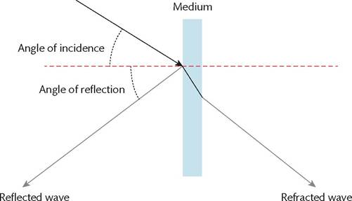
Figure 1.34 Wave front reflection and refraction.
Reproduced from Austin Ugwumadu, Medical Physics, in: Basic Sciences for Obstetrics and Gynaecology, Oxford University Press (2014) with permission from Oxford University Press.
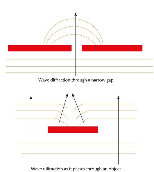
Figure 1.35 Wave diffraction.
Reproduced from Austin Ugwumadu, Medical Physics, in: Basic Sciences for Obstetrics and Gynaecology, Oxford University Press (2014) with permission from Oxford University Press.
Scatter is the combination of irregular reflection, refraction, and diffraction of a wave in multiple directions.
Absorption is the conversion of sound energy into heat as it travels through a medium.
Attenuation is a process whereby ultrasound signal strength is progressively reduced during transmission due to absorption of the ultrasound energy by conversion to heat. Attenuation is frequency and wavelength dependent.
Ultrasound uses the pulse echo principle. This is where a pulse of known speed is emitted and the time taken for the reflected echo to return is measured.
Artefacts: some images vary due to artefacts of ultrasound imaging. Examples of common artefacts encountered in obstetric and gynaecological ultrasound include the following:
• Shadowing occurs when there is strong reflection or attenuation leading to a diminished ultrasound beam with loss of imaging data distal to the reflector or attenuator.
• Increased through transmission is associated with lower attenuation than surrounding tissues, typically due to a fluid-filled structure such as a cyst.
• Reverberation artefacts occur when two or more intensely reflective interfaces cause the ultrasound beam to echo, resulting in echoes in cystic structures, appearing as solid elements.
• Refraction artefacts are caused by non-linear bending of the sound waves when there is a change in the tissue.
Radioactivity and X-rays
Radiation is broadly divided into ionizing and non-ionizing radiation. Ionization is a process whereby an atom or molecule is converted into an ion by adding or removing charged particles. Radioactive decay is the process by which an unstable atomic nucleus spontaneously loses energy by emitting ionizing particles and radiation. Ionizing radiation produced by radioactive decay includes:
• alpha radiation
• beta radiation
• gamma radiation.
Alpha particles are emitted from the nucleus of an atom. They consist of two neutrons and protons. They have a high mass and high energy but low depth of penetration. They are heavily ionizing.
Beta particles are divided into β- and β+ particles. The former is an electron that arises from beta minus decay of a neutron and the latter is a high-energy positron.
Gamma rays are high-frequency electromagnetic waves. They have a short wavelength. They are used for sterilization of medical equipment, gamma knife surgery, and in nuclear medicine.
X-rays are a form of electromagnetic radiation emitted by electrons with a wavelength in the range of 10 to 0.01 nm.
Ionizing radiation can be used for management of both malignant and non- malignant conditions in the form of radiation therapy. Radiation causes DNA damage by direct and indirect ionization of the atoms, which make up the DNA chain. Ionizing radiation is also used in the field of nuclear medicine where radioactive isotopes are administered internally. These radionuclides are atoms with an unstable nucleus and their radioactive decay emits ionizing radiation which is captured by a gamma camera. This is useful for both imaging and for treatment.
Magnetic resonance imaging
Magnetic resonance imaging (MRI) is an imaging technology using non-ionizing radiofrequency radiation inside a strong, constant, spatially homogeneous magnetic field. This process involves induction of protons by a magnetic field, followed by excitation with radiofrequency pulses and subsequent readout with receiver coils. Tesla (T) is the unit used in the International System of Units (SI) for magnetic fields. Most magnets in medical MRI produce a magnetic field of 0.5-3 T. Small differences in the microenvironment of different tissues can be detected by pulse sequences. The presence of multiple magnetic resonance properties of tissue leads to great flexibility in determining tissue contrast. This capability is superior to computed tomography (CT).
Limitations in the use of MRI include contraindications posed by metallic implants, and claustrophobia. Open MRI scanners and short-bore conventional scanners can address the issue of claustrophobia.
Intravenous contrast agents are often used to improve contrast between pathological and normal tissues or to perform angiography. Contrast agents carry a risk of inducing nephrogenic systemic fibrosis in patients with renal failure.
Lasers
Lasers are devices that emit electromagnetic radiation used to cut, coagulate, or ablate tissue in different clinical applications.
Lasers are commonly used superficially for cutaneous and ocular applications, as well as for minimally invasive procedures. The safe and appropriate use of lasers requires knowledge of laser delivery systems and laser-tissue interactions to achieve the desired clinical effect while minimizing complications.In the unexcited state, electrons orbit the nucleus at their lowest energy level, occupying orbits closer to the nucleus. Absorption of energy causes the electrons to become excited, moving to a higher orbit. As the electrons return from the excited state back to the ground state, they spontaneously emit photons of energy (electromagnetic radiation).
Laser devices produce a single, coherent wavelength within the ultraviolet, visible, or infrared portion of the electromagnetic spectrum. A laser beam can be continuous, pulsed, or quasi-continuous.
Concentration of energy into a small area in a very brief pulse causes production of high local temperatures and this in turn causes vaporization of tissues as well as cauterization action, which reduces blood loss.
Lasers are used predominantly as a source of energy to effect tissue destruction but also provide a coherent energy source for phototherapy or confocal microscopy, which allows for real-time imaging of tissues. Lasers destroy tissue through production of heat (photothermal), disruption of chemical bonds (photoablative), or reactions with a photosensitizer (photochemical). The interaction between the laser and tissue depends upon the wavelength, power, duration of exposure, and properties of the target tissue.
Gas lasers use noble gases (e.g. argon, helium) and other types of gases (e.g. CO2).
The CO2 laser is excellent as a cutting instrument because scattering is minimal, absorption in water is excellent, soft tissue vaporization is rapid, and the surrounding tissue damage is negligible. The CO2 laser permits coagulation of blood vessels smaller than 0.5 mm in diameter.
Neodymium-doped yttrium aluminium garnet (Nd:YAG) lasers emit light at mid-infrared wavelengths with pulse durations in the millisecond range. The longer wavelength of the Nd:YAG laser penetrates deeper into the tissue and can cause collateral thermal damage.
The eye and skin are the organs most susceptible to damage by laser radiation.
More on the topic Physics:
- Is Chemistry Reducible to Physics?
- The Politics of Fundamentality
- Of Bootstraps and Bohm
- References
- SOME (POTENTIAL) THEORIES OF EVERYTHING
- Philosophy of science is changing.
- Dispatches from the Science Wars
- Strong Emergence Doesn’t Work
- Dynamics and the Business Cycle
- References