Diagnostic Tests
Animals with PVD should be thoroughly examined for evidence of otitis media or interna. Diagnostic procedures for this condition are discussed in depth elsewhere; major points, however, are reviewed in this chapter.
It is important to note that any animal with PVD should have a differential diagnosis list that includes otitis media and interna, inflammatory polyps (especially in cats), neoplasia, trauma (fracture of the petrosal bone), ototoxicity (drugs), congenital disease (rare), generalized polyneuropathy, hypothyroidism, and idiopathic disease. Diagnostic procedures, other than a thorough otic examination, may include advanced imaging procedures with computed tomography (CT) or magnetic resonance imaging (MRI), bulla radiographs, the Schirmer tear test (STT), and/or myringotomy.Radiographs of the tympanic bulla (middle ear) may be used as a general screening test for middle ear disease. The presence of radiographically normal tympanic bulla, however, does not rule out the possibility of a middle ear infection because plain radiography is insensitive for detecting subtle soft tissue and bony abnormalities. Radiographs are most useful if oblique lateral, open-mouth, and ventrodorsal views are used and each side is examined for symmetry. Calcification of the external ear canals may be present in longstanding cases of otitis externa. Soft-tissue density within the bulla (such as exudates, granulation tissue, or neoplasia) is usually very difficult to visualize. However, in chronic cases of otitis media and interna, a thickening of the bulla or osteomyelitis of the petrous temporal bone or temporomandibular joint may be noted.
Advanced imaging should be performed in cases where otitis media or interna is suspected but not confirmed with the previous procedures and in all cases where CVD is suspected. Either CT or MRI can be used.
An animal with exudate in the middle ear will have a soft-tissue opacity easily visualized using CT (Figure 18-4).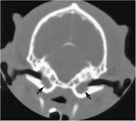
Figure 18-4
CT image of a dog with bilateral middle ear effusion (arrows). (From O'Brien DP: Comparative neurology, Columbia, MO, 2003, University of Missouri.)
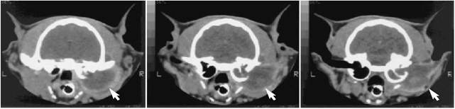
Figure 18-5
Serial CT images in a cat showing lysis of the right tympanic bulla and a large soft tissue mass associated with the right external ear canal (arrows). (From O'Brien DP: Comparative neurology, Columbia, MO, 2003, University of Missouri.)
Erosive bone disease, such as in an animal with a bony neoplasia of the middle ear, appears as a mixed lytic and proliferative lesion (Figure 18-5).
CT is generally considered to be superior in the imaging of bone and MRI better at imaging soft tissue. This is even more marked when trying to image parts of the brain because a beam-hardening artifact (due to the thick and dense petrous temporal bone) prevents adequate visualization of the brainstem using CT. Therefore, when imaging the brainstem is important, as in cases of suspected CVD, an MRI would prove superior (Figure 18-6).
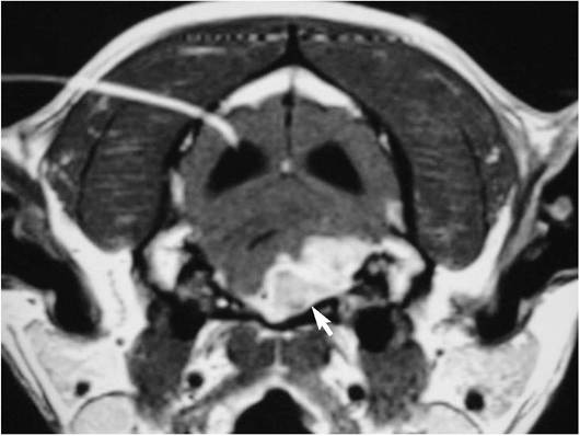
Figure 18-6
MRI of a dog with a tumor of the brainstem causing CVD (arrow).
Myringotomy, or aspiration of middle ear contents, can be a useful diagnostic tool. This test is usually performed when the tympanic membrane is intact, but there is direct visual or imaging evidence of fluid accumulation. This procedure is performed on a heavily sedated or anesthetized animal. In short, the technique involves visualization of the tympanum with an otoscope, rupture of the tympanic membrane with a blunt probe caudal to the malleus, insertion of a 20-gauge spinal needle through the hole, and aspiration and culture/cytology of the middle ear contents, followed by gentle flushing of the tympanic bulla with warm sterile saline.
Care is needed in this procedure to avoid damaging the auditory ossicles or nerves. This is best done by directing the flush caudally and ventrally.A Schirmer tear test (STT) should also be performed in all animals with otitis media or interna. The major petrosal nerve, a branch of the facial nerve (VII), runs through the middle ear and often is damaged by the inflammatory process. The lacrimal gland is innervated by the major petrosal nerve, and denervation results in decreased secretion and the potential development of keratoconjunctivitis sicca (KCS). Also, certain antibiotics used to treat otitis interna and media can result in decreased tear production. Therefore, a baseline STT identifies dogs that may develop KCS; it can be used as a benchmark to determine whether the antibiotic is causing decreased tear production.
Evoked-response audiometry can yield valuable information in animals with suspected hearing loss. Brain-auditory-evoked-response (BAER) is a test that records the electrical activity of the auditory nerve and brainstem time-locked with an auditory stimulus that is delivered to the ear. In general, hearing loss can be attributed to either a conductive or sensorineural problem. Conductive hearing loss is dysfunction in the peripheral hearing mechanism as a result of external and/or middle ear disease. Exudate or waxy buildup in the external ear canal or middle ear, a ruptured tympanum, or auditory ossicle damage results in decreased transmission of airborne sound waves to the cochlear apparatus. This is reflected on the BAER by increased waveform latency (longer transmission time) and decreased amplitude for a given sound intensity.
Sensorineural hearing loss is caused by dysfunction of the cochlear apparatus, cochlear nerve, or portions of the auditory pathway in the central nervous system. BAER findings are variable, ranging from a complete absence of all waveforms (such as in the case of congenital deafness in white-coated breeds of dogs) to changes in amplitude and latency. It is important to note that the BAER tests auditory pathways, not the sense of audition. It is possible for an animal to have a normal BAER and be deaf.
More on the topic Diagnostic Tests:
- Evaluation of Diagnostic Tests Under Local Conditions
- Application and Interpretation of Findings From PCR-Based Diagnostic Tests
- Immunity and Immune-Based Diagnostic Tests
- APPENDIX TWO Serological and Molecular Diagnostic Tests
- Simple and non-invasive tests, confirmatorytests, follow-up tests
- Tests Used in Prevalence Studies
- Commercial Diagnostic Kits
- DIAGNOSTIC TECHNIQUES
- Quality Control of PCR-Based Diagnostic Assays
- The Tests
- DIAGNOSTIC APPROACH IN CHD
- Diagnosis Based on the Use of Ante-mortal Tests
- DIAGNOSTIC APPROACH IN NEONATAL JAUNDICE
- Memory and Learning Tests