Components of the Vestibulocochlear System
The peripheral vestibular apparatus lies within the bony labyrinth of the petrous temporal bone. Important structures of the labyrinth include the utriculus and sacu- lus of the maculae, the semicircular canals, and the cochlea.
The maculae detect the pull of gravity and linear acceleration, whereas the semicircular canals detect angular and rotational acceleration. The cochlea houses the cochlear membrane, which is the receptor organ for the sense of audition. Information from the vestibular and cochlear systems leaves the peripheral apparatus in the vestibulocochlear nerve (cranial nerve VIII), which terminates in the brainstem. Neurons emanating from the cochlear apparatus course into nuclei within the brainstem and eventually project to the cerebral cortex, where the auditory information is processed, resulting in the sense of audition. The majority of the neurons from the peripheral vestibular system synapse in one of the four divisions of the vestibular nuclei that are located on both sides of the medulla oblongata, adjacent to the fourth ventricle. The vestibular nerve also projects directly to the cerebellum, which in turn sends projections back to the vestibular nuclei. Numerous projections from the vestibular nuclei are sent to different areas within the central nervous system.Projections from the vestibular nuclei to the motor nuclei of the oculomotor (CN III), trochlear (CN IV), and abducent (CN VI) nerves control reflex eye movement (Figure 18-1).
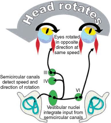
Figure 18-1
Vestibular control of eye movements. When the head rotates, the semicircular canals detect the direction and speed of the rotation. The vestibular nuclei integrate the information and direct the oculomotor (III), trochlear (IV), and abducens (VI) nuclei to move the eye in the opposite direction at the same speed. This maintains a fixed image on the retina even as the head moves. (From O'Brien DP: Comparative neurology, Columbia, MO, 2003, University of Missouri.)
Rotation of the head in one direction results invunjent of the eyes in the opposite direction at the same speedThis reflx maintains a ifed image on the retina as the head manimals with lesions affecting the cerebellar cortex, nuclei, or peduncles may show CVD. Such lesions may produce a paradoxical vestibular disease, in that the head tilt, ataxia, and strabismus are on the side opposite that of the lesion. In this instance the lesion can be localized by observing the side of the animal that shows proprioceptive deficits. For example, an animal with paradoxical vestibular disease may have a right head tilt and a left horizontal nystagmus, with prominent proprioceptive deficits on the left. The lesion would thus be localized to the left side of the brainstem. Other signs of cerebellar disease, such as dysmetria and intention tremors, would also be expected.
More on the topic Components of the Vestibulocochlear System:
- 44 Diseases of the Vulva and Vagina
- Index
- Chapter 46 Vulvar and Vaginal Disease and Neoplasia
- 30 Chronic Pelvic Pain
- Chapter 30 Pelvic Support Defects, Urinary Incontinence, and Urinary Tract Infection
- Liver disease