Abnormal Findings
Hyperintense thickening of the epithelium and a hypointense thick external ear canal have been reported in a dog with chronic otitis externa on both T1- and T2-weighted sequences.19 The two most commonly reported abnormalities in otitis media are the presence of hyperintense material within the bullae on T2-weighted sequences17,19 (Figure 4-21, A), which is compatible with accumulation of fluid, and enhancement of the inner surface of the bulla, likely due to inflammation, on T1-weighted sequences after injection of contrast material7 (Figure 4-21, B).
MRI is the first imaging modality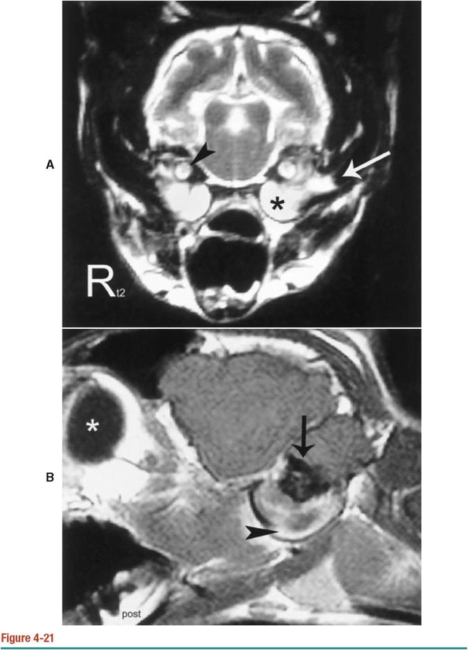
A, T2-weighted transverse image of a 13-year-old spayed female cat with a diagnosis of bilateral otitis. There is bilateral hyperintense volume of fluid associated with the tympanic bullae (asterisk). The fluid also fills the horizontal portion of the left external acoustic canal (arrow). The hyperintense signal of the intralabyrinthine fluid (arrowhead) is well visualized. B, T1-weighted sagittal image of a bulla of the cat depicted in Figure 4-21, A, obtained after injection of contrast material. There is a well-defined rim of enhancement (arrowhead) associated with the inner surface of the wall of the bulla. The curvilinear thin black signal void representative of the wall of the bulla is better defined because the air in the bulla has been replaced by isointense fluid. The globe (asterisk) and the petrosal portion of the temporal bone (arrow) are also noted.
C, Corresponding FLASH transverse image of the cat depicted in Figure 4-21, A. The fluid within the bullae is again noted. This sequence provides finer detail between the petrosal portion of the temporal bone and the intralabyrinthine fluid (arrows).
available to veterinarians that has the potential to diagnose otitis interna based on the detection of the intralabyrinthine fluid surrounding the semicircular canals. Preliminary studies suggest that MRI findings in otitis interna include absence of the hyperintense signal on T2-weighted spin-echo images of the intralabyrinthine fluid.16 The appearance of the intralabyrinthine fluid on additional sequences other than T1- and T2-weighted protocols needs further study before this sign can be used as a predictor of otitis interna in small animals.
MRI excels in depicting changes in the soft tissues surrounding the middle ear as well as in the caudal fossa because no artifacts hampering evaluation of the pons are produced. MRI has proven to be valuable in the diagnosis of dogs with vestibular disorders.16 By generating multiple sequences with different acquisition parameters, it is possible to further characterize the diseased tissue (Figures 4-22 and 4-23). Low-cellularity fluids, such as cerebrospinal fluid or true cystic lesions, appear hypointense (dark) on FLAIR sequences. Highly cellular or proteinaceous fluids, such as those present in inflammatory exudates in otitis, are hyperintense (white) on T2-weighted and FLAIR sequences. Blood and mineral appear as a signal void (black) on FLASH sequences.
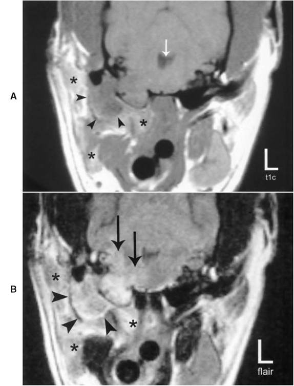
Figure 4-22
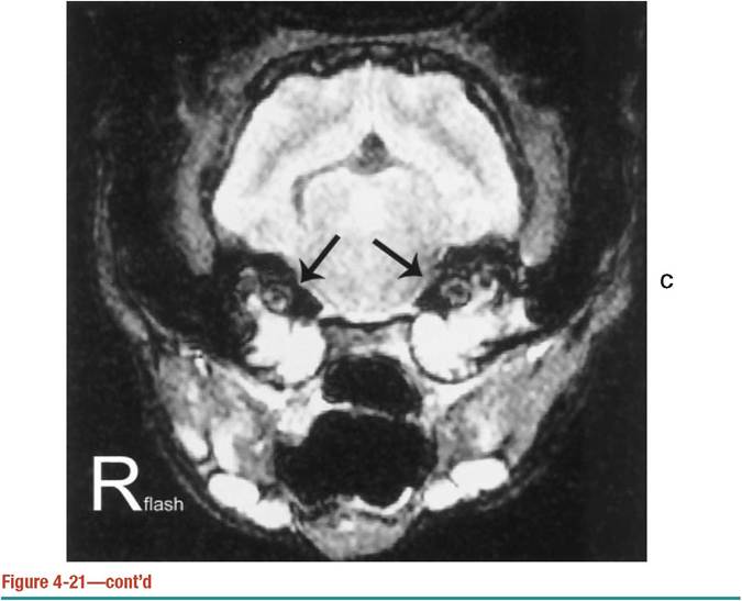
A, Transverse T1-weighted spin-echo image obtained after injection of gadolinium of a 7-year- old spayed female Chihuahua depicted in Figure 4-17 after an attempt to perform an ear canal ablation was aborted due to uncontrollable bleeding. The nonenhancing hypointense mass (arrowheads) is surrounded by heterogeneous enhancing tissue (asterisks) representative of inflamed muscle. The right bulla is completely obliterated by isointense tissue, and most of the petrosal portion of the temporal bone is absent. The mesencephalic aqueduct (arrow) is mildly distended.
B, Corresponding transverse FLAIR image of the dog in Figure 4-22, A. There is ill- defined perilesional edema associated with the right half of the pons (arrows). The mass (arrowheads) is a combination of fluid at the periphery and a more solid content in the center. Extensive inflammatory changes associated with the surrounding musculature (asterisks) are also noted.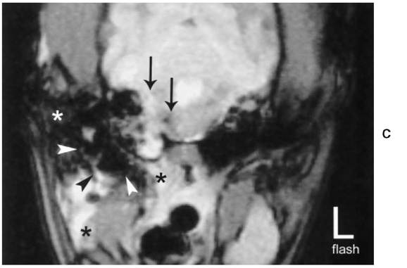
Figure 4-22—cont'd
C, Corresponding transverse FLASH image of dog depicted in Figure 4-22, A. The mass (arrowheads) is mostly blood, as indicated by the large signal void surrounded by a rim of fluid. The mass within the right bulla is also a combination of blood, solid tissue, and inflammatory fluid. The caudal fossa lesions (arrows) are representative of hemorrhage and inflammatory fluid. Inflamed surrounding musculature is again detected (black asterisks), with a more hemorrhagic component noted dorsolaterally (white asterisk).
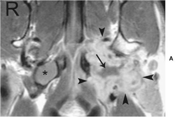
Figure 4-23
A, T1-weighted dorsal image at the level of the bullae of a Labrador-Golden Retriever mixed breed after injection of contrast material. There is a large mass of heterogeneous enhancement effacing the left tympanic bulla (arrowheads) and extending rostrally medial to the mandibular condyle. Nonenhancing areas within the mass suggest lack of blood supply to the center of the mass (arrow). There is a moderate volume of homogeneous fluid in the contralateral bulla (asterisk). The spatial and contrast tissue resolution of this image is higher than a similar CT dorsal plane reconstruction depicted in another dog in Figure 4-19, A.
Continued
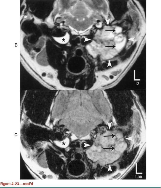
B, Transverse T2-weighted image of the dog depicted in Figure 4-23, A. The increased intensity in the right bulla (asterisk) is fluid, which is considered the result of otitis media. Within the mass (arrowheads) several foci of increased intensity (arrows) may also represent fluid or tissue. C, Transverse FLAIR image of the dog depicted in Figure 4-23, A. The fluid within the bulla (asterisk) remains hyperintense, which suggests fluid with a high cellular content. The mass (arrowheads) is of more homogeneous intensity than the corresponding T2-weighted image, as the intense foci noted on Figure 4-23, B, are now isointense (arrows). These foci are likely made out of more solid tissue than originally suspected. No biopsy of the mass was performed.
More on the topic Abnormal Findings:
- Case Summary
- Perioperative Care and Complications of Gynecologic Surgery
- Chronic Obstructive Pulmonary Disease
- index
- Imaging in Gynecologic Emergencies
- I PSYCHOSOCIAL ISSUES ^445
- Changes in the respiratory system
- Chapter 47 Cervical Neoplasia and Carcinoma
- Chapter 33 Disorders of the Breast
- Methods of intrapartum fetal monitoring