VD and DV Projections
Choosing between a DV and VD projection is more a function of hospital protocol or personal preference than clinical need. To obtain the VD view, the animal is placed in dorsal recumbency.
The primary beam enters the patient in a VD orientation at the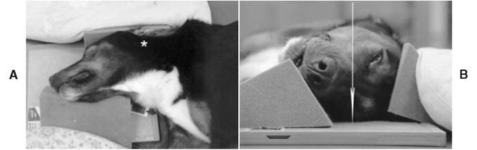
Figure 4-2
A, Patient positioning to obtain an oblique view of the middle ear. The resultant radiograph is named the left 20-degree ventral-right dorsal oblique view, which describes the entrance and exit points of the primary beam. The asterisk indicates the point where the primary x-ray beam enters the patient. B, Patient positioning to obtain a right 20-degree ventral-left dorsal oblique radiograph. The arrow indicates the trajectory of the primary beam.
Continued
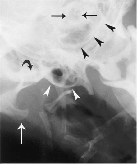
Figure 4-2—cont'd
C, Left 20-degree ventral-right dorsal oblique radiograph of the middle ear. The right bulla is displaced ventrally (white arrowheads). Only a faint outline of the wall of the left bulla can be seen (black arrowheads) with the left external acoustic meatus located dorsally (black arrows). There is superimposition of the right stylohyoid bone over the right tympanic bulla. The angular process of the right hemimandible (white arrow) is visible ventral to the right temporomandibular joint (curved arrow).
midline halfway between the external acoustic meatuses (Figure 4-3, A). The animal is placed in sternal recumbency to obtain a DV view. The primary beam is directed vertically centered at a point where an imaginary line connecting the bullae intersects with the midsagittal plane. The body of the mandible should be parallel to the cassette, to avoid distortion. The tongue should be pulled forward and maintained on the midline.
These views are used to assess the ear canals and to compare symmetry of the bullae and the petrosal portion of the temporal bones. The tympanic bullae cannot be evaluated fully in this view because they are superimposed over the petrosal portions of the temporal bones (Figure 4-3, B). There is no specific bone pattern associated with the petrous temporal bones, however; they should exhibit a symmetrical shape, size, and opacity on these projections. Reducing the milliamp seconds (mAs) by half to highlight the soft tissues allows visualization of the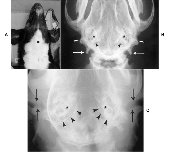
Figure 4-3
A, Patient positioning to obtain a VD view of the middle ear. The asterisk indicates the entrance point of the primary x-ray beam. Choosing between a VD and a DV view is more a function of hospital protocol or personal preference than clinical need. B, VD radiograph of the middle ear. The most caudal walls of the tympanic bullae (black arrowheads) are noted caudal to the more radiopaque petrosal portions of the temporal bones (asterisks). Rostral to the mastoid process (white arrowheads) lies the external acoustic meatus. The paracondylar process (white arrows) projects caudally. C, DV radiograph of the middle ear. The soft tissue exposure technique allows visualization of the external ear canals (arrows). The walls of the tympanic bullae (arrowheads) surround the petrosal portions of the temporal bones (asterisks).
horizontal portion of the external acoustic canals (Figure 4-3, C), which are noted as well-defined lucent structures. The canals tend to be wider laterally as the auricular cartilage expands to form the pinna. The average diameter of the proximal end of the annular cartilage is 4.1 ± 0.7 mm in dogs in which the tympanic membrane is visible otoscopically.1
More on the topic VD and DV Projections:
- Multiyear projections
- HOW ACCURATE ARE PROJECTIONS IN PRACTICE?
- Future Projections of Energy Demand
- PROJECTING FINANCIAL FLEXIBILITY
- The information age
- DIGESTIVE PROBLEMS
- Conventional Radiography
- Boon Andrew. The Ethics and Conduct of Lawyers in England and Wales. Hart Publishing,1999. — 808 p., 1999
- Griffiths-Baker Janine. Serving Two Masters: Conflicts of Interest in the Modern Law Firm. Hart Publishing,2002. — 227 p., 2002
- Grisso T.. Evaluating Competencies: Forensic Assessments and Instruments. 2nd edition. — Springer,2002. — 564 p., 2002
- Luban David. Legal Ethics and Human Dignity. Cambridge University Press,2007. — 350 p., 2007
- Ayupova Z.K.. Theory of state and law: textbook. - Almaty: Kazakh University,2015. - 192 pages., 2015
- Allen Danielle, Benkler Yochai et al. (eds.). A Political Economy of Justice. The University of Chicago Press,2022. — 416 p., 2022
- Barnes Rudolph C.. Military Legitimacy: Might and Right in the New Millennium.Frank Cass,1996. — 198 p., 1996
- Bedner Adriaan (ed.).. Real Legal Certainty and its Relevance: Essays in Honor of Jan Michiel Otto. Leiden University Press,2018. — 261 p., 2018
- Fridson M., Alvarez F.. Financial Statement Analysis. John Wiley & Sons, Inc.,2002. — 413 p, 2002
- Banking, Finance, and Accounting: Concepts, Methodologies, Tools, and Applications. IGI Global,2014. — 1593 p., 2014