Conventional Radiography
The five radiographic projections needed to visualize the soft tissue of the external acoustic canal and the bone of the tympanic bullae and petrous temporal bones are referred to collectively as a bulla series.
These views are: lateral, two opposite obliques (left 20-degree ventral-right dorsal oblique and right 20-degree ventral- left dorsal oblique), open-mouth (rostral 30-degree ventral-caudodorsal open-mouth oblique), and a ventrodorsal (VD) or dorsoventral (DV).Technique and Normal Radiographic Findings
High-detail film screen combinations (Kodak Lanex fine screens with Ektascan M film or 3M SE plus film with Assymetrix detail screens) are required to provide the contrast and spatial resolution needed to recognize the typical abnormalities seen in the canine and feline ear.
Lateral View
The animal is placed in lateral recumbency with the nasal septum parallel and the hard palate perpendicular to the tabletop cassette (Figure 4-1, A and B). The primary beam is centered on the external acoustic canal. A foam wedge or gauze roll placed beneath the rostral third of the nose is needed to maintain proper alignment. The head should be slightly extended to avoid superimposition of the bullae on the pharynx. Portions of the larynx and pharynx should be included in this view (Figure 4-1, C) to assess the temporomandibular joints and the nasopharynx because as some diseases, such as nasopharyngeal polyps and craniomandibular osteopathy, can also affect the middle ear. This author prefers a subtle rostral offset of one bulla from the other to compare them. To achieve this, the primary beam can be centered just rostral to the external acoustic meatus, or the rostral third of the nose can be elevated slightly from
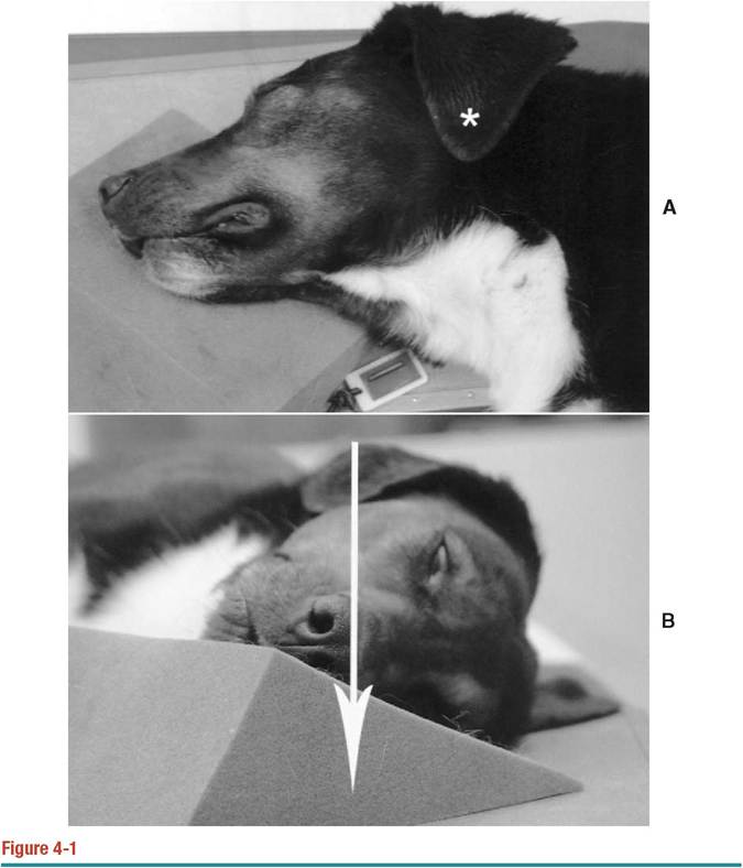
A, Patient positioning to obtain a lateral view of the middle ear.
The asterisk indicates the point where the primary x-ray beam enters the patient. A foam wedge under the maxillary bones ensures the nasal septum is parallel to the table. B, The arrow indicates the trajectory of the primary beam. The skull should be positioned with the hard palate parallel to the primary beam.Continued
parallel to the cassette (Figure 4-1, D). In animals with a large pinna covering the acoustic canal, such as hound or spaniel breeds, the pinna should be unfolded and placed dorsal to the skull to avoid allowing skin artifacts to obscure the area of interest. The endotracheal tube can be left in place. The external acoustic meatus is a circular to oval, gas-filled structure with well-defined inner borders. The tympanic bullae have smooth, thin-walled bone margins and a gas-filled lumen (see Figure 4-1, C
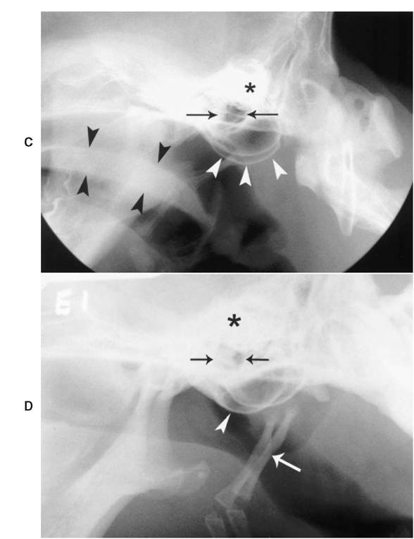
Figure 4-1—cont'd
C, Lateral radiograph of the middle ear. The left and right tympanic bullae are superimposed (white arrowheads). The petrosal portions of the temporal bones (asterisk) are located dorsal to the bullae. The soft palate is indicated with black arrowheads. The external acoustic meatus (arrows) is dorsal to the tympanic bullae. D, Lateral radiograph of the middle ear with one bulla rostrally positioned (arrowhead). The petrosal portions of the temporal bones (asterisk) are located dorsal to the bullae. The stylohyoid bones (white arrow) overlie the caudal aspect of the nasopharynx. The external acoustic meatus (black arrows) is also visible. and D). Thickness of the wall varies between breeds. There is less variation in the thickness of the bullae walls between breeds in cats. The petrosal portions of the temporal bones are highly radiopaque and superimposed on each other in this view; therefore, they cannot be fully assessed. In cats the bullae appear larger in proportion to the head than in dogs.
Oblique Views
Two opposite oblique views are taken.
With the animal in lateral recumbency, the bulla to be imaged is placed closer to the cassette. The thoracic limbs, sternum, nasal cavity, and mandible are rotated 20 degrees from the horizontal plane and are held in position with foam wedges (Figure 4-2, A and B). The mouth is closed to avoid superimposition of the mandible on the area of interest. The primary beam is centered at the base of the ear, ventral to the tragus. The primary beam travels through the patient in a lateral 20-degree ventral-left dorsal oblique direction. The tympanic bulla to be assessed is projected ventrally, while the contralateral bulla is superimposed over the caudal third of the calvarium and therefore cannot be assessed fully (Figure 4-2, C). Portions of the stylohyoid bone may be superimposed over the bulla of interest. The tympanic bullae have smooth, thin-walled bone margins and a gas-filled lumen. The external acoustic meatus is projected on the bulla as a circular to oval, gas-filled structure with well-defined inner borders.
More on the topic Conventional Radiography:
- Limitations of conventional historiography
- Chapter 38 Use and Management of Conventional ICT and Mobile Technology in Microfinance: A Bangladesh Perspective
- From Conventional Mortgages to Nonconventional Mortgages
- Selecting the appropriateimaging method, correctly applying the technique selected, and accurately interpreting the examination are the key steps in imaging ear disorders in dogs and cats.
- PCR Techniques
- Computed Tomography (CT)
- Conclusion
- Myringotomy
- CONTENTS
- Preface to Chapters 35—44
- Statement of the Problem
- Utilisation of Various Energy Sources
- HOW A REAL “WAR WITHIN ISLAM” WOULD LOOK
- Why Was There So Much Fraud?
- The Crisis As a Pivot: The Limits to Fiscal and Monetary Policy and the Need to Devise New Policy Tools
- Islamic banking emerged in Bangladesh in the mid-1980s with the establishment of the first Islamic bank in the capital city, fostering the subsequent formation of another seven full-fledged Islamic banks (IBS).[526]