DIGESTIVE PROBLEMS
The rumen is very small at birth and digestion of milk takes place in the abomasum. The calf quickly learns to eat solid food and by two weeks old ruminal movements and cudding should have started.
Continued development and expansion of rumen size are stimulated by propionic and butyric acids, the rumen fermentation products of the concentrate fraction of the diet. These substances also stimulate the development of papillae, small finger-like projections which increase the absorptive surface of the rumen. Rumen movements are stimulated by the presence of long fibre which physically ‘pricks’ the rumen wall. Straw is ideal for this, which is why so many farmers now rear calves on a straw and concentrate diet. The type of concentrate is important. The rumen is not fully developed until ten to twelve weeks old and so prior to this the calf is best fed fairly high levels of concentrate (2-3 kg/day) with ample high-quality protein (e.g. 20% CP). It is cost-effective to do so, because food conversion is much more efficient at the younger age, as shown in Table 3.1.The rumen of the young calf is also much more acid than in the adult, partly due to low saliva production by the calf. This high acidity, if not checked, can depress food intake. High- quality concentrates, containing a high level of digestible fibre, are therefore needed at this stage. Only after calves are 12 weeks old, when the rumen is fully developed, can concentrate quality be reduced and greater reliance placed on forages. Feeding straw stimulates rumen contractions and promotes cudding. This in turn increases saliva flow which decreases rumen acidity. High-starch concentrates without adequate balanced fibre should be avoided as they ferment rapidly, produce acidosis and retard rumen development.
Table 3.1. Food conversion ratio (FCR) is most efficient in the young calf and deteriorates with age.
| Bodyweight | FCR |
| 50 kg | 2:1 |
| 100 kg | 3:1 |
| 300 kg | 5.5:1 |
| 500 kg | 8.5:1 |
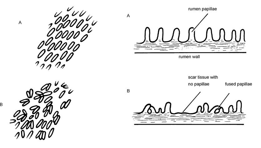
Figure 3.1. The development of the rumen wall is strongly influenced by diet: A - normal, high fibre diet B - high starch and no fibre
Figure 3.1. is a diagrammatic representation of the rumen wall of calves fed two different diets. Diet A was well balanced and produced good rumen papillae. Diet B was high in starch concentrates, leading to shorter papillae, some fusion of papillae and some areas so badly scarred by acidosis that they were totally devoid of papillae.
The physical form of the concentrate can also have an effect. Finely ground products (e.g. ground barley or maize) are bad because they ferment rapidly in the rumen and could cause acidosis. At the other extreme, a coarse mix is a big advantage because
• Calves eat coarse mix more slowly.
• They chew it more and in so doing produce more saliva.
• They often start eating it earlier than pellets.
Many farms feed pellets or pencils without any problems and I am certainly not recommending that everyone should change. However, if you are experiencing digestive problems like bloat or scour with your calves around weaning, I would certainly recommend changing to a coarse mix ration. This should be offered from one to two weeks of age and fed until at least one to two weeks after weaning.
One of the problems of ad lib feeding of milk, either cold or warm, is that although calf growth is very good, concentrate intake, and hence rumen development, is depressed and there is often a greater check at weaning. This is particularly the case if calves are abruptly weaned.
Gradually reducing milk over the two weeks prior to weaning helps to stimulate dry food intakes and ruminal development. This can be done by putting the milk bucket much lower than the teat, by placing a constricting clip on the pipe, or simply withholding access to milk a few hours each day. All three systems encourage concentrate intakes.Pot-bellies
At one stage it was thought that plenty of good hay was needed to stimulate rumen development. In fact this is not true and if concentrate is restricted (for example, either in the amount given or by inadequate feeding space) and the calves have free access to unlimited supplies of palatable hay, then the rumen becomes overstretched and a pot-bellied calf may result. This is because concentrates are needed to act as ‘food’ for the rumen micro-organisms which digest the hay. Absence of this ‘food’ leads to an accumulation of very slowly digesting hay in the rumen, eaten because the calf was hungry. In fact any rumen upset or poor rumen development could result in pot-bellied calves.
Chronic Diarrhoea
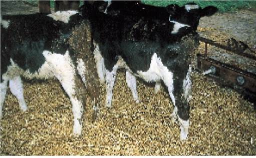
Plate 3.1. Chronic scour pre or post weaning leads to unthriftiness and, if not corrected, can even cause death.
It is not uncommon to see a dark, pasty grey chronic scour in calves either just before or just after weaning. The scour often has a ‘cakey’ appearance, looking as though it is associated with overfeeding of concentrates. In the majority of calves it simply causes stunting and poor growth. A typical example is shown in Plate 3.1. Probably the whole group will be affected to a greater or lesser extent and although the stunting is severe, deaths are rare.
I find it a particularly difficult condition to deal with and I suspect that we do not know the true cause.
Although digestive upsets are suspected, the rate at which the scour can pass through several groups of calves would suggest that infectious agents are also involved.
One suggestion is a protozoal infection of the large bowel, precipitated by dietary upsets, very similar to colitis in pigs. However, this has yet to be proven. Group-housed calves and calves which consume large quantities of concentrates are more likely to be affected. The condition is rare in individually penned calves or calves reared in hutches.Treatment
There does not seem to be an effective treatment and sometimes I wonder if it is better to let things run their course! However, I would suggest the following:
• Get your vet to test for coccidiosis, cryptosporidia and salmonella. Results are likely to be negative, but if positive, then specific treatment can be given.
• Try injecting with sulphonamides, which sometimes help.
• Badly affected calves must be taken off concentrates and put onto a generous and good-quality whole milk ration for two or three weeks; otherwise they will die. This suggests that the problem is in the rumen or large bowel and that the abomasum (where milk is digested) is unaffected.
• Try feeding yoghurt, for its probiotic effect (see Chapter 2). Badly affected calves seem to respond well to it.
• Change from ground concentrates to coarse mix and ensure that future batches of calves are reared on coarse mix.
• Check general feeding and husbandry procedures, especially in relation to oesophageal groove closure (see Chapter 2) and the type of concentrate and fibre being fed. The feeding of adult dairy cake to young calves is particularly dangerous, as part of the protein is likely to be indigestible for young calves.
Rumen Bloat
The mechanics of rumen function are described in Chapter 13 and the reader should refer to that section before reading the following. Rumen bloat can be caused in three ways:
• lack of rumen movements
• oesophageal obstruction, i.e. choke
• gas produced in the rumen forming a stable foam, known as frothy bloat, which cannot be released
In young calves the bloat is almost always due to lack of rumen movements, technically known as rumen atony, although I have occasionally treated calves which have had an apple stuck in their oesophagus.
Rumen atony is most commonly the result of a digestive upset and/or poor rumen development.Some of the more common causes of bloat are:
• Oesophageal groove failure, where milk falls into the rumen, rather than passing into the abomasum. The milk sours and ferments, producing gas which cannot be released because the immature rumen does not contract. A typically affected calf is shown in Plate 2.15. This is seen mainly in young calves.
• Poor rumen development. Calves may develop bloat one to two hours after a feed of concentrates. Some may be normal again by the next feed, whereas in others the rumen stays dilated. The discomfort associated with this prevents the calf chewing the cud, or eating straw or other long fibre, and this makes the problem worse.
If there is a high incidence of bloat in your calves, check that:
• Their concentrate is not too finely ground.
• It does not contain excessive levels of starch and inadequate digestible fibre.
• Good-quality palatable straw is available at all times. This may simply mean that the calves are freshly and liberally bedded with clean straw every day, as in Plate 2.2.
Treatment
The treatment given must depend largely on the severity of the bloat. If it is only mild and disappears two to three hours after feeding, then putting the calf onto reduced quantities of a coarse mix may be adequate. More severely affected calves, or calves which repeatedly blow up, will need additional treatment. This could be one or more of the following, again depending on the severity of the bloat:
• Return to a whole milk diet.
• Deflate the calf with a stomach tube, trocar or needle.
• Surgically prepare a permanent rumen fistula.
Return to a whole milk diet Sometimes the removal of all concentrates, dosing with an oral antibiotic for three to four days (to suppress rumen fermentation) and returning the calf to a whole milk diet for
two to three weeks will work. Then slowly reintroduce concentrates.
This is laborious, but works well inmany calves. However, a proportion continue to get blown.
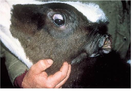
Deflate the rumen Although many people use a needle or a trocar and cannula and mechanically puncture the calf's skin, I think that a stomach tube is the best option. It is less traumatic for the calf and carries less risk. A length of flexible 15 mm garden hosepipe will suffice, provided it does not have sharp ends. If you reverse the calf into a corner, stand beside it and hold its mouth upwards (as shown in Plate 3.2) the stomach tube can be used quite easily.
From Figure 2.1 it can be seen that when the tube is inserted into the calf's mouth it must pass over the top of the larynx and trachea before it can be swallowed and fed down into the oesophagus. If you are in any doubt about whether the tube is in the oesophagus or the trachea, simply place your ear over the end of the tube and see if you can feel or hear air moving in and out in parallel with the breathing. If the answer is yes, the tube is in the trachea and it needs to be withdrawn and reinserted.
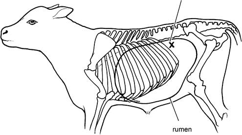
If a large-bore needle or trocar and cannula is to be used, it should be inserted on the left side of the calf, at a point mid-way between the last rib and the edge of the spine. The correct position is shown in Figure 3.2 and more details are given in Chapter 13. The needle or trocar should be pushed downwards and forwards, towards the calf's elbow on the opposite (right) side. Hold the needle or trocar in position, pushed firmly into the rumen, while the gas escapes and, if possible, at the same time squeeze the rumen by lifting it upwards with your knee to expel all the gas. Injectable antibiotic cover should be given for several days afterwards, to avoid the risk of peritonitis.
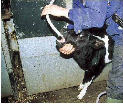
Plate 3.2. Passing a stomach tube is an easy and effective way of relieving rumen bloat.
Figure 3.2. The correct position for inserting a needle or a trocar and cannula is on the LEFT side, at a point in a triangle mid-way between the last rib and the spine.
Point of insertion of trocar and cannula
Permanent rumen fistula For calves which repeatedly develop ruminal bloat, construction of a permanent hole, opening the rumen onto the skin wall on the left side is by far the best option. It is one of the most common operations I perform and the improvement in the calf in terms of growth and food intake is dramatic. It is a simple (and therefore inexpensive) procedure, with an almost 100% success rate.
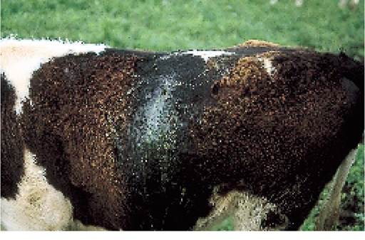
Plate 3.3. Construction of a permanent rumen fistula (a hole directly into the rumen) is the safest way to deal with a chronically bloated calf.
Plate 3.3 shows a calf after the operation. Although rumen contents may spill down over the side of the calf, it usually worries the calf less than the owner! Sometimes the hole slowly closes on its own, but more often it is necessary to have it closed with sutures when the calf is 12 months old or more. Beef animals may be sold for slaughter with the hole still discharging.
Colic
The word ‘colic’ simply means severe abdominal pain; it does not give any indication as to what is causing the pain. It may be due to a twisted gut or an intussusception or one of the other serious conditions discussed in Chapter 13. On occasions calves may be seen kicking at their stomachs or even rolling on the ground and bellowing with pain, due simply to a spasm, that is an excessive contraction, of the intestine. They can make a rapid recovery, less than one to two hours following the administration of drugs to relax the intestine. A similar syndrome may be seen in calves still on liquid diets.
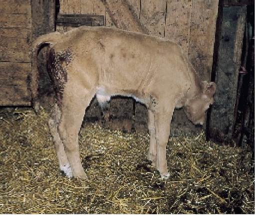
Plate 3.4. Calf continually straining due to coccidiosis. Note the raised tail, protruding rectal mucosa and bloody scour.
Coccidiosis
This is caused by small protozoan parasites, Eimeria zurnii and E. bovis, which burrow into the wall of the lower gut. Typically, affected calves pass semi-solid or very loose faeces, usually containing variable quantities of chocolate-brown blood. Scouring is a feature and the calf’s tail becomes soiled, but the faeces are not as liquid as in some cases of diarrhoea in younger calves. If the faeces are examined carefully, small lumps of a fawn-coloured gelatinous material may be seen. This is the mucosa, or lining, of the intestine.
Probably the most characteristic clinical sign of coccidiosis is straining, technically known as tenesmus: the calf stands with its tail raised and appears to be continually trying to force out small quantities of blood, mucus and faeces. A typical example is shown in Plate 3.4. After a few days the calf is dull, and it runs a moderate temperature and loses weight rapidly. Death can occur in untreated cases. You will need your vet to confirm the diagnosis so that specific anticoccidiosis therapy (e.g. sulphonamides, to toltrazuril or amprolium) can be given. Affected calves should be dosed at treatment level and any in-contact animals at preventive level, because it is likely that all calves are exposed to the same source of infection and the condition can rapidly spread. The drugs monensin and lasalocid can be used for prevention.
The coccidian life cycle
This is shown in Figure 3.3. The coccidiosis eggs or oocysts are taken in by mouth, pass through the acid barrier of the abomasum and hatch out in the large intestine (the colon, caecum and rectum), where they invade the cells lining the gut wall. Once inside the cell the oocyst repeatedly divides, to produce thousands of vegetative forms, the mero- zoites. These rupture the cell and are liberated into the intestine to infect and destroy adjacent healthy cells. Resistant forms, the oocysts, are produced sexually at a later stage and are passed out in the faeces. The oocyst has a very thick wall and as such it can survive in the environment for many months, waiting to be eaten by another calf so that its life cycle can start again.
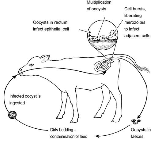
Figure 3.3. The life cycle of Eimeria zurnii and E. bovis, the coccidiosis parasites.
The initial source of infection is most probably the cow, since many adult animals carry the infection at low levels without showing any symptoms. Affected calves excrete large numbers of oocysts which can pass to their pen-mates, however. This occurs especially where hygiene is poor and there is an increased risk of faecal contamination of the feed, for example when water and food troughs are dirty, or inadequate bedding is used. The oocyst is an extremely resistant form and is not affected by many of the standard disinfectants. Dirty pens should be thoroughly cleaned, washed and then soaked with an ammonia-based product or a specific proprietary anticoccidial disinfectant, before a new group of calves is introduced. Hygiene is important in control although the ideal way of stimulating immunity is for calves to ingest oocysts during the first few days of life when they are still receiving colostrum.
Salmonellosis
The disease in young calves, usually involving S. typhimurium, has been described in Chapter 2, and the overall problem of salmonellosis is dealt with in Chapter 11, where the wide range of other species of salmonella, called serotypes, is described. A less common type in weaned calves is S. dublin. Infection is contracted from symptomless carriers (see Chapter 2), either the dam or from other calves when groups are mixed at weaning. One of the peculiar and often frustrating aspects of S. dublin is that infection may exist within the herd for several years without ever being seen as disease. When disease appears, and no stock have been purchased, it is difficult to explain why the outbreak has occurred. In the carrier animal, infection persists in the mesenteric lymph glands, the small ‘drainage' organs associated with the intestine.
Excretion of infection, i.e. the passing of salmonella in the faeces of carrier calves, is likely to be very intermittent and may not occur at all for quite long periods. This means that it is difficult to identify carrier animals simply by taking faecal swabs and trying to isolate S. dublin in the laboratory. A positive result shows that infection is present and action can be taken. However, a negative result can either mean that the calf is not a carrier, or it may simply mean that the calf was not shedding infection when the swab was taken. Serial swabbing of a group of calves, e.g. at weekly intervals, would give a better chance of identifying carriers, but even then a negative result would not be conclusive proof of absence of infection.
Clinical signs
S. dublin can cause scouring, and in this respect it resembles S. typhimurium, but it can also cause other clinical signs such as septicaemia, pneumonia, joint ill or meningitis, and these may occur without any obvious change in the faeces. The affected calf will run a high temperature in the early stages and scouring may occur, but it may not be particularly severe. Sometimes scouring is profuse, however, and lumps of intestine wall, blood and mucus are passed, viz the calf has dysentery. The calf will be dull, its coat bristling rather than smooth, its appetite reduced and there may be some coughing. On occasions a group of
Plate 3.5. Necrosis and loss of the ear tips can follow a Salmonella dublin infection.
calves may simply appear unthrifty and sudden deaths occur, but following post-mortem examination and bacterial culture, salmonella can be isolated from throughout the carcase.
Some calves recovering from septicaemia develop necrosis of the extremities. The calf in Plate 3.5 had shown an unidentified illness two months previously; then its ear tips were found to be falling off. S. dublin was isolated from the faeces of several other calves in the group, even though many had apparently recovered fully. Two others were not so lucky. Although their ears were not affected, their feet developed necrosis and started to fall off. Obviously these calves had to be culled.
Salmonella in weaned calves is often an extension of the disease which has been present earlier in life. The acute phase is over and the calves remain unthrifty with pneumonia and/or arthritis.
S. typhimurium and other salmonella serotypes can also cause scour, dysentery, pneumonia and death in weaned calves.
Treatment and control
Diagnosis is difficult and your vet will need to examine the calves and take faecal samples to the laboratory for culture. Once the presence of S. dublin has been confirmed, antibiotic therapy may be administered to affected calves and, whenever possible, the calves should be isolated, to reduce the weight of challenge of infection to the remainder of the group. A good dead vaccine is available, and other calves can be vaccinated to give them protection before entering the infected area.
Although antibiotics need to be given to avoid fatalities, there is now some evidence that they in fact prolong the period of excretion of the organism, and in so doing they reduce the chances of self-cure and increase the risk of an animal becoming a carrier. Treatment needs to be considered very carefully, therefore, since many animals will throw off the infection themselves and achieve a full cure.
S. dublin can infect adult cows (see Chapter 11), so careful hygiene is needed to prevent the spread of infection. When the infected group has left the building, clean out and rest as described in Chapter 2.
Ideally, soiled bedding needs to be stacked and heated to avoid pasture contamination, since it has been shown that faeces may remain infectious for up to three months even when spread onto pasture.
Necrotic Enteritis
This condition primarily affects home-bred single-sucked beef calves around six to eighteen weeks old. Affected calves often have bloody diarrhoea, due to the presence of ulcers throughout the intestinal tract. Ulcers may also be seen in the mouth, and necrotic enteritis can therefore be confused with BVD/mucosal disease. Most affected calves die, although fortunately the incidence in any one herd is likely to be fairly low. The cause remains unknown.
More on the topic DIGESTIVE PROBLEMS:
- Problems of the Digestive System
- THE DIGESTIVE TRACT
- Chapter 13 MISCELLANEOUS DIGESTIVE, RESPIRATORY AND OTHER CONDITIONS
- Behavioral Problems
- Eye Problems
- Problems of the S-view
- Environmental problems
- Gynecological Problems
- Conclusions: Some Problems of Classification
- MINOR PROBLEMS IN NEWBORNS
- Problems in practice
- Specific periods and problems
- Skin and mouth problems
- Head and Nerve Problems
- Management of IDU-related problems
- Problems of Intensionality
- Problems for Hobbes