CALF PNEUMONIA
Calf pneumonia (virus pneumonia or enzootic pneumonia) must be the most common of all the diseases of the weaned calf and it undoubtedly causes the highest losses in this age group in terms of both mortality and reduced growth rates.
Pre weaned calves can also be affected, but generally they are protected by antibodies obtained from the colostrum. Passive colostral antibody levels drop significantly by two to four months old, however, and it is at this age that pneumonia starts to be seen, although it may occur in housed animals of any age, including adult dairy cows.Clinical signs
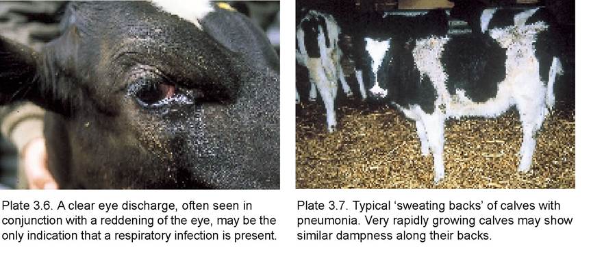
Probably the first indication of the presence of pneumonia will be that a few calves have a slightly red eye and there is a clear discharge, making a wet mark over the calf’s face (Plate 3.6). This should not be confused with New Forest disease or a foreign body in the eye. Both of the latter conditions produce a discharge, but they are also painful and the calf keeps its eye tightly closed. With calf pneumonia, the eye remains open.
Soon after, or maybe at the same time, coughing will be heard and the cough is generally a deep ‘chesty’ type, almost as if the calf is trying to bring up phlegm. Some calves may then develop a noticeably faster breathing rate, while more severely affected animals will be standing with their heads down, backs arched and breathing very heavily, finding it difficult to get enough air. The hair on their backs often stands up with a ‘spiky’ ungroomed appearance as in Plate 3.7, the result of sweating from a high temperature. These calves will not be eating and are likely to be standing apart from the rest of the group. Even in the early stages, affected calves may be off their food and running a high temperature (40.5-42.0°C), and sometimes acute outbreaks may occur, with fatalities, before any significant coughing has been heard.
Causes
Calf pneumonia is known as a multiple aetiology syndrome, that is it is caused by one or more of a whole range of organisms, including bacteria, viruses and mycoplasmas. Environmental factors are also extremely important.
Some of the more commonly found infectious agents involved in pneumonia are:
Viruses
• respiratory syncytial virus (RSV)
• para-influenza type 3 (PI3)
• infectious bovine rhinotracheitis (IBR)
• bovine viral diarrhoea (BVD)
• coronaviruses
Bacteria
• Pasteurella multocida and P. haemolytica
• Haemophilus somnus
• Actinomyces (Corynebacterium) pyogenes
Mycoplasmas
• Mycoplasma dispar, M. bovis, ureaplasmas
• Acholeplasma laidlawii
It is almost impossible to diagnose the different causes of calf pneumonia on clinical signs alone. Swabs, blood tests or post-mortem tissues are needed for a full diagnosis. However, a few of the infectious agents have some specific properties which are worth noting. For example:
• RSV can be involved in acute outbreaks of pneumonia, leading to sudden death. The calf in Plate 3.8 was one of a group of eight calves which surprisingly developed pneumonia while outdoors. Despite treatment the calf died within 24 hours. A postmortem examination showed the typical swollen emphysematous ‘burst lung’ appearance of RSV (Plate 3.9) and the virus was demonstrated in the tissues. Emphysema can sometimes be detected with a stethoscope as crackling and squeaking sounds in the chest.
• IBR can cause very red eyes (as in Plate 4.7) and a severe infection of the trachea (Plate 4.6). The disease is described in detail in Chapter 4.
• Pasteurella haemolytica (especially serotypes A1 and A2) is often present in the noses of healthy cattle, and it is only when animals are stressed that it invades the lungs to cause pneumonia. Stresses include sudden temperature or other environmental changes, concurrent infection or even calving.
Shipping fever is the name given to acute pasteurella pneumonia seen in cattle after a long journey. Typical signs are a high temperature, panting and appearing very sick, with minimal coughing.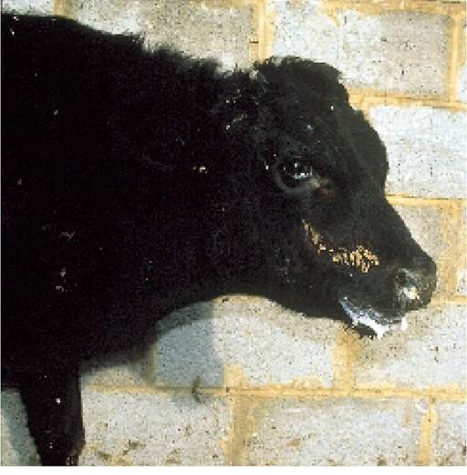
Plate 3.8. Calf coughing badly, caused by respiratory syncytial virus (RSV) infection.
However, pasteurella most commonly invades after viral infections. For example, it has been shown that the lungs are particularly susceptible to pasteurella infections for up to 30 days after an RSV infection. This is one reason why repeat treatments are commonly needed in outbreaks of calf pneumonia. It is not that the antibiotic is ineffective; it is simply because the damaged lung tissue is highly susceptible to reinfection by pas- teurella living in the nasal passages. Recently pasteurella has become an increasingly common cause of acute toxic pneumonia, producing severe lung damage and even death in adult dairy cows. Toxins produced by Pas- teurella haemolytica damage the phagocytic cells in the lungs, allowing some strains of the organism to multiply almost unchecked. It is highly probable that pasteurella would be isolated from the dark purple consolidated areas (A) of lung seen in Plate 3.10. The small remaining area of pink lung (B) is normal tissue.
• Haemophilus somnus may cause a ‘sleepy’ pneumonia.
• Actinomyces pyogenes produces chronic abscesses in the lung tissue, which may persist for the whole of the animal’s life. Typical examples are shown in Plate 3.11.
All calves, on every farm, will be carrying some of these infectious agents, but disease occurs only when natural defences are low or when there is an excessively high load of infection in the environment. The latter is known as the atmospheric load. These two aspects will be discussed separately. Some of the points were covered in Chapter 1 in discussion of the ‘balance’ of disease.
Natural defences
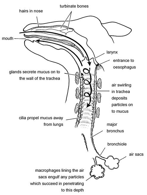
The defence mechanisms of the respiratory tract are shown diagrammatically in Figure 3.4.
They consist of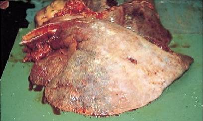
Plate 3.9. Burst lungs, typical of RSV infection. Note how the lung tissue is grossly swollen, with free air producing grey areas just under the lung surface. These are technically known as emphysematous bullae.
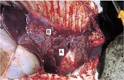
Plate 3.10. Typical lung seen in calf pneumonia. The dark consolidated areas (A) could be infected with Pasteurella, and it is unlikely that any air is flowing through this part of the lung. The pink area (B) is normal lung.
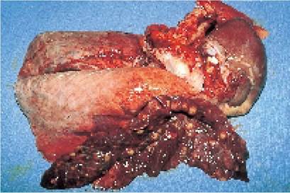
Plate 3.11. Small white abscesses scattered throughout the lung are a common consequence of inadequately treated pneumonia. They may remain with the animal for the rest of its life.
• hairs in the nose, which prevent entry of large particles
• nasal turbinate bones with a covering of mucus. The bones warm the in-coming air and the mucus traps air-borne particles
• mucus glands lining the trachea. These secrete a sticky mucus which traps particles as they swirl past in the air
• cilia, which moving in a
wave-like motion, propel the mucus and any entrapped
particles back up the trachea and into the mouth, from where it is either swallowed or coughed back into the environment
• alveolar macrophage cells line the terminal air sacs (respiratory alveoli) and continually patrol the area. They engulf any particles which succeed in penetrating to this depth.
• antibodies
It is only when infection has penetrated all of these defences that respiratory disease, e.g. pneumonia, occurs.
Antibodies to pneumonia organisms are obtained via the colostrum, and
calves receiving inadequate colostral Figure 3.4. The defences of the respiratory system.
intakes are therefore more susceptible.However, very high levels of infection and an unsuitable environment can overcome even good levels of immunity.
The activity of the cilia can be damaged by high levels of ammonia and other gases (e.g. from wet bedding and poor ventilation), by dust, by certain viruses and by chilling. Once lung damage has occurred, it is easier for infection to gain entry and cause pneumonia. In this state we say that the defence mechanisms have been compromised and the calf is more susceptible to disease.
Atmospheric load
This is the term given to the combined amount of infection and particulate matter carried in the air. Probably 99% of the different bacteria, viruses, dust and fungi present in the air could not cause disease on their own, but if present in sufficiently high numbers they overload, and therefore compromise, the calf's defences. The lung macrophages and other defence systems do not distinguish between dust, ‘normal' bacteria and pathogenic bacteria and viruses. They attempt to remove all of them. Hence if the defence systems are over-loaded dealing with dust, this may allow the few disease-forming agents to cause problems. A good example of this is RSV. It is difficult to produce RSV pneumonia experimentally by infecting calves with RSV, but if the same calves are first exposed to a high level of dust, then pneumonia develops.
Outdoors there are probably only 150 particles (dust, bacteria, fungi and viruses) per cubic metre in the air, whereas in a typical enclosed calf house this can rise to 400,000 per cubic metre! Where do all these particles come from? There are several sources:
• the bedding, especially if the straw is mouldy from poor harvesting or storage
• the calf’s skin
• breathed out from the calf’s lungs. The mucus and cilia are a very efficient filtering system, however, and probably some 95% of all inhaled particles are retained. Infection breathed out in expired air is, therefore, not as important as we once thought
Surprisingly, the disease-causing organisms which the calf does breathe out do not live very long.
For example, RSV survives for only 40-60 seconds in the air! This very short survival time for disease-causing organisms has two important consequences:• Calves have to be in very close contact in order to pass pneumonia from one to another. This is one reason why stocking density is so important and why calves licking one another is thought to be an important way of spreading infection.
• Once a building has been depopulated, it is highly unlikely that it will retain pneumonia infections for very long.
The size of the atmospheric load at any one time (i.e. the number of particles present in the atmosphere) will depend on:
• the amount being given off by calves and bedding
• the amount being removed by
- death of the organisms
- sedimentation (falling to the ground)
- inhalation by calves (and therefore filtration by mucus and cilia)
- ventilation (carried out of the building)
Humidity is a vitally important factor for both processes. If a house is humid and the bedding is wet, far more infection will be given off from the straw. Secondly, humid air can support considerably more micro-organisms than dry air, because their death rate is lower. Increasing humidity from 60% to 90% is said to lead to a ten-fold increase in the atmospheric load. Thirdly, the high ammonia often associated with humid buildings reduces the action of cilia, leads to chilling and generally lowers the calf’s defences. This is why we see far more pneumonia in damp, humid and foggy weather. In fact in one trial a sudden change from cold and dry to warm, humid conditions was the only environmental factor which precipitated disease when calves were experimentally exposed to infection.
Prevention
From the above, it can be seen that the quality of the calf’s environment is vitally important in the prevention of pneumonia, the main factors being:
• The calf should have a warm, dry bed.
• Calf pen floors should have a slope of 1:20 to facilitate drainage.
• All surface water should be swept or drained into channels and taken out of the building
• Stocking density must be kept low. For example, a decrease in stocking density by 50% is equal to increasing the ventilation rate by 20 times!
The approximate target stocking densities for calves up to ten weeks old are 1.5-2 square metres per calf of floor area and 8-9 cubic metres air space. If your house only allows 1-4 cubic metres per calf, then pneumonia is almost a certainty. In the face of a pneumonia outbreak it is worth consider-
Some important factors in the prevention of calf pneumonia
• minimise dust (including moulds)
• ensure adequate ventilation, which
- removes dust and noxious gases
- removes respiratory pathogens
- reduces humidity
• ensure a dry bed, which
- reduces chilling
- reduces noxious gases
- improves humidity
• avoid mixing
- differing age groups
- animals from different sources
• minimise
- group size and stocking density
- stress and intercurrent disease
• maximise immunity
- colostrum
- vaccination
• medicate with antibiotics
- inject whole group when 20% are affected
- put into milk for pre weaned calves
ing a reduction of the number of calves in a house. This is especially so for intensively reared calves on high concentrate diets, which breathe faster and need more air space anyway. Pneumonia sometimes occurs in outdoor calves, especially in large groups during hot weather. This is thought to be because the calves lie quite close to one another in a group and this allows the rapid spread of infection from one calf to the next. If faced with an outbreak of pneumonia in a large group of calves, whether housed or outdoors, it is a good idea to subdivide them into smaller groups, ideally in separate air spaces, to reduce both the severity and the rate of spread of infection.
• Provide adequate ventilation but freedom from draughts. The lying area for calves should have a wind-speed barely detectable on your face (this is approximately 0.2 metre per second). Provided that calves have a dry bed and are free from draughts, temperature is not too important, particularly for weaned calves. Given the option, many calves will lie outside even on quite cold days, which is why I think that open-fronted, naturally ventilated, monopitch buildings are ideal housing.
• Regularly clean out the shed, for example, every four to six weeks, depending on the number of calves present. If the straw bedding is allowed to build up and compost, this will have several effects:
- it may significantly decrease the air space in the building.
- it produces ammonia and other noxious gasses which reduce the effectiveness of the calf’s
respiratory defence mechanisms.
- it generates warmth and humidity, both of which favour the survival of bacteria and viruses in the
air and both of which make calves uncomfortable.
• Avoid mixing calves from different sources, since they may well be carrying different infections and have differing levels of antibody protection (see Figure 1.6).
• Avoid mixing different age groups, as they will have different antibody levels. In this context mixing means sharing the same air space.
• Straw used for food or bedding should be free of moulds and stored under cover.
Treatment
The first factor one should consider when faced with a group of coughing calves is whether any treatment is necessary. If coughing and eye discharge are the only symptoms, all the calves are eating and none have a significantly elevated temperature, I would not treat. The infection should spread and the calves should develop their own active immunity without any disease. On the other hand, if a proportion are panting, off their food and have elevated temperatures, I would treat the whole group.
• Isolate individually sick calves. Not only is this good nursing which aids recovery, but it also reduces the challenge dose for other calves.
• Give medication. Although mainly used for treatment, antibiotic medication of a whole group of calves will reduce the spread of bacterial causes of pneumonia and decrease the effects of viral agents. Antibiotics may be given:
- as long-acting injections for older calves
- in the milk of pre weaned calves, because milk passes directly into the abomasum
It is best not to give oral antibiotics to weaned animals as they may interfere with rumen function, although there are some exceptions to this.
Antibiotics only provide protection during their period of activity. They do not give the longer term protection given by vaccines. Some of the newer antibiotics reach high concentrations in the lungs and are particularly effective against pneumonia. Tilmicosin is especially interesting. It reaches high concentrations in lung tissue, it is effective against bacteria and mycoplasmas, it decreases toxin production and a single injection gives five days of ‘bacterial killing’ in lung fluid, followed by two to three weeks of antibiotic persistence in the lung macrophages, the bacterial killing cells (see Chapter 1 and Figure 3.4). As lung damage from RSV will render the lungs susceptible to infection for the next 30 days (see previous section), this degree of persistence is ideal.
New products are being developed all the time and your vet will advise you on the most suitable drug for your circumstances. Remember also that antibiotics do not have any effect against viruses. Treating the whole group may help to lower the overall level of bacteria and mycoplasmas in the environment to the extent that the calf’s own defences will then be able to cope with the reduced challenge and develop an immunity.
Severely ill animals will need respiratory stimulants, anti-inflammatory drugs and other supportive therapy, and veterinary attention should be sought for these. Others may develop chronic lung infections, leading to poor growth and intermittent bouts of pneumonia for weeks after the initial outbreak. I have found prolonged antibiotic cover to be well worthwhile in such cases; for example giving daily antibiotic or a long-acting penicillin or tetracycline injection twice weekly for three weeks or more. It eventually worked on the Charolais heifer in Plate 3.12.
Vaccination
This is a complex area and definitely needs veterinary advice. The type of infection involved in a particular pneumonia outbreak can be assessed by:
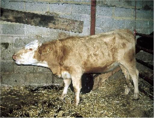
Plate 3.12. Long-standing chronic pneumonia. Note the open mouth breathing as this Charolais heifer fights for breath. A three-week course of antibiotic by injection eventually produced a cure.
• nasal swabs
• tissue from calves which have died
• blood samples taken when the calf is first affected and again two to three weeks later, looking for a rising antibody titre (see Chapter 1). Unfortunately in calves less than four months old, the presence of colostral antibodies may interfere with the interpretation of results.
Good live intranasal vaccines are available against IBR and PI3 and a live intramuscular vaccine for RSV. These are the three most common causes of pneumonia. IBR and PI3 vaccines are temperature attenuated strains, i.e. the virus can multiply in the lower temperatures of the nose (and this stimulates the calf to develop an immunity), but the normal body temperature of the calf’s lungs inhibits growth of the vaccine and so disease cannot develop. With a special applicator, 2 ml of the vaccine is sprayed into the animal’s nose (Plate 3.13). There is also evidence that these vaccines stimulate the production of interferon and as such can be used during an outbreak of pneumonia to give protection against a whole range of viruses.
DEFICIENCY DISEASES
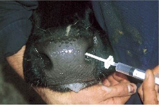
Plate 3.13. Administering an intranasal vaccine, for example against IBR or PI3 viruses. The applicator on the syringe produces a spraying effect.
Because growing animals generally have a higher requirement than adults for minerals, vitamins and trace elements, it is in this age group that nutritional deficiencies are most likely to be seen. Weaned calves are
normally reared indoors on a forage and concentrate diet, and problems may occur towards the end of the
winter, especially when feeding and management are poor. The mineral and vitamin requirements of the
animal and the effects of the various deficiencies are given in detail in Chapter 12, and in this section
white muscle disease. CCN, which is an induced deficiency, is listed under nervous diseases later in this
I shall be dealing with only one of the slightly unusual deficiencies, that is muscular dystrophy or chapter. Deficiencies of copper and vitamin A are common in calves and dealt with in Chapter 12.
Muscular Dystrophy (White Muscle Disease)
The name muscular dystrophy means ‘abnormal development and function of the muscles’ and it is a muscular abnormality which causes the clinical signs and the white areas seen in the muscles at post-mortem. Disease generally occurs at turnout in the spring and can be precipitated by the stress of bad weather. Calves which have been fed rations containing inadequate levels of vitamin E and/or selenium during the winter develop muscles which are weak and have areas of degeneration. Often no clinical signs are seen indoors, where the calves are relatively inactive. A few days after turnout, however, when they have been running around, a stiffness of gait may be noticed, with the legs unusually rigid. Some animals may be so badly affected that they become recumbent, while others may be found dead from heart failure, the heart muscle having degenerated. Muscle degeneration leads to release of the pigment myoglobin and this is occasionally seen as a red discolouration in the urine. If the chest muscles are involved there will be difficulty in breathing and affected calves may appear to have pneumonia.
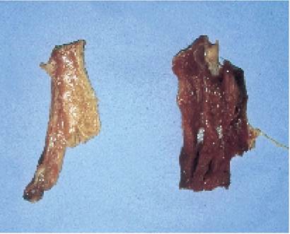
Plate 3.14 shows a piece of pale, white muscle (left) taken from a calf which died from muscular dystrophy, compared with a normal coloured piece
Plate 3.14. Muscular dystrophy caused by a deficiency of vitamin E and/or selenium. The
dark-coloured muscle on the right is normal. of muscle on the right. In addition to being very pale,
the affected muscle has a ‘gritty' appearance, due to precipitation of calcium. Only very limited areas of the carcase will be affected and so it is essential that a thorough post-mortem is carried out. Blood samples from live, affected calves can be tested for selenium, vitamin E or muscle degeneration to assist in the diagnosis. Occasional animals exhibit what is known as the ‘flying scapula' effect. The muscle attachment between the shoulder blades and the chest degenerates, the spine drops and the shoulder blades start to protrude above the backbone, as shown in Figure 3.5.
Occurrence
With improvements in testing procedures for selenium and vitamin E, surveys have shown that deficiency is very common. Deficiency does not always seem to be associated with disease, however, and the cost benefits of treatment must be carefully evaluated before embarking on any control programme. Traditionally disease occurred in beef suckler herds which had been over-wintered on a diet of straw and turnips. Many pastures have now been found to be deficient, however. Table 3.2 gives an idea of the feedingstuffs which are good or bad sources of vitamin E.
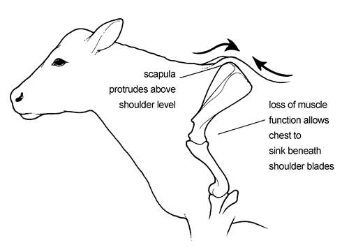
Figure 3.5. ‘Flying scapula'. Degeneration of the muscle attachment between the shoulder blades and chest leads to the shoulder blades protruding over the spine.
Table 3.2. Some dietary sources of vitamin E.
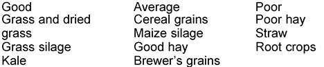
Total dietary selenium requirement = 0.1 ppm in dry matter.
Vitamin E and selenium
Although they are two totally unrelated chemicals, they act on similar mechanisms (involved with the metabolism of unsaturated fatty acids) within the animal. In intensively fed animals, an increase in the oil content of the concentrate, and particularly in the proportion of unsaturated fatty acids in the oil, leads to an increase in the vitamin E requirement. Animals with a marginal selenium status can be precipitated into disease by vitamin E deficiency and vice versa. Selenium deficiency is closely related to soil type and hence all crops grown in certain areas may be deficient. On the other hand, vitamin E levels are more related to the type of plant, its stage of growth and the method of conservation. For example, vitamin E levels are very low in hay which has been badly weathered in its making and in grain which has been stored using propionic acid as a preservative.
Treatment and control
This is an area where you will undoubtedly need veterinary advice, since an excess of selenium is extremely toxic. There is a wide range of possible courses of action, however. For example:
• Inject a selenium/vitamin E preparation. This is essential in the treatment of affected animals to achieve a rapid effect. A long-acting product is also available as a control measure.
• Selenium bullets. These are given by mouth and slowly dissolve in the reticulum. A variety of products are available, some containing selenium only and others combined with additional trace elements such as copper and cobalt. Consult the manufacturer’s instructions for dosage rates.
• Add sodium selenite to the ration to produce a final dietary concentration of 0.1 ppm. This is an extremely low level however, only one-tenth of a gram in one ton of mix, and it is impossible to achieve an even distribution of selenium unless specialist mixing facilities are available.
• Water-soluble preparations are available, which can be placed in the drinking water and slowly dissolve at a specified rate to provide the animal’s selenium requirement. One such system is known as Aquatrace. The pellets are formulated so that when the selenium concentration in the water reaches a level which will satisfy the animal’s requirements, no further selenium will dissolve. If some of the water is drunk, fresh water flows into the tank, the selenium concentration falls and this then permits more pellets to dissolve.
Tendon Rupture
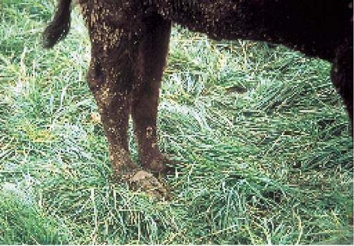
Not all calves which go lame immediately after turnout have white muscle disease. Plate 3.15 is a good example. This nervous Limousin heifer ran wildly around the field for three to four hours when first turned out, eventually stopping because of exhaustion. The next day her lower limbs were badly swollen and her fetlocks drooped towards the ground - in fact the fetlocks were almost resting on the ground. Although a blood sample would show severe muscle damage, her vitamin E and selenium status was normal. She was simply suffering from tendon rupture associated with gross over-exercise. She was returned to the farm and kept in a loose-box with plenty of feed, but took three to four months to recover.
Plate 3.15. Tendon rupture, caused by excessive exercise at turnout, led to the fetlock collapsing onto the ground.
I have seen the same syndrome several times. On another occasion a group of calves were repeatedly chased around a field by a donkey when they were turned out!
More on the topic CALF PNEUMONIA:
- OTHER CALF DISEASES
- DISEASES OF THE CALF
- This chapter deals with the health of the calf from birth to weaning, that is until approximately six weeks old.
- INFECTIVE PNEUMONIA
- Chapter 3 THE WEANED CALF
- Chapter 2 THE YOUNG CALF
- Pneumocystis carinii pneumonia
- Community-Acquired Pneumonia
- Hospital- and Ventilator-Associated Pneumonia
- NON-INFECTIVE PNEUMONIA
- Eosinophilic Granulomatous Pneumonia in Brown Norway Rats
- Pneumocystis Jirovecii Pneumonia
- Pneumonia Virus of Mice Infection