NERVOUS DISEASES
There are several diseases which can produce nervous signs in calves and these include lead poisoning, cerebrocortical necrosis (CCN), meningitis, tetanus and hypomagnesaemia.
Hypomagnesaemia can occur in milk-fed or suckler calves of three to six months old where little or no supplementary concentrates are being fed, and it may produce sudden death or nervous signs.
Milk is a poor source of magnesium and any reserves in the skeleton are quite quickly depleted. Hypomagne- saemia in calves occasionally results from scouring or from excessive drenching with liquid paraffin, both of which prevent the absorption of magnesium from the gut. Further details of hypomagnesaemia are given in Chapter 6.Meningitis is caused by a wide range of different infectious agents, but as it is most commonly seen in the young calf it is discussed in Chapter 2.
Tetanus is the least likely of the disorders. Of the few cases I have seen, one was the result of a rubber castration band being applied to an excessively large calf. Tetanus then developed in the necrotic stump. The clinical signs and methods of control are similar to those for adult animals and are dealt with in Chapter 4.
Lead Poisoning
Lead is still the most common cause of poisoning in farm animals and it is usually young calves or heifers which are affected, probably because of their inquisitive nature and tendency to lick and chew at unusual objects. There are several possible sources of lead, the most common being paint. Old doors are still used in the construction of calf pens. This is extremely dangerous, since old paint invariably contains large quantities of lead and calves tend to chew at woodwork. The door shown in Plate 3.16 was the cause of lead poisoning in a Gloucester suckler calf. Although the condition was diagnosed and treatment given, the calf did not recover.
Other possible sources of lead include:
• putty and traditional ‘liniment’ or ‘white lotion’
• golf balls and lead shot
• lead plates from batteries (Plate 3.17)
• pasture contamination, e.g.
beside motorways (from petrol), near lead mines and from certain types of industrial workings. The latter are now very carefully controlled.• contaminated food. In 1989 a batch of lead-contaminated rice bran became incorporated into compound animal feed and was quite widely sold to farms in the south-west of England. There was an outbreak of animals showing signs of lead poisoning, but perhaps economically more significant was the fact that Food Safety legislation was brought into operation. This prohibited the sale of milk or beef from those herds which had consumed the food until blood levels of lead in the affected animals had fallen to below acceptable values. Some herds were unable to sell stock for several months. Not surprisingly, the compensation claims were enormous!
Clinical signs
The signs of the disease vary, depending on whether there has been a high intake of lead over a short period (acute poisoning), or a lower intake over a more prolonged period
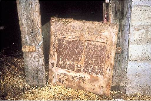
Plate 3.16. The paint from this old door was the source of lead for a Gloucester calf, which eventually died from lead poisoning.
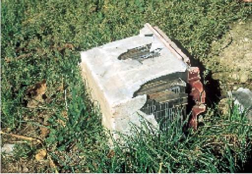
Plate 3.17. Cattle licking the plates of old discarded batteries is another common cause of lead poisoning.
(chronic poisoning). Acute poisoning is more common and calves may show symptoms a few days after eating the lead. The affected animal is blind and experiences periods of extreme excitement, bellowing, frothing from the mouth and trying to run up the wall. Quiet periods may follow, when the calf stands almost motionless, often pushing its head into a corner or against the feeding trough. It stops eating, it will probably be constipated and it may run a temperature. Death may occur in as little as one to three hours after the onset of symptoms, with the calf finally lying on its side, kicking with its legs and bellowing, as if it has severe abdominal pain.
Chronic lead poisoning produces a dull animal, which may be reluctant
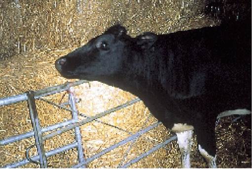
Plate 3.18. Chronic lead poisoning. Some animals are dull and simply push their head against a wall, as in this case. Acute poisoning produces more violent nervous signs.
to move or simply stands pushing its head against a wall or bales of straw, as in Plate 3.18. This is probably a sign of a severe headache.
Treatment
Lead poisoning is a serious condition and you should consult your veterinary surgeon if you suspect it. He will most probably take blood and dung samples to confirm the diagnosis and then administer calcium disodium versenate by intravenous injection. This chemical combines with the lead in the animal’s blood, producing an inert form which is readily excreted from the body. The affected calf, and the others in the group, should be given 100 g of magnesium sulphate (Epsom salts) by mouth. This has the double action of producing insoluble lead sulphate in the gut, thus reducing the rate of lead absorption and also acting as a purgative to quickly carry ingested lead out of the intestine. All possible sources of lead should be carefully considered and removed.
Cerebrocortical Necrosis (CCN) or Polioencephalomalacia (PEM)
The name means degeneration of the grey matter of the brain. Disease can occur in any age of calf, although it is most commonly seen at three to nine months old and especially in housed calves on a fairly high concentrate diet. The cause is an infection by two bacteria, Bacillus thiaminolyticus and Clostridium sporogenes, both of which live in the rumen and produce a substance, thiaminase, which destroys thiamine (vitamin B1). Although there is plenty of thiamine in the rumen initially (partly from the diet and partly from thiamine-producing bacteria), with CCN large quantities are destroyed before it can be absorbed and thiamine deficiency results.
Thiamine is needed for the manufacture of glucose, and as glucose is essential for brain function, lack of thiamine leads indirectly to degeneration of the grey matter in the brain.
CCN is most commonly seen on low fibre/high concentrate diets. Not only do these diets produce an acid rumen, which favours the growth of B. thiaminolyticus and Cl. sporogenes, but they also increase the requirement for thiamine in ruminal metabolism.There are other syndromes in calves which produce almost identical polioencephalomalacia changes in the brain and similar nervous signs. These include:
• bracken poisoning. Bracken contains thiaminase
• molasses toxicity, due to decreased propionate levels in the rumen and consequently low glucose
• water deprivation, especially if it is combined with high sodium and/or sulphate intakes, e.g. brackish water.
• lead poisoning
• sulphate toxicity. Ammonium sulphate has been incorporated into animal feedingstuffs at 0.5% to prevent urolithiasis. If high levels are fed, toxicity, seen as a form of CCN, has been reported. Ammonium chloride is now more commonly used and is safer, although it is more expensive
Clinical signs
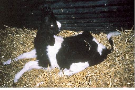
Plate 3.19. Calf with CCN. It is blind and pushing its head backwards.
Blindness is a common clinical sign and the affected calf tends to wander around the pen with its head held up and nose forward, often walking into things. Its temperature will probably be normal, although it may stop eating and soon develop a very hollow appearance. If the disease is allowed to progress, the calf becomes recumbent, often with its head pushed over its back, as in Plate 3.19. This is known as opisthotonos. Eventually the animal rolls onto its side and may die following bouts of kicking and struggling.
Treatment and prevention
Treatment consists of giving large doses of thiamine and it is surprising how quickly quite severely affected calves recover. The first dose will most probably be administered by your veterinary surgeon as an intravenous injection, to obtain rapid action. He may use a multivitamin complex or a simple thiamine solution. If several cases have occurred in a group of calves, it may be worth supplementing their ration with thiamine-rich sources, e.g. 60 g of brewer’s yeast per calf per day, or 20-30 mg of synthetic thiamine per kilogram of concentrate, although it has been suggested that feeding additional thiamine simply encourages the proliferation of B. thiaminolyticus and Cl. sporogenes and makes the situation worse. Sometimes CCN is preceded by, or associated with, a bout of scouring. As diets leading to an acid rumen may be involved, sodium bicarbonate added to the concentrate at 1.5-2% or the addition of chopped straw to the concentrate may be useful preventive measures.
More on the topic NERVOUS DISEASES:
- P Keerthi Kundana, Mona Gajre, Alpana Kondekar, Mukesh AgrawalNeurological disorders account for ~15-20% of hospitalizations, which may be divided into three major categories: (a) central nervous system disorders, involving brain and spinal cord, (b) neuromuscular disorders involving peripheral nerves and muscles, and rare disorders of autonomic nervous system.
- The various cardiovascular diseases observed in HIV-infected patients and widely described in the literature have been predominantly coronary and peripheral arterial diseases (PAD) and remain poorly known.
- Degenerative Changes in the Nervous System
- Central Nervous System Tumors
- ASSOCIATED CENTRAL NERVOUS SYSTEM MALFORMATIONS
- Disorders of Central Nervous System
- Central Nervous System Infections Meningitis
- Central Nervous System and Retinal Disease
- HIV-1 GENE EXPRESSION IN THE NERVOUS SYSTEM
- 22 HIV-1 Infection and Cell Death in the Nervous System
- HIV-1 SANCTUARIES AND THE ROLE OF BLOOD-TISSUE BARRIERS: CENTRAL NERVOUS SYSTEM, TESTES, AND RETINA
- Spontaneous Hemorrhagic Necrosis of the Central Nervous System of Fetal Hamsters
- OTHER CLOSTRIDIAL DISEASES IN WILDLIFE
- BIBLIOGRAPHY FOR NONINFECTIOUS DISEASES
- INTERSTITIAL LUNG DISEASES
- THE CLOSTRIDIAL DISEASES
- WHY STUDY WILDLIFE DISEASES
- VECTOR-BORNE VIRAL DISEASES
- BIBLIOGRAPHY FOR PARASITIC DISEASES