OTHER CALF DISEASES
Meningitis
The meninges are the fibrous layers which surround the brain and separate it from the skull. Meningitis simply means inflammation of the meninges and may be caused by a range of bacteria including streptococci, salmonella species and E.
coli.The initial source of infection is often the navel, although meningitis can also be secondary to scouring and enteritis. Colostrum-deficient calves are much more susceptible. The clinical signs depend on the nature of the infection invading the meninges and on the part of the brain affected. The calf may or may not be blind, but often the pupils are dilated and the eyes move from side to side in a jerking movement known as nystagmus.
The calf in Plate 2.36 was a typical case. It walked around the pen with its head on one side, continually pushing against the wall. Some calves appear to have an intense headache, in that they stand apart from the others with their head down, possibly pushing it against a feeding trough or into a corner. In this respect the symptoms resemble lead poisoning. More severe cases tremble and eventually fall to the ground with fits and spasms. There is usually a raised temperature and some calves develop a white cloudy debris inside the eye (Plate 2.37). This is panophthalmitis and shows that the whole eye is affected. It is not commonly associated with meningitis.
For treatment your veterinarian will prescribe an antibiotic which can pass across the blood-brain barrier. This is a physiological barrier which normally protects the brain from large molecules and which certainly prevents many drugs from entering. Antiinflammatory drugs and painkillers will also help. Nursing is very important. The affected calf should be penned on its own and if it has stopped eating, it should be given milk to drink to maintain its strength and/or drenched with electrolytes to prevent dehydration.
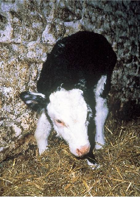
Plate 2.36. Calf with meningitis. It was standing with its head on one side, continually walking around the pen, pushing its head against the wall. There may also be a middle ear infection present.
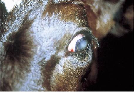
Plate 2.37. Calf with panophthalmitis, i.e. infection of the whole eye resulting from meningitis.
Middle Ear Disease
This can be confused with meningitis but it is a much less severe condition. Affected calves hang their head to one side (because they have earache), but otherwise they continue to feed, eat and grow normally. In some animals the ear-drum eventually bursts and a moist purulent discharge oozes from the ear canal. This assists recovery.
Injectable antibiotic for four to six days is needed to overcome the infection.
Calf Diphtheria
This occurs mainly in the mouth and may be seen in calves both before and after weaning and occasionally even in yearlings. Although the name is identical to the condition in man, the disease has a different cause and is far less serious. The bacterium concerned, Fusobacterium necrophorum, gains entry to the soft tissues after the thick epithelial lining of the mouth has been damaged. Once established, the infection forms an ulcer covered by a layer of thick pus and this can be seen inside the calf’s mouth.
The common sites affected are the inside of the cheek and at the back of the tongue. The cheek form is probably caused by the calf accidentally biting the inside of its mouth and it is seen as a swelling of the skin between the upper and lower teeth (Plate 2.38). This often causes little adverse effect on the calf, whereas the tongue form leads to difficulty in swallowing and affected calves drool and often froth at the
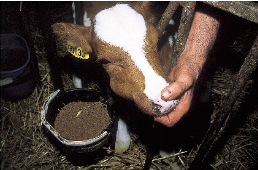
Plate 2.38.
Calf diphtheria: the cheek is swollen. This usually responds well to a few days of injectable antibiotic.mouth. When examined, they may have a mass of partially chewed food at the front of the tongue and this needs to be removed in order to see the pus and blood associated with the diphtheria ulcer. These calves will also have a high temperature and often a foul-smelling breath. If left untreated, infection can pass down into the lungs and cause a fatal pneumonia, or into the rumen to produce digestive upsets.
Laryngeal diphtheria
Occasionally the larynx (voice box, Figure 2.1) is the primary site of infection. Affected calves breathe extremely noisily, a ‘roaring' or ‘snoring' breathing, but they are not particularly ill and their respiration rate may be normal. This syndrome should not be confused with calf pneumonia, where breathing will be quieter but faster, and the calf will be very sick.
Antibiotic therapy by injection is needed and your vet will prescribe a suitable drug. If the calf is badly affected, it needs to be fed liquids, preferably three or four times daily, and removed from the rest of the group, since it could act as a source of infection to the others. Laryngeal diphtheria is slow to respond, and may need continual antibiotic treatment for three to four weeks.
The calf in Plate 2.39 had already been given a long course of antibiotic, but the infection on its larynx was so severe that its breathing was almost totally obstructed. A tube was inserted into the trachea (an operation known as a tracheostomy) and the calf breathed through the tube until the larynx healed.
For prevention of diphtheria, avoid dirty feeding troughs and hay containing thistles or other sharp material which might damage the lining of the mouth. If there is an infected calf, make sure that it has its own bucket and does not suckle from the same teat as the other calves in the group.
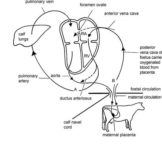
Figure 2.13.
Blood circulation in the cow, showing the attachments of the placenta. Pathways indicated by the broken line apply to the developing calf only, although sometimes the foramen ovale (RAto LA) fails to close at birth.Heart Defects
Figure 2.13 shows the normal circulation of blood in the body. The heart is divided into four compartments, two atria and two ventricles. In the adult animal the left ventricle (LV) pumps blood around the body and back into the right atrium (RA). The right atrium drains into the right ventricle (RV) which then pumps blood through the lungs. From the lungs the blood returns into the left atrium (LA) and then into the left ventricle, where the whole circulation starts again.
The developing calf in the uterus has two modifications to this, shown as dotted lines in Figure 2.13:
• From its body there is a direct flow through its navel to the placenta of its mother and back again.
• Because the placenta supplies the calf with oxygen its lungs are not needed and so there is a bypass or ‘shunt' mechanism whereby blood can flow directly from the right to the left atrium (RA to LA). This is called the foramen ovale.
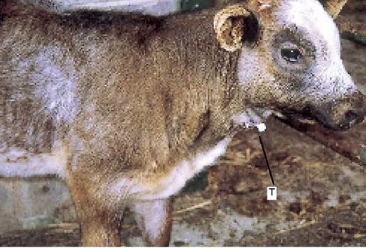
Plate 2.39. Laryngeal diphtheria. This calf had such badly obstructed breathing that a tube (T) had to be inserted into the trachea to allow airflow while the treatment was taking effect. The tube was removed two weeks later.
At the point of birth the foramen ovale should close so that as soon as the calf draws its first breath, all of its blood can be pumped through its lungs to collect oxygen. Unfortunately in some calves the foramen fails to close. Insufficient blood is pumped to their lungs and they are, in effect, short of oxygen. Such calves will appear to be short of breath and they will pant and have a racing pulse after relatively mild exercise. They will also be much more susceptible to pneumonia. There is no specific treatment, although heart stimulants and antibiotics to prevent or treat the pneumonia will help. In some calves the foramen slowly closes and by three to six weeks old they will have fully recovered. Others remain permanently affected and become so stunted that they have to be put down.
Similar syndromes are caused by other heart defects; for instance occasionally there is an interseptal ventricular defect (a connection from LV to RV) or a patent ductus arteriosus (a blood vessel connecting the aorta (A) to the pulmonary artery (B).
More on the topic OTHER CALF DISEASES:
- DISEASES OF THE CALF
- CALF PNEUMONIA
- This chapter deals with the health of the calf from birth to weaning, that is until approximately six weeks old.
- Chapter 3 THE WEANED CALF
- Chapter 2 THE YOUNG CALF
- The various cardiovascular diseases observed in HIV-infected patients and widely described in the literature have been predominantly coronary and peripheral arterial diseases (PAD) and remain poorly known.
- RESPIRATORY DISEASES
- BIBLIOGRAPHY FOR NONINFECTIOUS DISEASES
- INTERSTITIAL LUNG DISEASES
- THE CLOSTRIDIAL DISEASES
- WHY STUDY WILDLIFE DISEASES
- VECTOR-BORNE VIRAL DISEASES
- OTHER CLOSTRIDIAL DISEASES IN WILDLIFE
- BIBLIOGRAPHY FOR PARASITIC DISEASES
- Bibliography for parasitic diseases
- BIBLIOGRAPHY FOR PARASITIC DISEASES