NAVEL PROBLEMS
Navel Structure
During pregnancy the navel is the calf’s lifeline, supplying it with nutrients from the placenta and removing its waste products. The navel is a complex structure, as shown in Figure 2.12.
Plate 2.26 is the post-mortem appearance of a calf which died as a result of navel ill. Fresh blood (which carries food and oxygen) enters from the placenta via the two umbilical arteries (A) which feed into the calf’s aorta, the main blood vessel running along its back under its spine. The blood then passes around the body and eventually reaches the liver. ‘Used’ blood (low in oxygen and carrying waste products) exits from the calf’s liver and back out into the placenta via a single umbilical vein (V). The calf produces urine and this is passed back to the placenta via a single tube, the urachus, which exits from the tip of the bladder. The whole structure (two umbilical arteries, one vein and the urachus) is covered by a layer of peri-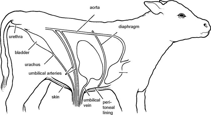
Figure 2.12. The structure of the navel
toneum which becomes continuous with the placenta. There is a hole in the muscle and skin of the body wall (the umbilical ring) to allow unrestricted passage of the navel cord.
At birth the navel cord is the only external part of the animal not covered by a protective layer of skin. It is therefore very susceptible to infection, especially when damp, since bacteria much prefer moist conditions.
Navel Ill
Navel ill is generally seen in calves in their first week of life and, despite very simple control measures that are effective, it is still an extremely common condition. In the early stages the calf may show no generalised signs of illness, but the navel cord feels enlarged and painful and the tip is generally moist and has a purulent smell.
Plate 2.27 shows a typical example.
The calf’s temperature will be raised. More advanced cases will be dull, reluctant to move and the pain makes them stand with an arched back. In most cases the infection is localised at the tip of the cord and generally responds well to treatment. However, pus may track up any of the internal structures shown in Figure 2.12 and particularly along the umbilical vein. This is because the arteries have strong muscular walls which contract and expel any remaining blood, whereas residual blood may be left pooling in the vein. Internal abscesses are therefore more common in the umbilical vein and they may even track up into the liver. The calf in Plate 2.28 was four weeks old when it was found dead one morning, having been off-colour for only two days. Note the large abscess in the navel cord tracking up towards the liver. Infection may also track up along the urachus and into the bladder, where it causes cystitis.Treatment Daily antibiotic injections should be given for several days, depending on the severity of the condition, and it is useful to keep the moist end of the cord bathed in warm dilute antiseptic to encourage pus to drain. Sometimes a persistent navel discharge continues. In these cases there is probably a deeper internal abscess which needs to be flushed out. Introduce a catheter through the discharging sinus (as in Plate 2.29) and gently syringe in 5-10 ml of dilute antiseptic solution. This will either run back out again on its own or it may have to be sucked out with the syringe.
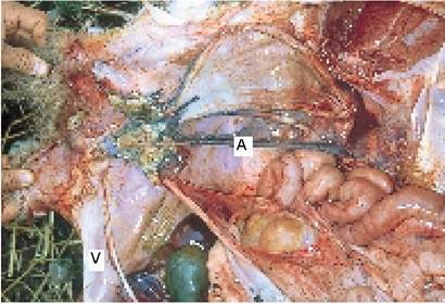
Plate 2.26. Navel III at post-mortem, showing the umbilical artery (A) and vein (V). A matchstick has been placed at the exit of the bladder into the urachus.
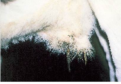
Plate 2.27. Atypical case of navel ill. Note the enlarged fleshy navel with a wet tip, which will be painful and have a purulent smell.
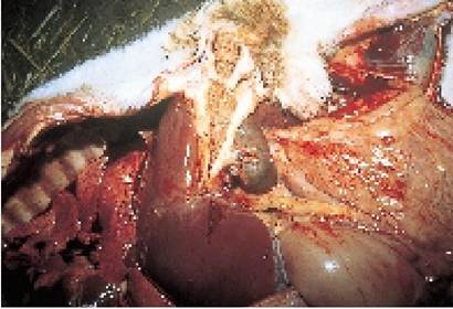
Plate 2.28. Navel abscess extending into the liver from a four-week-old calf which died from navel ill.
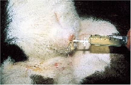
Plate 2.29. Deeper navel infections can be treated by flushing out the internal abscess with a mild antiseptic solution.
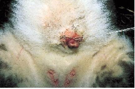
Plate 2.30. Afleshy lump of granulation tissue (proud flesh) protruding from the navel. This needs to be ligated.
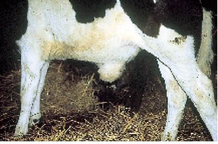
Plate 2.31. A navel (umbilical) hernia is soft and fluctuating and can be gently pushed back into the abdomen.
Repeat daily for four to six days. Do not attempt this procedure when you first see a case of navel ill. Wait four to five days until the internal abscess has been reasonably well walled off. Pumping in fluid too aggressively or too early can lead to internal rupture of the abscess, resulting in peritonitis and death.
Sometimes a red fleshy lump is left protruding from the end of the navel as in Plate 2.30. This should be tied off flush with the skin, using a piece of nylon. Tighten the nylon every three to four days and the lump will fall off, usually in seven to ten days.
Prevention Ensure that cows calve down in a clean environment and immediately after birth thoroughly spray the fleshy navel cord with an antibiotic aerosol, or dip it into iodine. This has two functions. Firstly it dries the cord, making it less attractive to bacteria, and secondly it destroys any bacteria which may have already become established.
It is probably the drying of the cord which is the most important part in the control of navel ill and hence alternative navel dressings, for example copper sulphate crystals, which produce a drying effect, may be beneficial.
Calves with large fleshy navels (more common in beef breeds) are particularly susceptible to navel ill.
Umbilical Hernia (Navel Rupture)
The blood vessels from the placenta pass through a small hole in the skin and muscle of the calf’s abdomen, and this should close at birth. Sometimes the hole in the muscle is larger than necessary however, and, after birth, this allows a length of small intestine to prolapse through and lie between the skin and the muscle, producing a swelling in the navel region (Plate 2.31). The condition is correctly termed a hernia and should be differentiated from a rupture:
• hernia: a prolapse of intestine or some other body organ through a natural opening in the body wall, e.g. a scrotal hernia or umbilical hernia
• rupture: a split in the body wall at an unnatural site, due to injury or excessive strain, and the prolapse of intestine or some other organ through this artificial opening.
An umbilical hernia may not be noticed until the calf is two months old or more, often after weaning, when the abdomen becomes full of solid food.
In the younger calf hernias have to be distinguished from navel ill. The main differences between a hernia and navel ill are:
• A hernia is soft, fluctuating and painless. In a calf with navel ill the swelling is hard and the tip is likely to be damp and foul-smelling.
• A hernia can normally be pushed back into the abdomen manually, or it will disappear when the calf sits upright on its tail.
• With a hernia the calf has no temperature and is not ill.
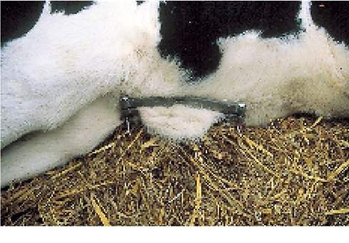
If a hernia is small it will eventually resolve, since the hole in the body wall remains the same size, but the intestine enlarges with age until it is eventually too big to pass through the opening in the body wall. Other cases require surgery. On smaller hernias some veterinary surgeons apply a metal or plastic clamp (Plate 2.32) to the loose fold of skin covering the hernia.
The calf must first be anaesthetised, then rolled on to its back so that its belly is facing upwards. The intestine then falls back into the abdomen and the clamp is applied, making sure that all the loose skin is pulled through. The screws can be tightened every two to three days and the clamp eventually falls off in one to two weeks, during which time the skin has healed at the base. Continuous antibiotic cover should be given to prevent infection. The procedure does not seem to be particularly stressful for the calf, presumably because by the time it has recovered from the anaesthetic the segment of skin protruding through the clamp has lost its nerve supply.Strangulated hernia
This is a rare phenomenon, but is one good reason why large hernias should be treated. Sometimes a segment of
Plate 2.32. A metal hernia clamp.
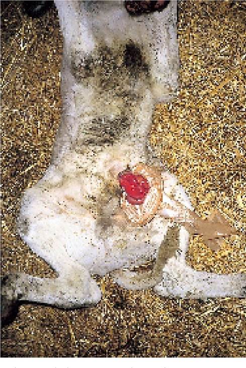
Plate 2.33. Intestines prolapsed through the navel of a newly born calf.
intestine in the hernia sac twists over on itself, leading to a blockage. The blocked intestine dilates, producing enormous pressure and pain inside the hernia, until it eventually ruptures. Death is due to peritonitis.
Intestinal prolapse
On occasions, large quantities of intestine prolapse through the fleshy navel cord immediately after birth and lie exposed on the ground. Plate 2.33 shows a good example. This is a serious condition, but if the calf is operated on promptly it can be saved. Keep the calf warm and still, the intestines clean and covered, and call for veterinary assistance. Sometimes it can be very difficult to decide if a large sac hanging from the navel contains intestines or simply fluid.
Joint Ill
We have already said that infection entering the navel at birth can pass along the cord to the liver and then spread around the body via the blood stream, and this is especially so in colostrum-deficient animals. The bacteria often localise in the joints, and this produces joint ill (Plate 2.34).
Joint ill is seen at a later stage than navel ill, probably at two to four weeks old, and although the infection may have originally entered at the navel, calves with joint ill do not necessarily have an associated navel ill.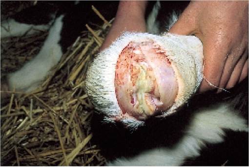
Plate 2.34. Joint ill. When pus has filled the joint, as in this calf, treatment is very difficult.
Plate 2.35. Joint ill. Note the very swollen knee. The calf is not taking weight on this leg.
Some calves which recover from a severe bout of scouring may also develop joint ill. This is caused by bacteria which have leaked into the circulation from a damaged and inflamed intestine. A range of different bacteria may therefore be involved.
The first sign ofjoint ill is likely to be lameness. The calf in Plate 2.35 has an obviously swollen right knee and is not taking its full weight on that leg. If more than one joint is involved, the calf may be seen as generally lethargic and reluctant to move. It will have a high temperature. Later, heat and a fluid swelling appear in one or more joints, and it is the hocks and knees in particular which are commonly affected. Because there is no blood flow into the joints, the condition is difficult to treat. If a case is caught in the early stages, then a prolonged course of antibiotic, for example up to three weeks, may produce a cure.
If a fluid swelling can be palpated around the joint, then an improved response may be obtained by getting your veterinarian to drain pus from the joint and inject antibiotic into the joint space. However, great care with hygiene is needed; otherwise more infection may be introduced, making matters worse.
Once a calf has become totally recumbent from joint ill it is not worth treating. Probably half of the calves developing serious joint ill die. Early treatment is essential. Prevention simply consists of dressing the navel at birth, as described for navel ill, and ensuring adequate colostrum intake.
More on the topic NAVEL PROBLEMS:
- Behavioral Problems
- Eye Problems
- Problems of the S-view
- Environmental problems
- Gynecological Problems
- Conclusions: Some Problems of Classification
- MINOR PROBLEMS IN NEWBORNS
- Problems in practice
- Specific periods and problems
- Skin and mouth problems
- Problems of the Digestive System
- Head and Nerve Problems
- Management of IDU-related problems
- Problems of Intensionality
- Problems for Hobbes
- Lung Problems