Pneumonia Virus of Mice Infection
Pneumonia virus of mice (PVM) was originally discovered following serial blind lung passages in mice, which ultimately produced an agent capable of causing pneumonia. Under natural conditions, PVM is relatively innocuous in immunocompetent mice, but it has increased in significance with the advent of immunodeficient mice, in which it causes significant disease.
Epizootiology and Pathogenesis
PVM is a highly labile virus with a low degree of contagion, requiring close contact between mice. PVM infection occurs in laboratory rodents throughout the world, but its prevalence is declining. Clinical disease caused by natural infections with PVM has never been reported, except in immunodeficient mice. Among BALB/cBy, DBA/2, 129Sv, SJL, C3H/HeN, and B6 mice that were experimentally inoculated with a pathogenic strain of PVM, SJL mice were highly resistant, BALB/c and B6 mice displayed intermediate susceptibility, and DBA/2, C3H/ HeN, and 129Sv mice were the most susceptible, based upon virus titer and pulmonary disease. Intranasal inoculation of immunocompetent BALB/c mice with a pathogenic PVM strain results in clinical illness with nonsuppurative perivascular and interstitial inflammation as well as bronchiolar desquamation and inflammation, which peak within 2 weeks and undergo resolution within 3 weeks. Virus replication occurs in alveolar lining cells, possibly alveolar macrophages, and to a lesser extent in bronchiolar epithelium. Natural isolates of PVM are typically nonpathogenic. Replication is restricted largely to the nasal mucosal epithelium, with minimal pathology. The low pathogenicity of PVM allows immunodeficient mice to develop progressively severe interstitial pneumonia with wasting syndrome without succumbing acutely. In these mice, PVM antigen is confined to alveolar type 2 cells and occasionally bronchiolar epithelial cells.
SCID mice that were naturally infected with P. murina, then inoculated with a normally nonpathogenic isolate of PVM, developed more severe Pneumocystis pneumonia and higher Pneumocystis cyst counts, whereas PVM-infected, but Pneumo- cystis-free, SCID mice survived for 2 months despite high PVM titers in lung. Thus, PVM-related pneumonia in immunodeficient mice is often complicated by Pneumocystis, and vice versa, since both may be common agents in some mouse colonies.Pathology
Clinical signs of disease and gross lesions are absent in natural PVM infections of immunocompetent mice. Microscopic lesions in intranasally inoculated experimental mice consist of mild necrotizing rhinitis, necrotizing bronchiolitis, and nonsuppurative interstitial pneumonia. Infiltrating leukocytes include neutrophils, but lymphocytes and macrophages predominate. Residual perivascular infiltrates of lymphocytes and plasma cells can persist for several weeks after virus is cleared. Immunodeficient mice manifest chronic wasting with cyanosis and dyspnea. Lungs are pale, fleshy, and firm and do not collapse. Microscopically, alveolar septa are thickened with edema and infiltrating macrophages and leukocytes, and alveolar spaces are collapsed and filled with fibrin, blood, macrophages, and large polygonal mononuclear cells, representing detached alveolar type 2 cells (Fig. 1.34). In sections stained by immunohistochemistry, viral antigen can be demonstrated within infected pneumocytes.
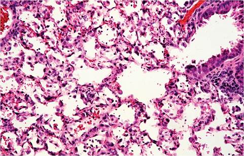
FIG. 1.34. Progressive interstitial pneumonia in a SCID mouse infected with pneumonia virus of mice (PVM).
Diagnosis
Most PVM infections are detected retrospectively by seroconversion. Because PVM is not highly contagious, the number of seropositive mice within a colony can be small. Seropositive mice often have mild pulmonary perivascular lymphoplasmacytic infiltrates. Differential diagnoses for pulmonary disease and wasting syndrome in immunodeficient mice include Sendai virus and Pneumocystis murina infections, which also cause progressive pulmonary disease. PVM lesions are similar to Sendai viral lesions microscopically, but PVM tends not to induce bronchiolar hypertrophy like Sendai virus. During active infection, PVM can be identified by immunohistochemistry, virus isolation, PCR, or MAP testing of suspect tissues. Nude mice do not seroconvert to PVM.