Otoscopy
The conventional otoscope is configured to project a bright light through a conical tube. Objects in the ear canal reflect the light back to the examiner’s eye. A large magnifying lens at the eyepiece focuses on the end of the otoscope cone and aids in enlarging the image.
For procedures using an otoscope, an “operating” head is available with a very small lens at the eyepiece. This allows the examiner to place small instruments through the cone without having to move the lens. The disadvantages of this system are that (1) the light may not be strong enough to transilluminate the eardrum, and (2) when instruments are used, the view of the examiner is often obliterated.The Video Otoscope
A video-based otoscope (MedRx Video Vetscope, MedRx, Inc., Largo, Florida) has been designed to overcome the shortcomings of the hand-held otoscope. This is a very useful instrument for examining, cleaning, and drying the ear canal because it gives the examiner a clear real-time image on a video monitor (Figure 2-10). The high-quality glass lens pack of the Video Vetscope probe begins with a 2-mm lens at the tip of the 4.75-mm diameter probe. This lens configuration provides a 110-degree angle of view and an infinite focus to the image ahead of the probe tip. Even in small patients in which the ear canal diameter is smaller than the tip of the probe, the wide
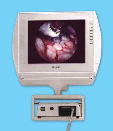
Figure 2-10
Video otoscopy allows the client and veterinarian to view the patient's ear canal on a video monitor. (Courtesy MedRx, Inc., Largo, Florida.)
viewing angle and the long focal length of this instrument allow examination of the ear canal. Light fibers within the tip of the probe are connected to a powerful halogen light source, providing excellent illumination. A 2-mm working channel is built into the Vetscope probe, separate from the lens system.
Instruments can be placed through the working channel without obscuring the view of the procedure. The entire instrument is attached to a miniature color video camera (Figure 2-11), which is connected to a video monitor and a video printer.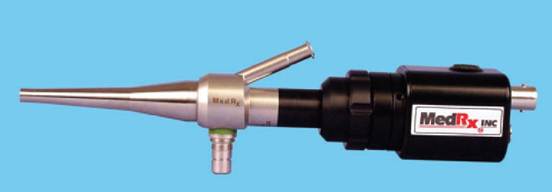
Figure 2-11
Video Vetscope probe attached to a miniature video camera. (Courtesy MedRx, Inc., Largo, Florida.)
A
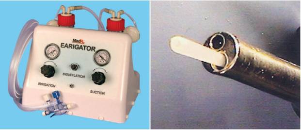
Figure 2-12
MedRx Earigator with independent flushing and suction. A, The free end of the trumpet valves connects to the flared end of a catheter. B, The catheter is extended through the 2-mm working channel. (Courtesy MedRx, Inc., Largo, Florida.)
For flushing and suctioning of the ear canals, a 5 Fr feeding tube or a polypropylene urethral catheter can be inserted through the video otoscope’s working channel. Use of an irrigation/suction machine (Earigator, MedRx, Inc., Largo, Florida) (Figure 2-12) makes this procedure very efficient. The catheter is inserted into the working channel with the otoscope positioned in the ear canal. The catheter can be advanced into the ear canal with clear visualization (Figure 2-13). Small pieces of epithelium, wax, hairs, and pus can be flushed away from the ear canal epithelium and then suctioned out of the canal while the examiner views the procedure on the video monitor. In this manner, even the smallest pieces of detritus can be removed. The examiner can extract large pieces of debris and concretions of wax and medications from the ear canal under visualization with the Video Vetscope by using a grasping type of endoscopic forceps inserted through the 2-mm working channel or an ear curette (Figure 2-14).
The video otoscope also has documentation capability. When a lesion is encountered, the image on the video monitor can be “frozen” and then printed out as a 4- ? 5-inch color glossy photograph with the use of a video printer.
Thus, there is a photographic hard copy document of the ear disease to place in the medical record for comparison on rechecks. These photographs are also helpful in counseling owners and showing them the severity of the ear disease; they make the veterinarian’s medical or surgical recommendations more valid. Documentation capability is very useful in a referral practice. The referring veterinarian can receive a color photograph of the ear canal in addition to the written report. The photographs can also be used to illustrate contributions to veterinary medical literature. The video otoscope can be coupled to a videocassette recorder so that the examination and surgical procedures can be videotaped and kept for documentation. A computer-based database system is also available to catalog and store video examination records as both digital photos and 30-second digital video clips (Figure 2-15).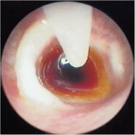
Figure 2-13
The open-ended tomcat catheter is advanced through the 2-mm channel of the Video Vetscope toward the flush solution. Advancing the catheter allows visualization of the suctioning process. Always keep the end of the catheter in view to prevent iatrogenic myringotomy.
After cleaning and examining the ear canal, the examiner can gain a better understanding of the ear disease and better formulate therapeutic protocols.