Physical Injury
Many of the cattle ailments we have to deal with are the result of physical trauma. The best example is lameness. Hoof and limb disorders are frequently associated with either trauma to the foot, for example, prolonged standing on a hard surface producing sole ulcers, or damage to the leg, for example resulting in dislocation of the joint or even fracture of a bone.
Examples of other physical injuries include:
• teat damage
• haematomas (blood blisters under the skin)
• skin cuts
• foreign bodies, e.g. barley awn or grass seed in the eye
• burns, either caused by fire or sunburn
• chemical injury, e.g. tank cleaner mistakenly used for teat dip
• abscesses (following penetration of the skin by thorns, nails, fragments of metal etc.)
Congenital Disorders
Congenital diseases are abnormalities which are present at birth. They may be caused by genetic factors or by ingestion of teratogens during pregnancy.
Genetic defects
The incidence of inherited genetic defects in cattle is said to be quite high (one in 500 births), but as at least half of the calves are stillborn, they do not represent a major problem to the cattle industry. Typical examples include cleft palate (Plate 1.2), harelip (Plate 1.3), spina bifida (Plate 1.4), a very small tail or no tail at all (hypoplastic tail, Plate 1.5), contracted tendons (Plate 1.6), hydrocephalus, brachygnathia (parrot mouth, Plate 1.7) and umbilical hernias. If it is found that a particular bull is throwing a high incidence of calves with genetic defects, he should be culled. Sometimes the defect is only seen when a bull mates with a particular cow. In this case the defect is said to be caused by a recessive gene, in that the defect will only appear if both the sire and dam are carrying the gene for that defect.
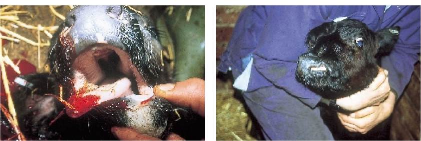
Plate 1.2.
Cleft palate. This calf could not even suckle and was destroyed. Where the defect in the hard palate is smaller, the cow's teat may cover the hole during suckling and it is only when the calf starts eating solid food, or drinking from a bucket, that a severe nasal discharge indicates that something is wrong.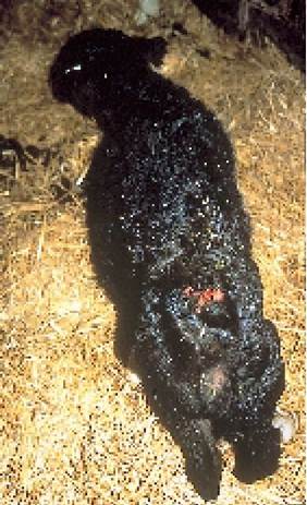
Plate 1.4. Spina bifida. The tops of two lumbar vertebrae have not closed, leaving a hole into the lumbar spine, seen here as a red area. This calf was partially paralysed in the hind legs.
Plate 1.3. Harelip (also called cleft lip or primary cleft palate). This can also make suckling difficult, although this calf was able to drink from a bucket.
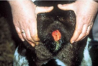
Plate 1.5. No anus and minimal tail (anal atresia and coccygeal hypoplasia). Calves with a totally blind anus rapidly develop abdominal distension, severe pain and colic. This calf was lucky and faeces were passed through the vagina. No tail or a shortened tail is commonly seen on its own without anal atresia.
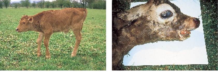
Plate 1.6. Contracted tendons at the fetlock, as in this calf, are common, particularly in larger calves. They will correct in time without treatment.
Plate 1.7. Brachygnathia (parrot mouth or overshot upper jaw) is most commonly seen in stillborn calves.
Teratogenic defects
Teratogenic defects result from the ingestion of toxic agents during pregnancy. The most publicised example of this must be the effect of thalidomide in man, which resulted in the birth of deformed children. In cattle, ingestion of some species of the plant Lupinus can produce crooked calf disease, where calves are born with malpositioned legs, either excessively flexed or excessively extended. Oral dosing of early pregnant cattle with the ringworm treatment griseofulvin should be avoided, because of the risk of producing deformed, full-term calves.
Viral and other infections can also produce congenital defects. The best examples are BVD virus and Neospora caninum. Infection of mid to late pregnant cows with either agent can result in defects such as cerebellar hypoplasia. In this condition the part of the brain known as the cerebellum is far too small (hypoplasia, Plate 1.9) and the affected calf is unable to stand. The calf in Plate 1.8 is a typical example. At birth it was unable to stand or suckle and when lifted it pushed its head back over its back (opisthotonos). Mid pregnancy infection with Akabane virus or BVD can cause arthrogryphosis (Plate 1.10), a condition in which the hind limbs become fused or deformed.
Not all congenital abnormalities are immediately apparent at birth. Strabismus is a good example. Plate 1.11 shows a Hereford heifer with bilateral convergent strabismus,
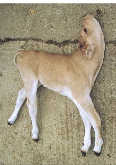
Plate 1.8. Cerebellar hypoplasia. Although this calf appeared normal and healthy at rest, as soon as it tried to feed or stand it fell over and its head went into spasm over its back (opisthotonos). BVD and Neospora are possible causes.
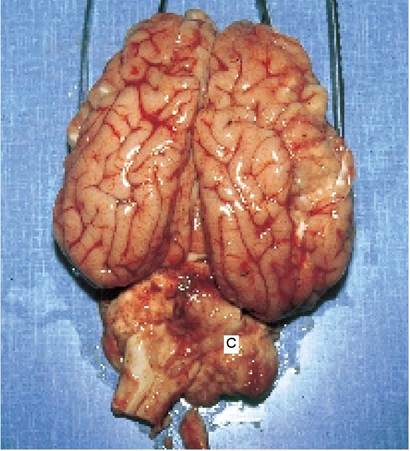
Plate 1.9. Cerebellar hypoplasia. The cerebellum (C) is the part of the brain which controls balance.
that is both eyes (bilateral) point inwards (convergent) with a squint (strabismus). This condition gets progressively worse as the calf gets older and in some instances results in almost total blindness. In one unfortunate incident I dealt with, a dairy farmer purchased a freshly calved heifer with strabismus. A few weeks after purchase she had a bad fright and ran off. Because she could not see very well she became totally disoriented and finished up by drowning in the slurry pit. Fortunately, relatively few congenital defects have such a dramatic ending!
Other congenital defects causing blindness include cataracts (Plate 1.12) and microphthalmia or anophthalmia (very small or no eyes, Plate 1.13).
Cataracts are an opacity of the lens. They can be hereditary or caused by BVD infection of the dam during late pregnancy. Most cataracts are left untreated and although calves seem to manage with very limited or zero vision, treatment is possible. A very fine knife is inserted between the cornea and sclera (the clear and white parts of the eye) and one or two cuts are made across the front of the lens. The aqueous humour (the liquid in the front part of the eyeball) then slowly dissolves away the lens until sight has been restored.See Chapter 4 for other eye disorders.
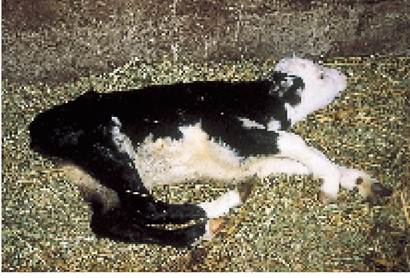
Plate 1.10. Arthrogryposis. The hind legs are fixed in this extreme flexion position and cannot be moved. Often the pelvis is also involved (when a caesarean birth is necessary). This calf also had spina bifida. Arthrogryposis can be either teratogenic in origin or inherited.
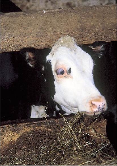
Plate 1.11. Strabismus (squint). Note how the eyeball is protruding and pointing in towards the nose. Both eyes were affected. This is a progressive condition which can eventually lead to blindness.
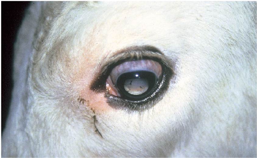
Plate 1.12. Acataract is an opacity of the lens. The centre of the eye has a blue appearance, as in this calf.
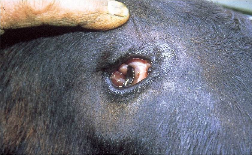
Plate 1.13. Microphthalmia. This calf was born with the eye almost totally missing.
DEFENCES AGAINST DISEASE
Animals (and also man!) are continually exposed to a range of infectious agents and physical factors which could potentially cause disease, but fortunately disease occurs relatively rarely. This is because we all have a range of excellent defence mechanisms which afford a degree of protection against moderate challenge.
These defences will be outlined briefly in the following section and can be subdivided into:• Physical barriers
- skin
- respiratory passages
- digestive tract
- eye mechanisms
- commensal bacteria
• Chemical barriers
- acid in the stomach
- alkaline in the intestine
• Immunity
The body has an excellent ability to recognise materials which are ‘foreign’, that is materials which are not part of itself, and to control them. At the same time it has to recognise those tissues which are part of itself and leave them alone. The mechanism for dealing with ‘foreign invaders’ or ‘non-self’ materials has two components, namely:
- cellular mechanisms: certain cells are able to recognise and engulf infectious and other foreign agents
- humoral mechanisms: a system of ‘active’ proteins, most commonly known as antibodies, assist in the detection and destruction of non-self tissue
In addition, there are two categories of both cellular and humoral defence systems:
- innate systems: these exist in all animals and do not rely on previous exposure to an infection
- induced or acquired systems: these come into play after an animal has been exposed to an infection.
Following exposure, the precise characteristics of the invading organism are remembered and the body produces specific defences against it, using both cells and antibodies. These are then ready to attack the invader if it manages to gain entry into the body for a second time. This is the nature of vaccines. They give the unexposed animal a mild or dead form of the infection. The animal then manufactures huge quantities of specific defence materials (both antibodies and cells) and is able to repel an invasion by that infection. Different vaccines are needed for each disease.
More on the topic Physical Injury:
- Children and adolescents with spinal cord injury (SCI) must deal with the multisystem involvement imposed by the injury that is compounded by physical and psychological growth and development, which cause complications not seen in the adult.
- Werner Reiss, author of the most detailed recent discussion on the subject of violence in the Greek world, defined violence as ‘a physical act', stating further that it is a ‘process in which a human being inflicts harm on another human being via physical strength’.1
- 3. Injury (Iniuria)
- Cause of Injury
- Spinal Cord Injury
- INJURY SEVERITY
- ACUTE KIDNEY INJURY
- Drug-Induced Liver Injury
- Morbidity Related to Age at Time of Injury
- Immersion Injury (Trench Foot)
- Childhood-Onset Spinal Cord Injury
- Level of Injury-ASIA Impairment Scale
- Costs of Injury